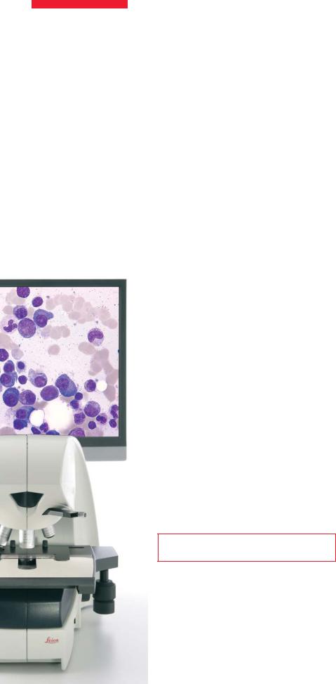
Leica DMD108 Manual

A P P L I C AT I O N R E P O R T
Leica DMD108 in Remote Breast Cancer Diagnosis
Digital Microscopy
Proves its Value
Anja Schué, Leica Microsystems
When the surgeon in Varberg Hospital in the South of Sweden takes a tissue sample from a patient’s lymph node before a breast operation, a frozen section is immediately produced for clinical diagnosis. But the pathology unit that makes the diagnosis is 70 kilometres away, in Halmstad County Hospital. Nevertheless, the pathologist there has the high resolution microscopic image before his eyes immediately, as soon as the specimen preparation is complete.
That this is possible today can be attributed first and foremost to an innovative technology from Leica Microsystems. The Leica DMD108 digital microscope was integrated into the videoconferencing system of the two hospitals for that purpose in 2007. Dr. Tomas Seidal, Head of the Pathology and Cytology Department at Halmstad County Hospital, talks about his experiences with the Leica DMD108.
Dr. Seidal, how successful has the introduction of digital microscopy been?
In our breast cancer diagnosis, the Leica DMD108 has proved excellent. We are extremely satisfied with the system. The remote diagnosis saves us from losing valuable time transporting samples. Our pathologists are very pleased with the quality of image resolution and colour, and also with the easy, user-friendly handling. And I would like to stress that Leica Microsystems provided excellent support for the installation of the Leica DMD108 equipment and during the start-up phase.
“The Leica DMD108 is at the forefront of digital microscopy techniques by now.”
The Leica DMD108 is also a great help to us in our conferences with surgeons, pathologists and oncologists. I well remember how surprised and impressed all those involved were the first time they saw the excellent quality of the microscope image on the monitor. Another advantage is that microscope stage movements are immediately visible on screen and easy to follow for the viewer.
In the days when we were still using several microscopes with discussion bridges and someone kept moving the sample extremely quickly by hand, it almost made us seasick to watch.
Is remote microscopic diagnosis here to stay in pathology?
The subject of telepathology has been discussed for many years and is an innovation driver. The reasons for the interest in telepathology are the increasing specialisation of medical centres and also the specialisation of pathology itself – which entails a growing need for a second opinion. Digital microscopy is a significant development in this context. It offers fantastic image quality today and saves time, although it is not used everywhere yet.
Some pathologists are still sceptical and reluctant to trust new technologies. Nevertheless, I think the future belongs to virtual microscopy, even if it takes a few years before it is fully accepted. It can lead to shorter surgery times – not only in cases of breast cancer, but also for other forms of cancer, where biopsies are made during the operation. The digitisation of specimens and the integration of the data in the patients’ files offer tremendous potential for innovations and improvement of work routines.
10 reSOLUTION
 Loading...