


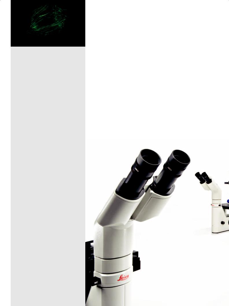
Leica DM IL LED
Brilliant Optics Combined with Innovative Illumination
The New Inverted Microscope for Routine and Laboratory Microscopy
in Cell Biology and Medicine
Compact and stable
•Lean and sturdy design
•Plenty of space for operation
•Low stage height
•Large dimensions and low center of gravity of microscope
•Large working distances
Wide variety of possible applications
•Cell biology and medicine
•Micromanipulation (injection, IVF, ICSI)
•Medicine
•Biotechnology
•Developmental biology
•Transgenics
•Molecular biology
•Fluorescence applications
Inverted Routine
Microscopy in a New Light
Optical performance and illumination are key elements in microscopy. Both characteristics are unified in the new design of the Leica DM IL LED. As the first inverted routine microscope, the Leica DM IL LED is not only equipped with outstanding Leica HC optics, but also features innovative LED illumination. The transmit- ted-light illuminator including optimized condensers and improved contrast methods are adapted specifically for cell biology applications. High stability, plenty of space for operation, large working distances, illumination without heat development and the separately accommodated electronics provide optimum conditions for microscopy. The Leica DM IL LED is exceptionally well-suited for uses ranging from various cell and tissue culture examinations in life sciences, developmental biology studies or micromanipulation in cell biology to living cell examinations in transgenics or electrophysiology.
2
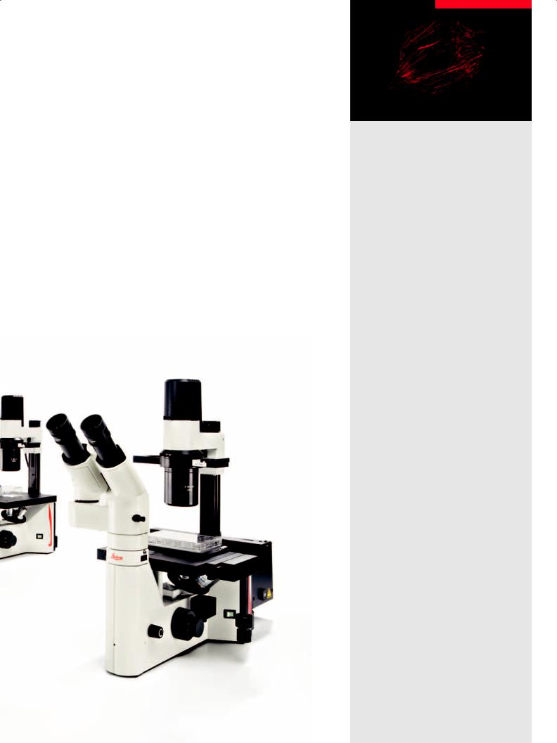
The fluorescence version, the Leica DM IL LED Fluo, also offers a variety of possible applications. Optionally, it is also available with the new LED illumination.
Heatable microscope stages and 3-plate cross-stages provide great flexibility for experiments on living cells under physiological conditions.
The Leica DM IL LED has a further advantage that distinguishes it from other microscopes in its class: The stand is highly compatible with components of the Leica research microscopes. Objectives, eyepieces, tubes, camera ports, contrast methods. Additionally, special tubes and condensers have been developed for the Leica DM IL LED.
Integrated fluorescence
•Manual fluorescence with three filter cubes
•Integrated shutter
•Optionally LED, classic mercury illumination or fiber optic coupling
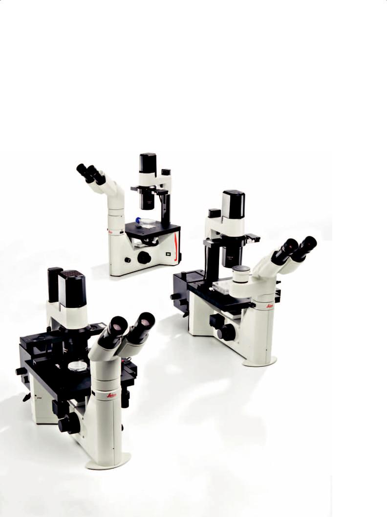
Flexible and modular
•A full range of optical components
•Compatible with research stands
•Unheated and heated stages
•Large selection of tubes
•Comprehensive range of accessories for special applications
3
The Most Comprehensive Array
of Contrast Methods
All available contrast methods can be adapted to individual applications easily and quickly. Two condensers have been developed specifically for the Leica DM IL LED, which can be used for the entire magnification range of the respective contrast method. The high-resolution S40/0.45 condenser makes even tiny details of a specimen visible. Both condensers, the S40/0.45 and S80/0.30, allow for use of phase contrast up to the 63x objective as well as Integrated Modulation Contrast (IMC) up to the 40x objective.
Fatigue-free operation
The ergonomic arrangement of all controls such as the focus dial, brightness controller, condenser height adjustment, objective nosepiece and XY stage adjustment allow users to be relaxed while working with the microscope – even for hours. The heightadjustable stages, Ergo tubes with variable tube height, flexible viewing height, and the interpupillary distance and diopter setting enable each user to configure his or her personal Leica DM IL LED. The large working distances provide sufficient room for large culture flasks, and the unobstructed view of the specimen area facilitates handling more difficult specimens.
4
 Loading...