


Zeiss KINEVO 900 User Manual
KINEVO 900 from ZEISS
Advancing Surgical Certainty

Mastering the complex.
ZEISS KINEVO 900
// INNOVATION
MADE BY ZEISS
KINEVO 900 – The Robotic Visualization System
Just like you, we love challenging the status quo.
The result? Over 100 innovations to perfect the already acclaimed surgical visualization platform. KINEVO® 900 from ZEISS is designed to deliver more functionalities than any surgical microscope today.
ZEISS KINEVO 900 combines digital and optical visualization modalities, offers a unique Micro-Inspection Tool and will impress you with its Surgeon-Controlled Robotics. All to enable you to gain greater certainty in a virtually disruption-free workflow.
Designed to meet real needs. To make a real difference!

A lot more. And, a lot less too.
When treating complex vascular conditions, you typically work at high magnification. Even the slightest vibrations can cause disruptions. And constant manual repositioning to better visualize structures or precisely approach deep-seated lesions can become extremely tedious. Not anymore! ZEISS KINEVO 900 delivers a lot more positioning precision with a lot less effort.
PointLock
|
|
|
|
|
Focus |
Activate |
|
Swivel |
|
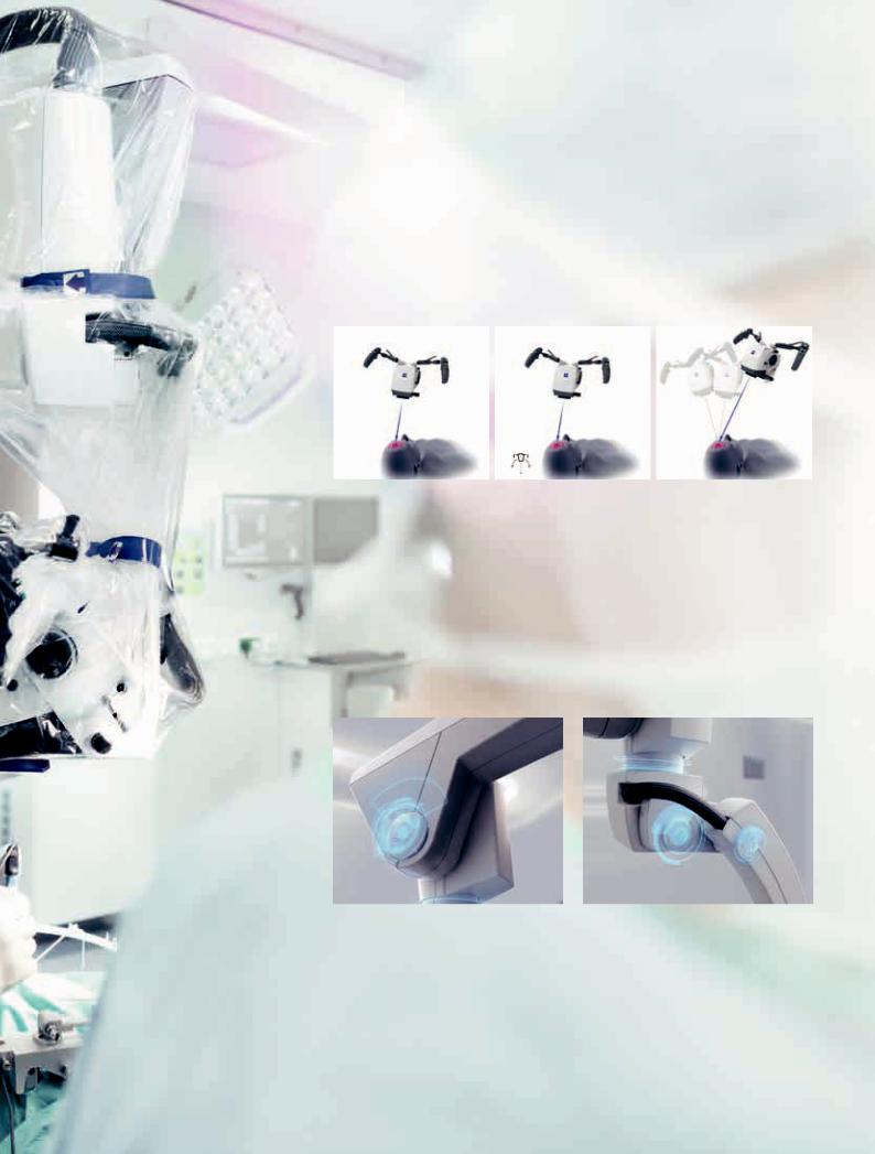
Surgeon-Controlled Robotics adds a complete new level of ease to precise positioning. Imagine being able to focus and move around a structure to visualize the targeted anatomy – reducing any manual hassle. In addition, PointLock enables you to do a KeyHole movement to observe a larger area inside a cavity – a particular benefit in areas with narrow access. Simply put:
Focus. Activate. Swivel.
Active vibration damping
You know the problems that can be created by the tiniest vibrations. The active damping provided by ZEISS KINEVO 900 minimizes collateral system vibrations, ensuring rock-solid stability. Enabling you to completely, and steadily, focus on what matters most: your treatment.
5

When you need it. Where you need it.
The new navigation interface of ZEISS KINEVO 900 is designed to work in concert with your navigation device. When you require precise repositioning to reexamine previously visualized structures or when you need to align with a pre-mapped trajectory, making use of all six axes, the Robotic Visualization System™ delivers precise positioning at the push of a button. Putting you exactly where you need to be – when you need to be there.
PositionMemory
|
|
|
|
|
Save |
Move |
|
Recall |
|
When working on a tumor case, you may already have identified regions of concern where you want to protect the functional structure. After storing these in PositionMemory, you can come back and visualize them at the exact same magnification, working distance and focus – without losing time for manual repositioning. In a nutshell: Save. Move. Recall.
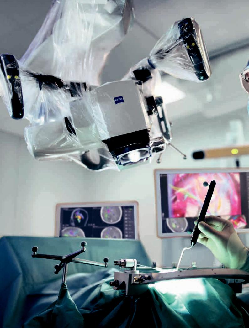
Image-guided surgery
Image with Brainlab Microscope Navigation Software
Minimize time-consuming efforts in approaching challenging neurosurgical pathologies. Combine the Surgeon-Controlled Robotics of ZEISS KINEVO 900 with navigation interface to approach deep-seated pathologies in cranial surgery, brain stem or skull base tumor removals – right when you need it.
7
 Loading...