How it Works
Log In / Sign Up
Buy Points
How it Works
FAQ
Contact Us
Questions and Suggestions
Users
Zeiss
Loading...
#
ΣIGMA VP-FE-SEM
V
Victory 56 T* FL
VICTORY 8x20 T* COMPACT
Victory 8 x 26 T* PRF
2
Victory 8 x 45 T* RF
3
Victory 8x56T*RF
Victory DC4
Victory FL 10x42
Victory FL 10x56 T
Victory FL 8x32
Victory FL 8x42
Victory FL 8x56 T*
Victory Harpia 22-65x85
VICTORY HARPIA 85
VICTORY HARPIA 95
Victory HT
VICTORY HT 10x42
VICTORY HT 10x54
VICTORY HT 8x42
Victory HT 8x54
4
Victory NV 5.6 x 62 T*
Victory PhotoScope 85 T* FL
Victory Pocket
2
Victory Pocket 10x25
2
Victory RF 10x42
Victory RF 10x54
Victory RF 8x42
Victory RF 8x54
Victory SF
Victory SF 8x42
VICTORY V8 1.1-8x30 M
VISU 200
Visualas 532s
Visucard
2
VISULAS 532s
3
Visulas YAG II
VISULAS YAG III
3
VISUPHOR 500
VISUSCREEN 100
VISUSCREEN 500
VR ONE brille
VR ONE headset
VR ONE Plus
3
X
XBO 75
Z
ZEISS VICTORY 10x32 T* FL
ZEISS VICTORY 10x42 T* FL
ZEISS VICTORY 10x56 T* FL
ZEISS VICTORY 7x42 T* FL
ZEISS VICTORY 8x42 T* FL
ZEISS VICTORY 8x56 T* FL
ZEN2.5
ZEN3.1
ZX1
Loading...
Loading...
Nothing found
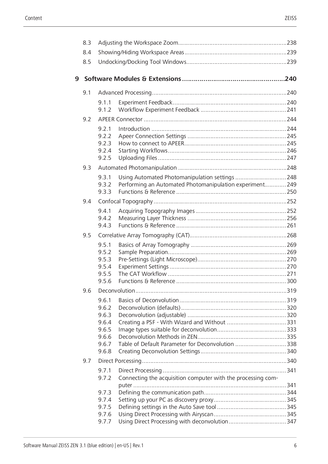
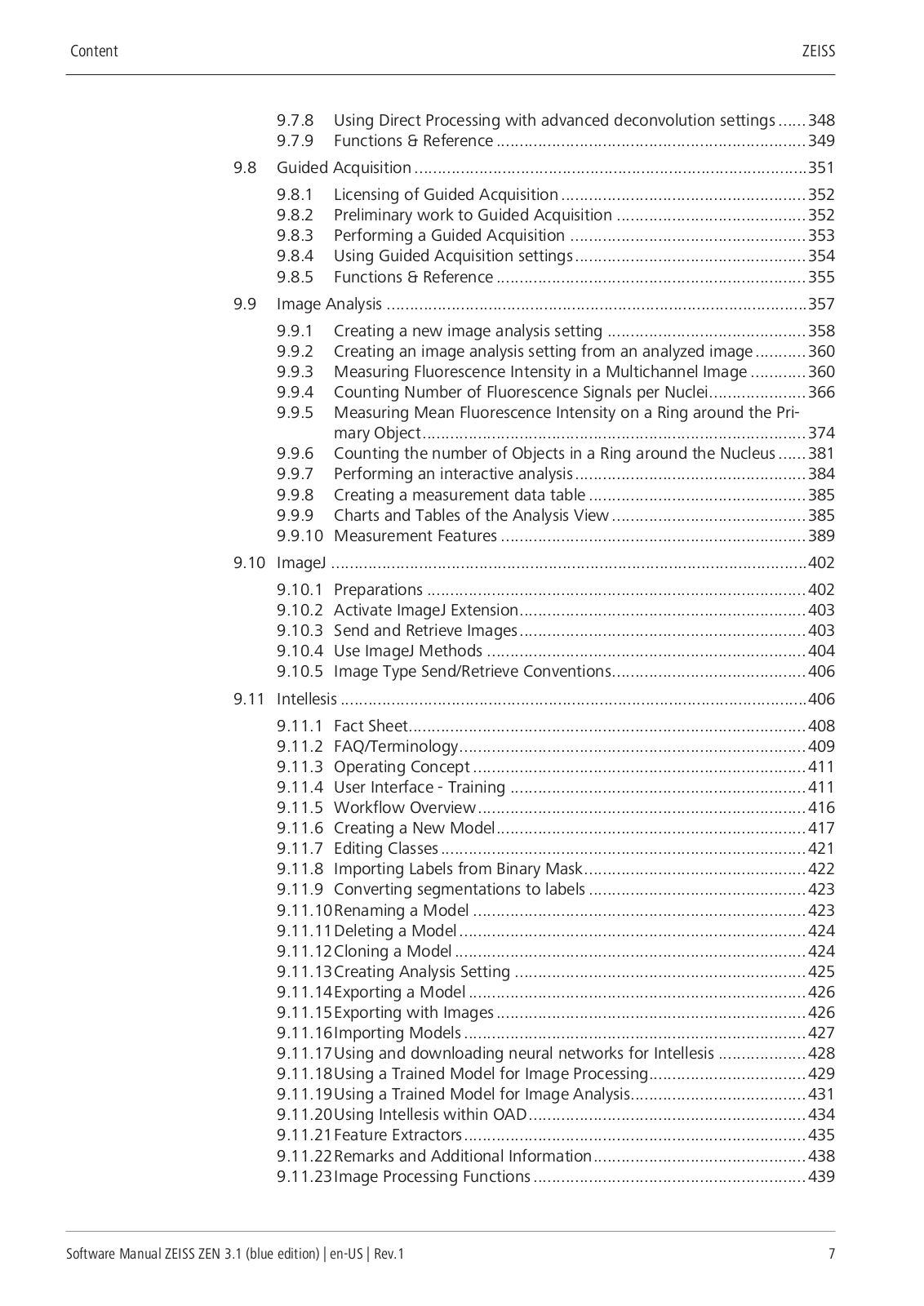
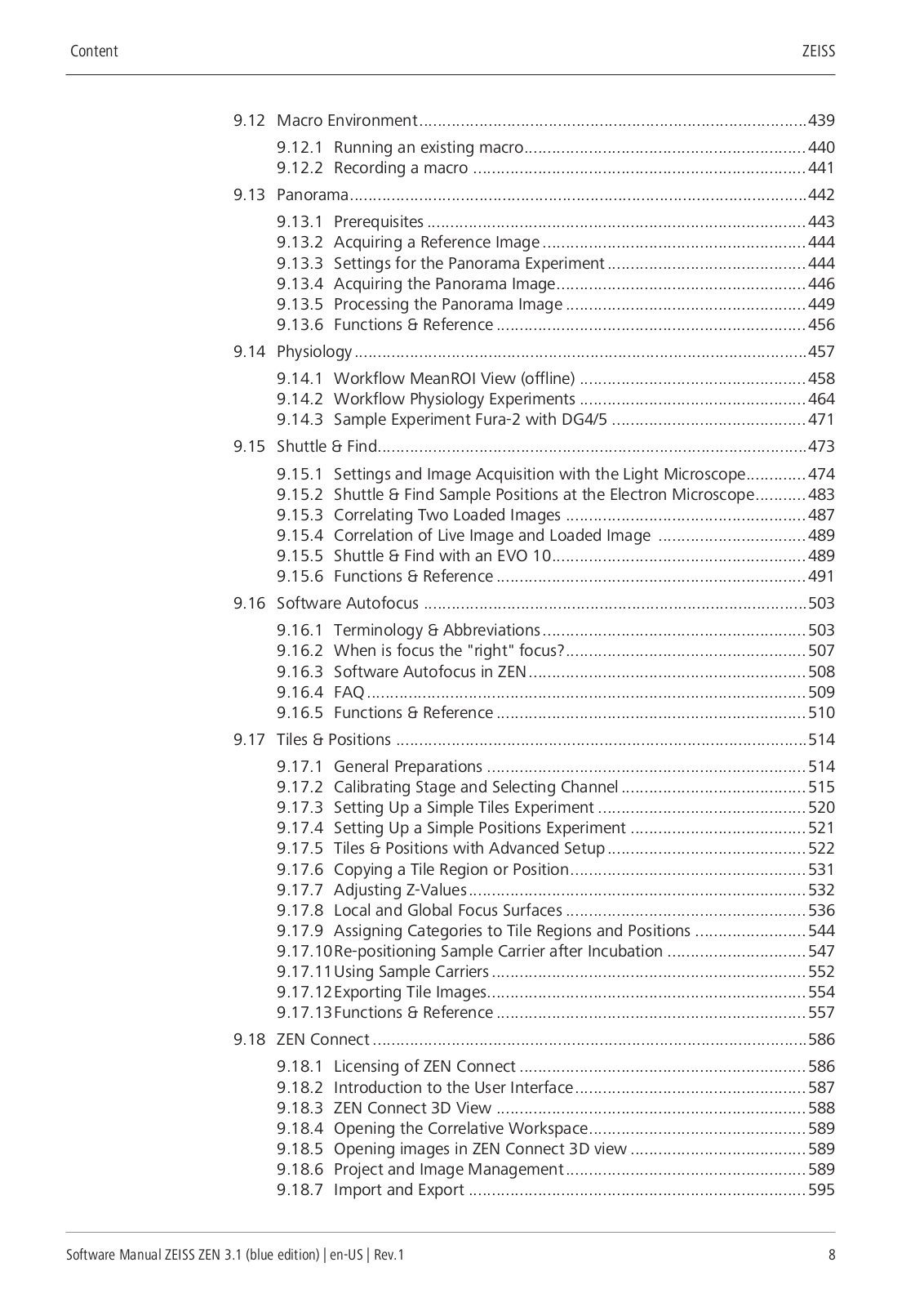
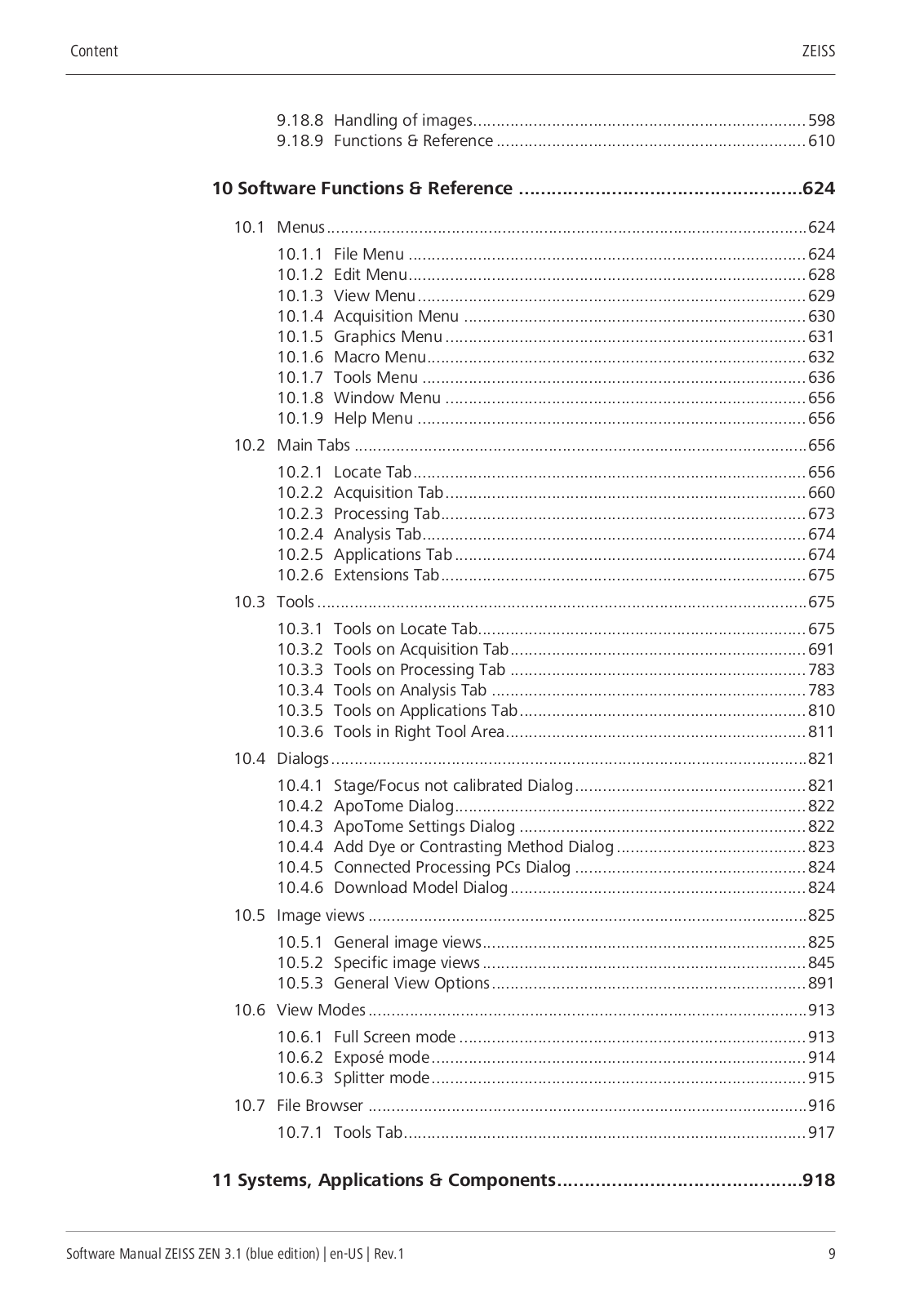
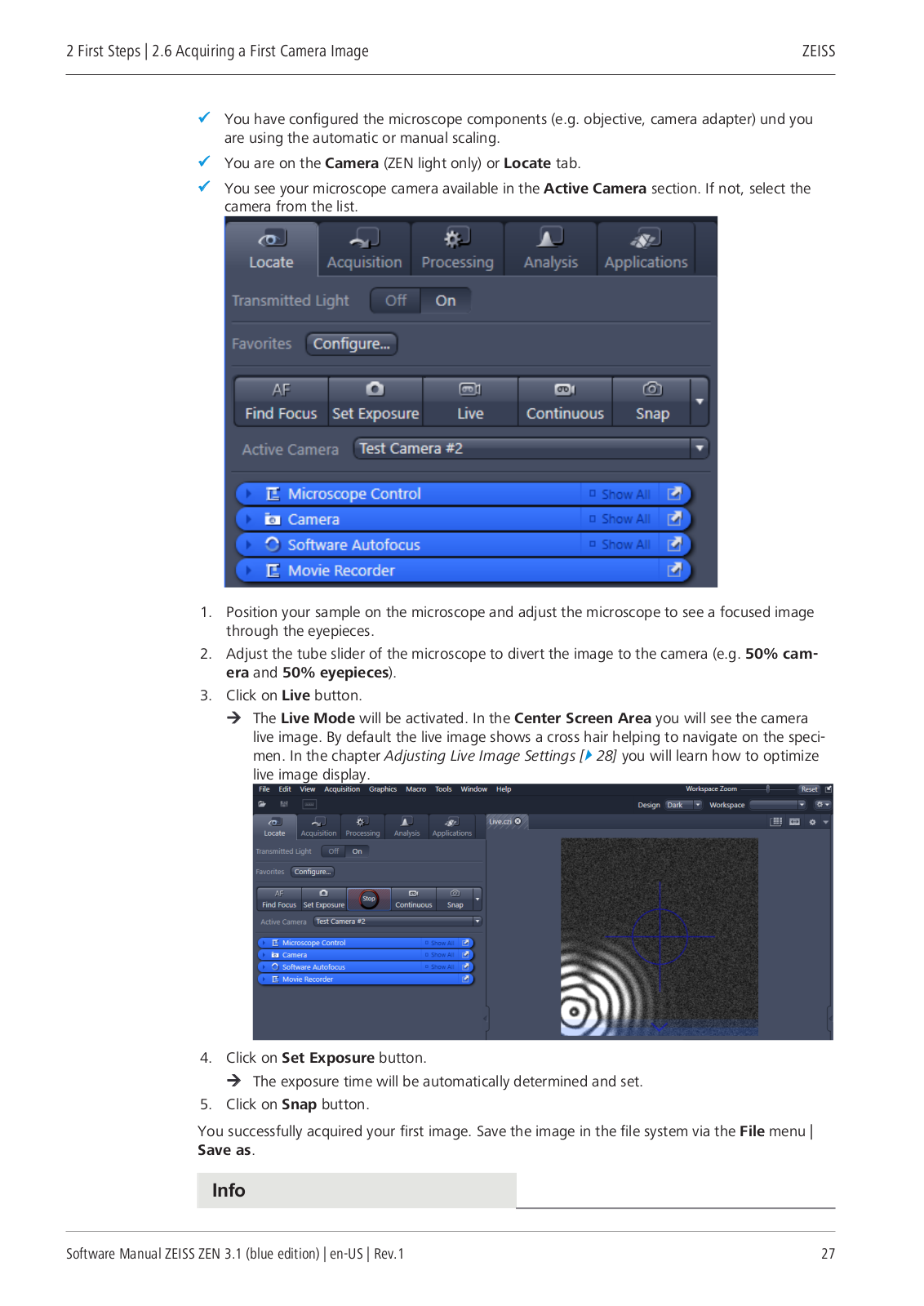
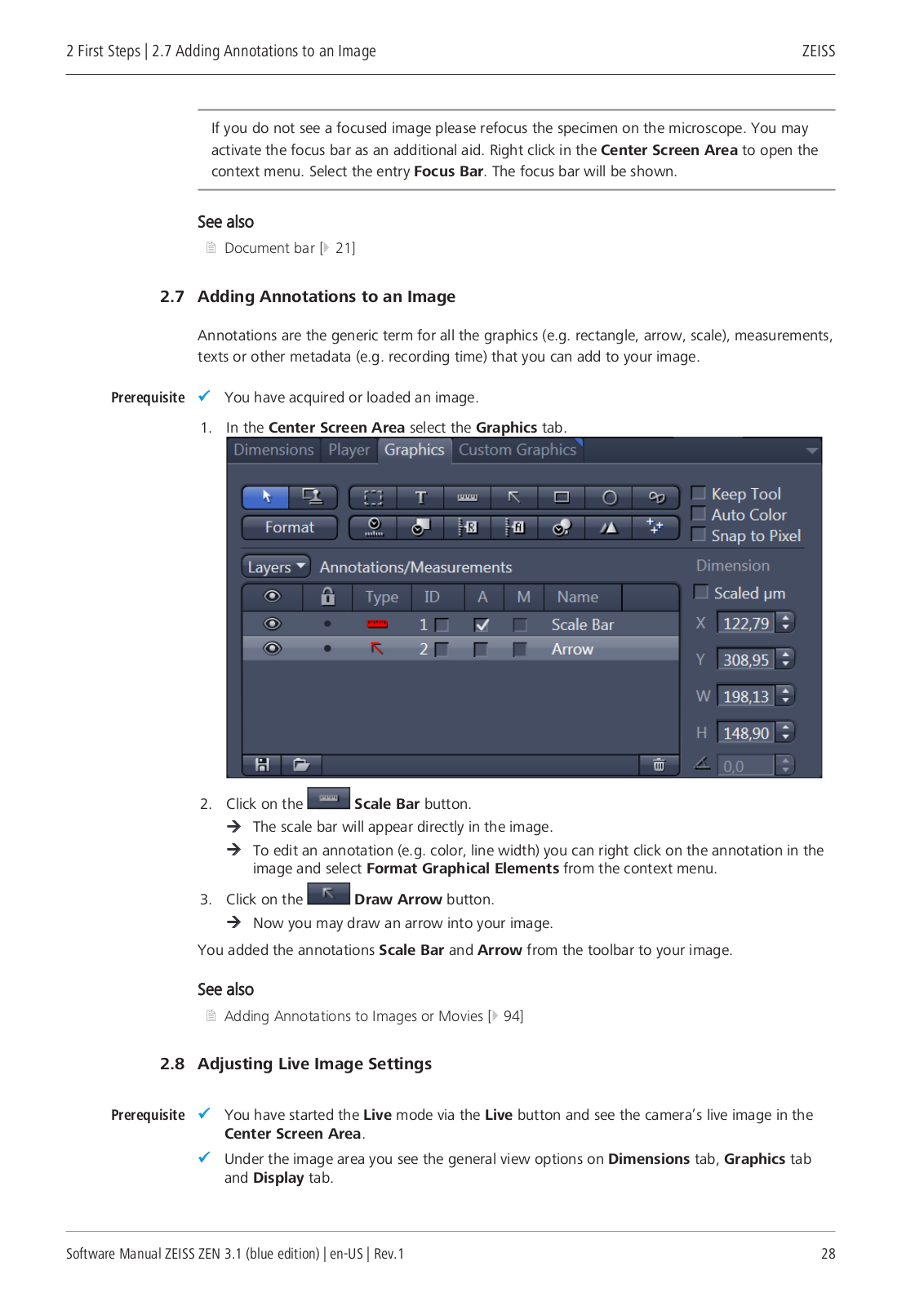
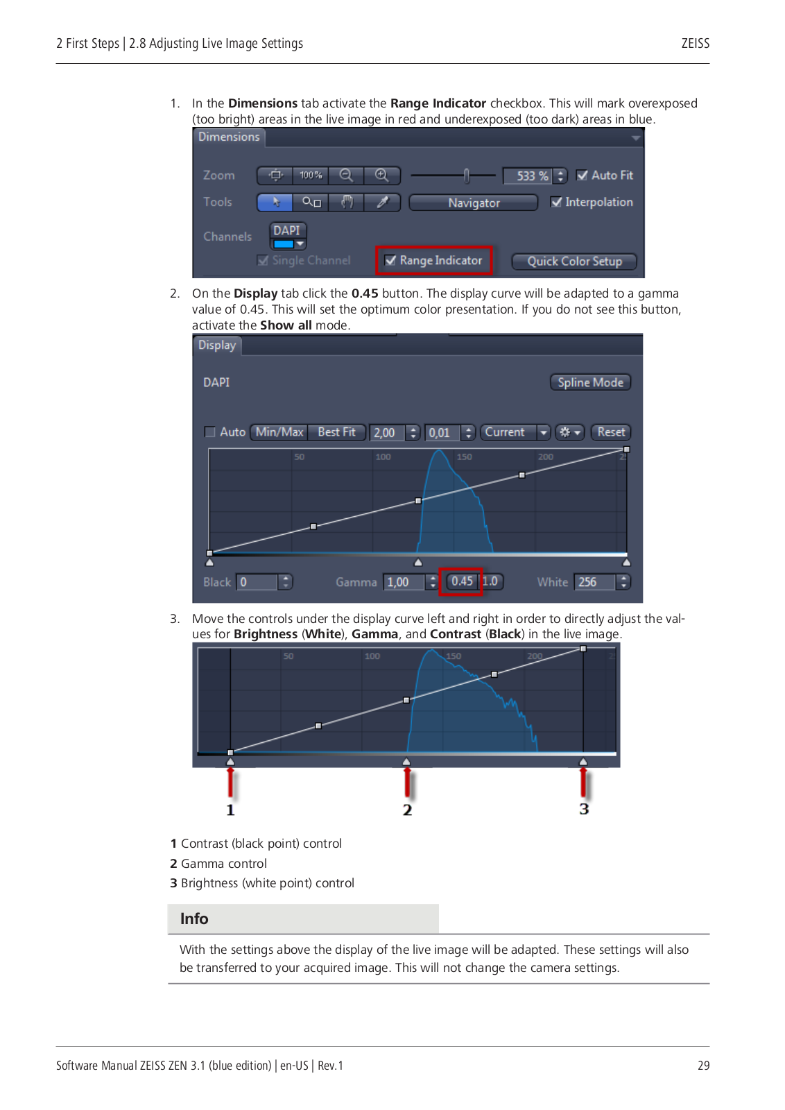
ZEN3.1
User Manual
1124 pgs
109.03 Mb
0
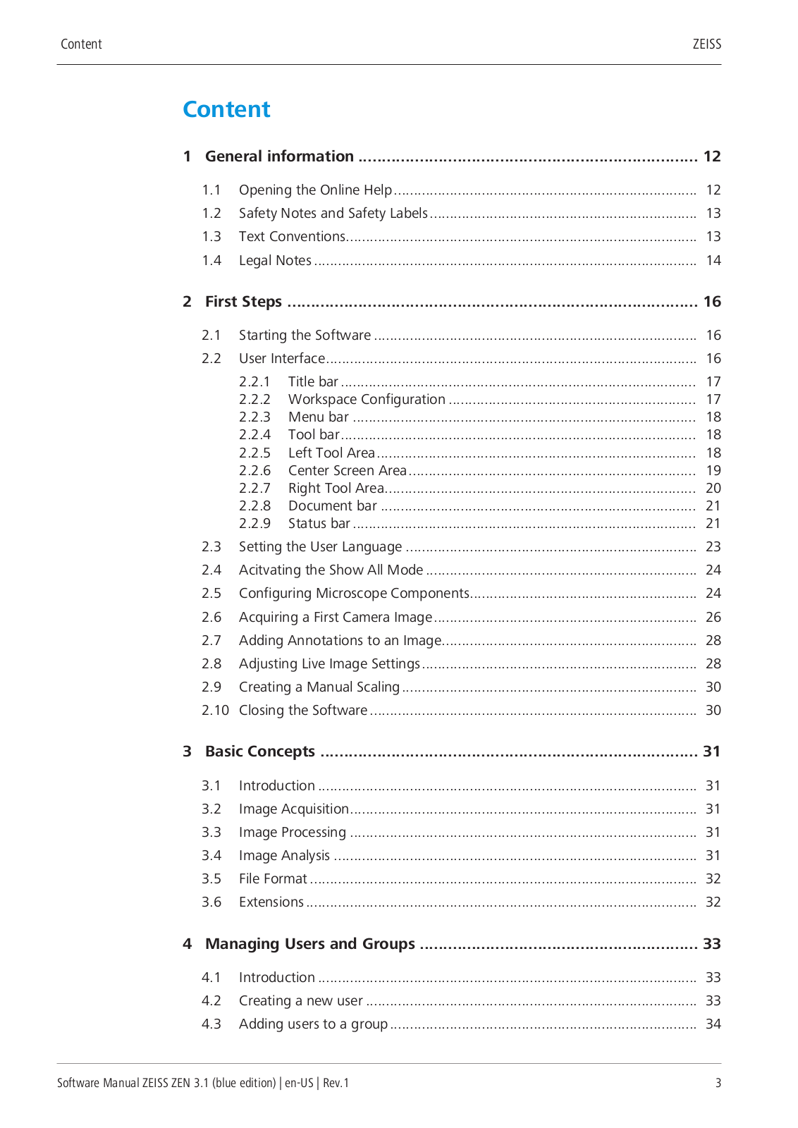
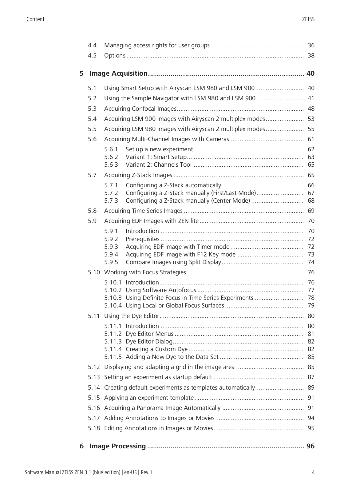
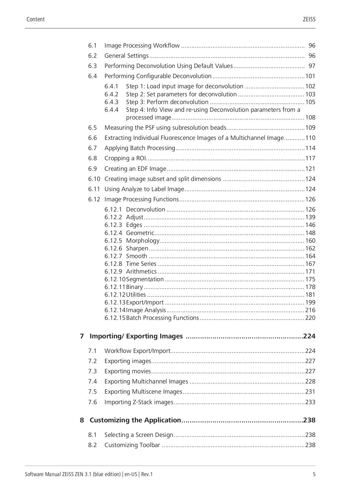
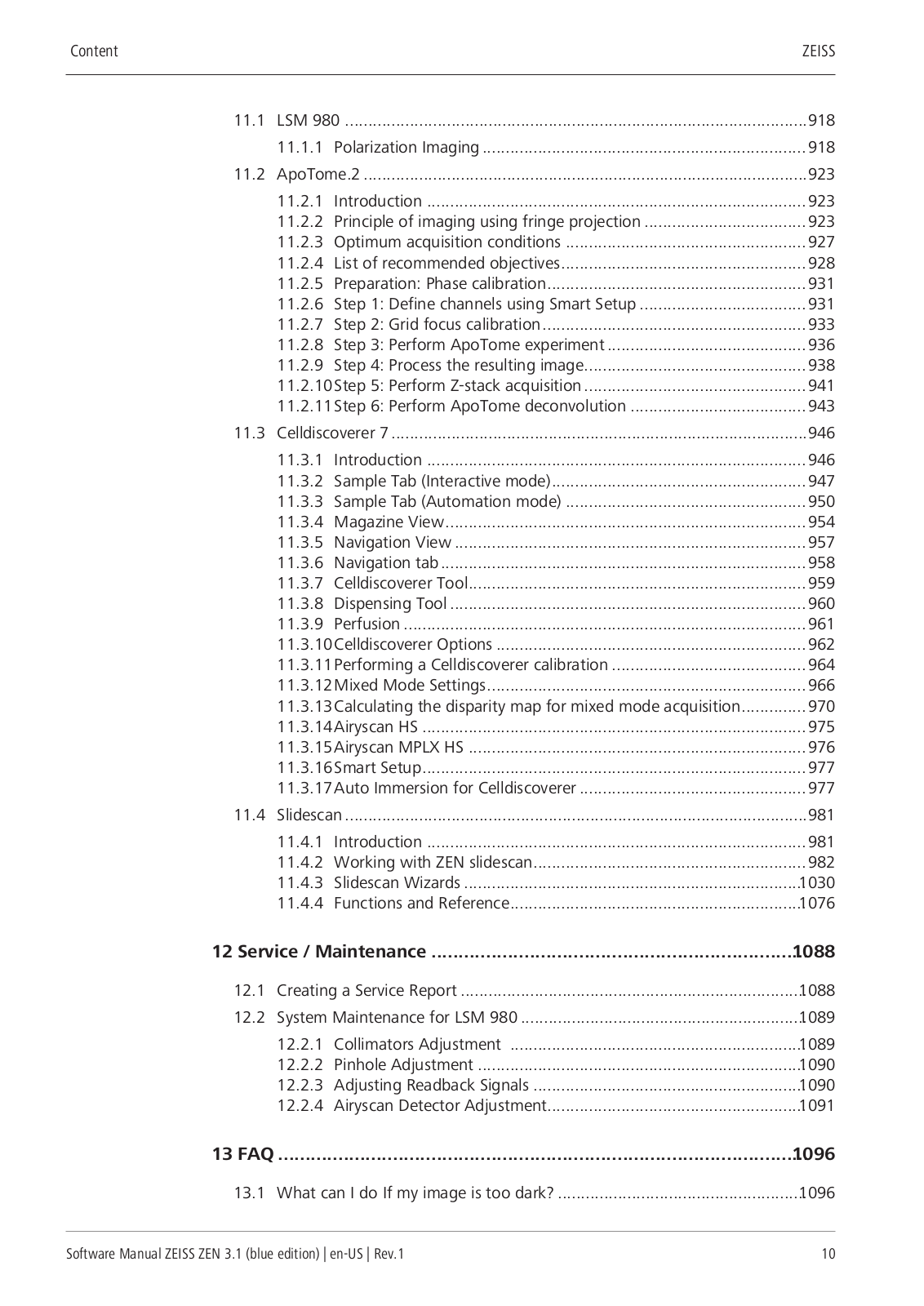
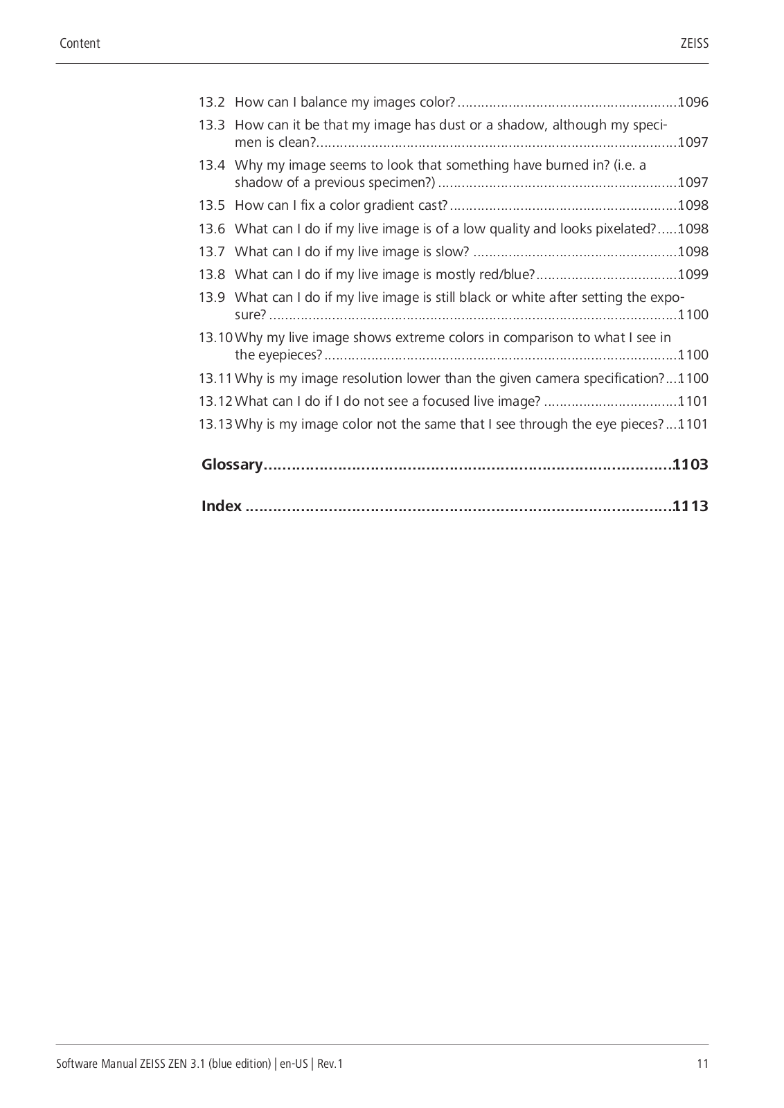
Table of contents
Loading...
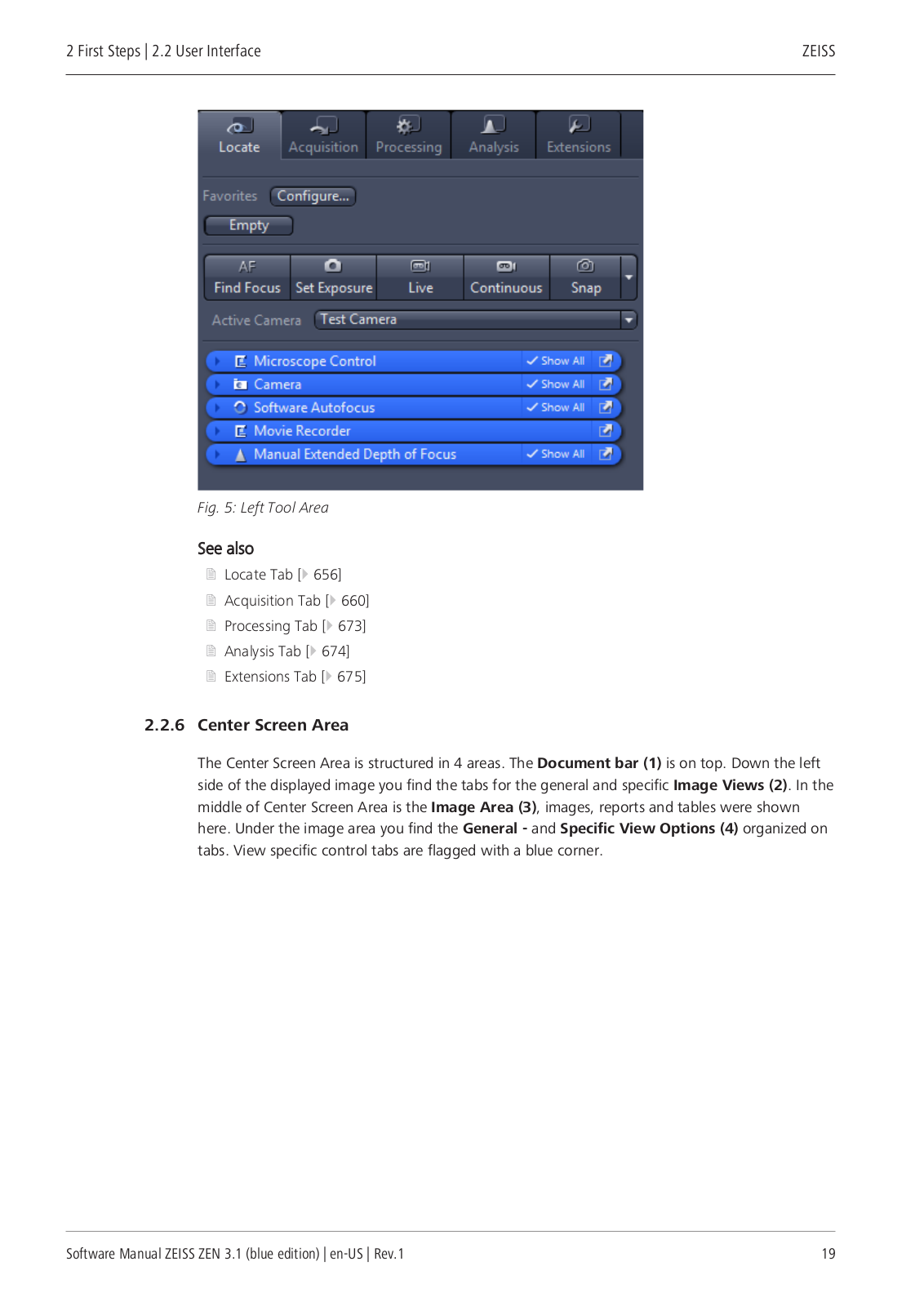
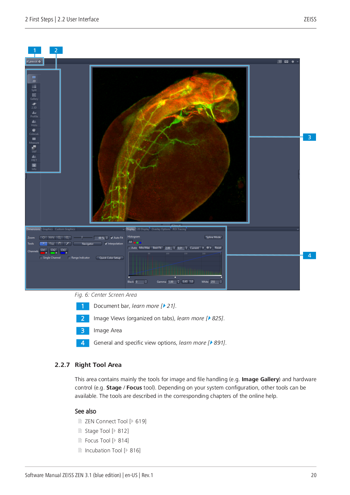
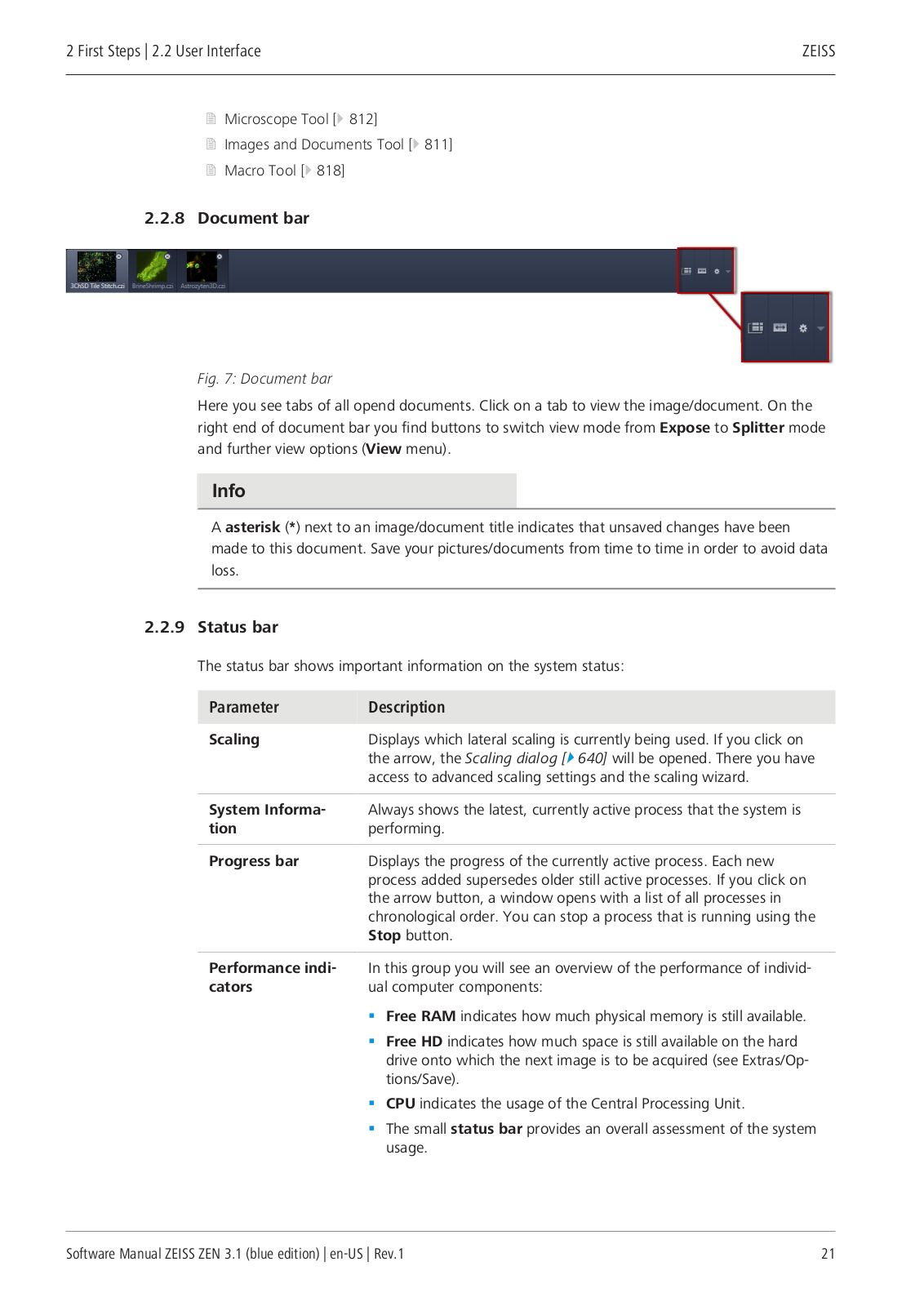
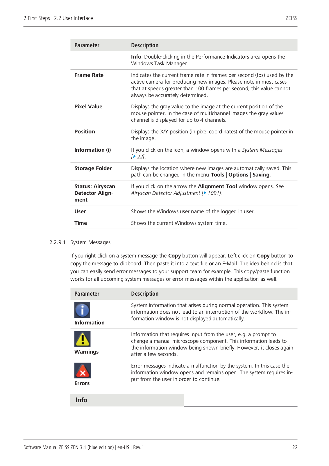
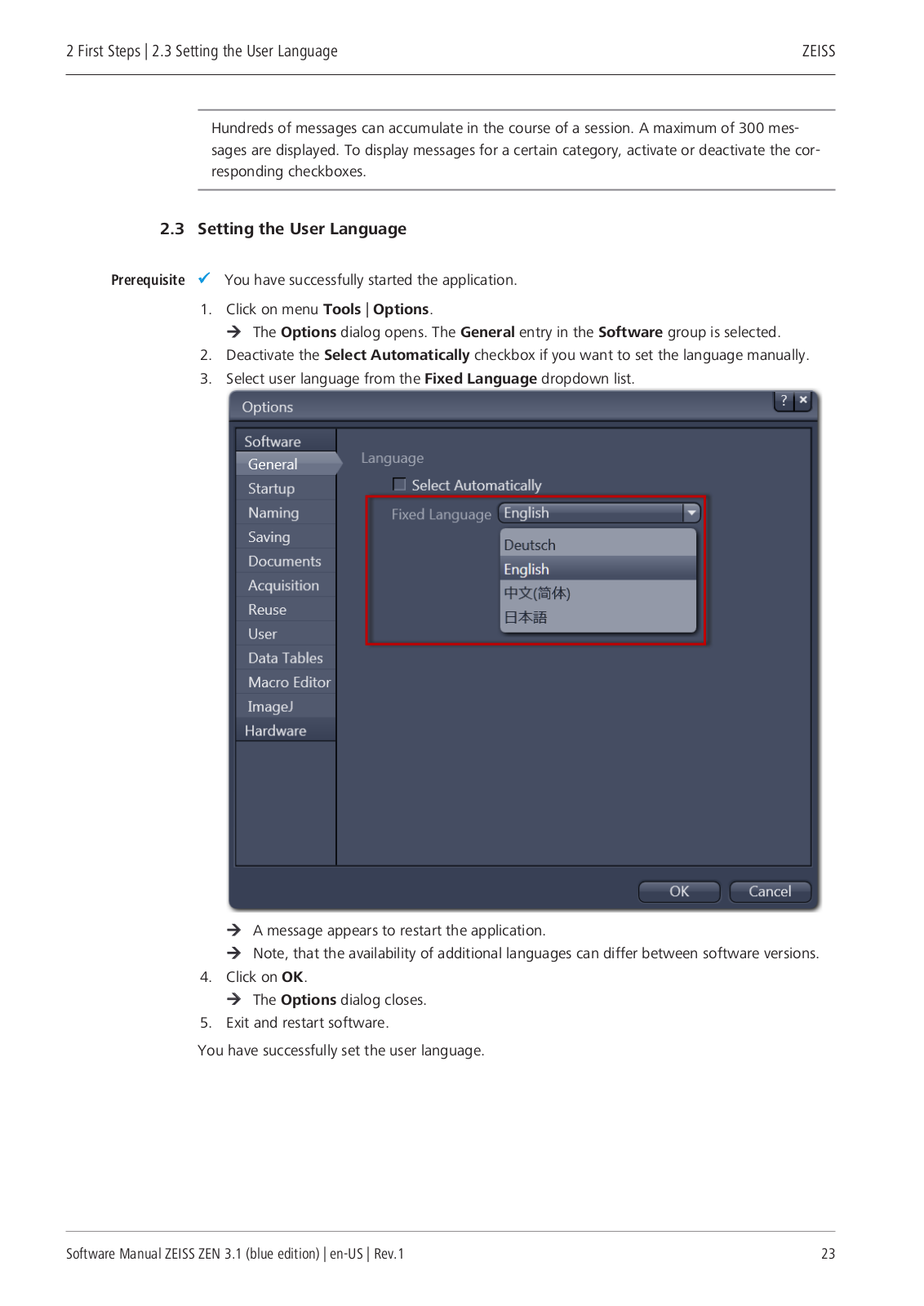
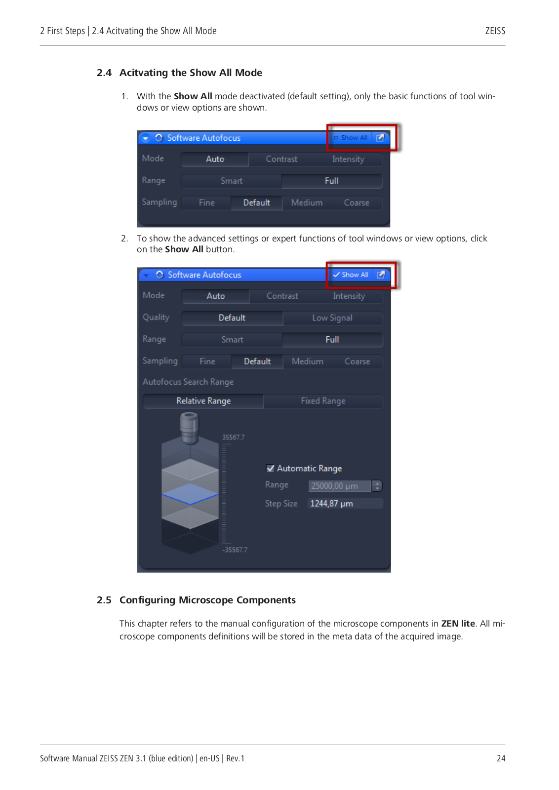
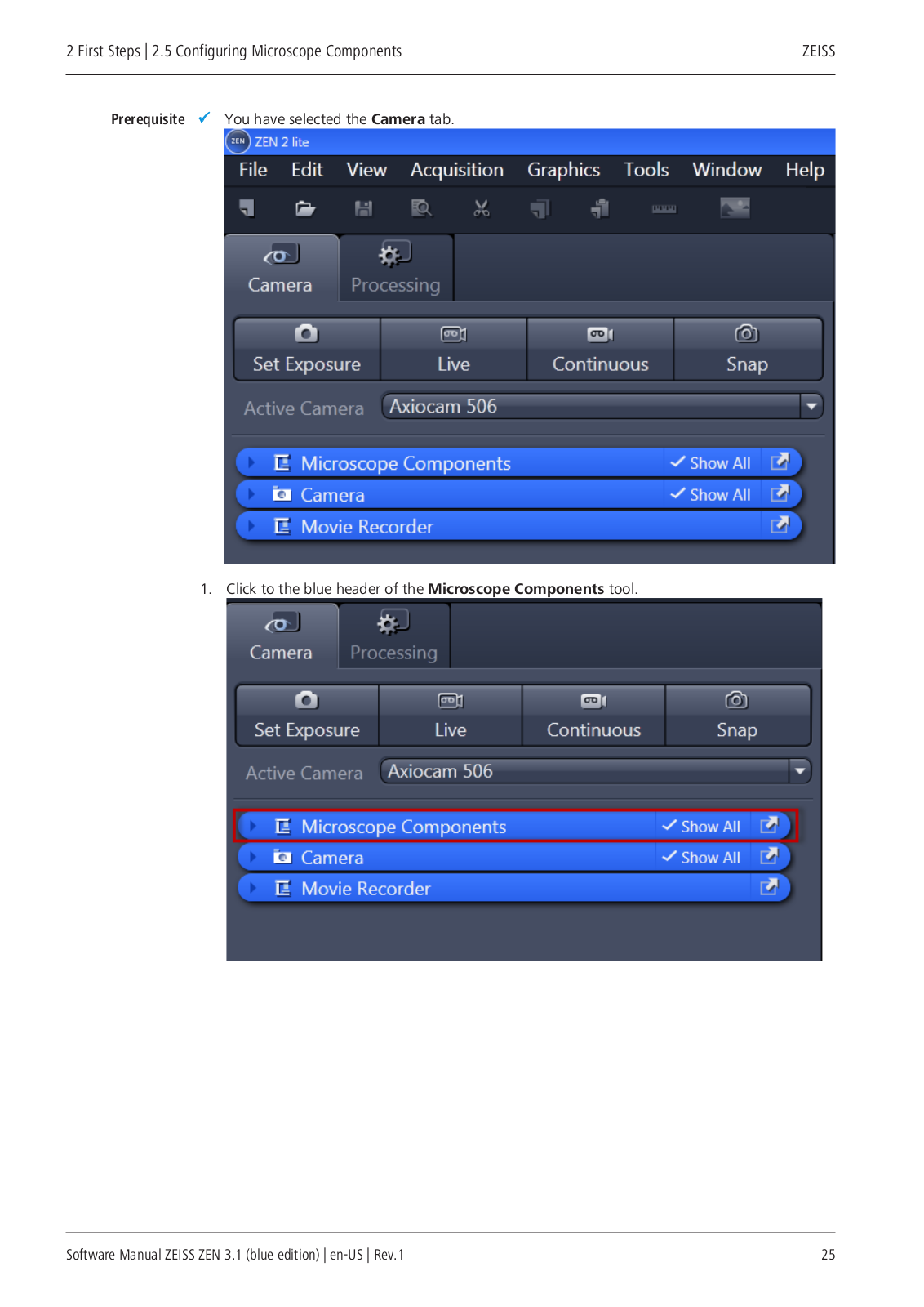
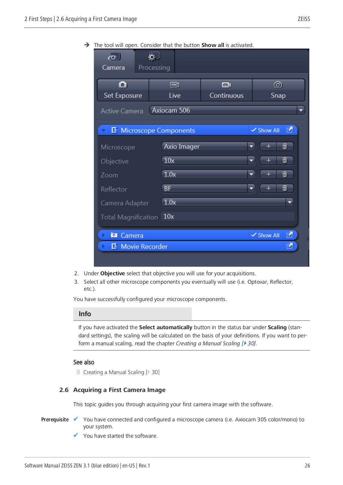
Zeiss ZEN3.1 User Manual
...
Zeiss User Manual
Download
Specifications and Main Features
Frequently Asked Questions
User Manual
Download
Loading...
+
hidden pages
Unhide
You need points to download manuals.
1 point = 1 manual.
You can buy points or you can get point for every manual you upload.
Buy points
Upload your manuals
 Loading...