


For over 130 years, Toshiba has led the world in developing technology to improve the quality of life. This Made for Life™ commitment is reflected in our family of leading-edge imaging systems for MRI, CT, ultrasound, cath labs, X-ray and nuclear medicine. From creating our first X-ray tube in 1915 to introducing the first Dynamic Volume CT Scanner in 2007, Toshiba continues to build upon our legacy with technological innovation that improves patient care while providing lasting quality for a lifetime of value.
Toshiba — A History
of Leadership
1875 • Founding of Toshiba
1915 • First X-ray Tube
1973 • First Real-time Ultrasound Scanner
1989 • First Helical CT Scanner
1990 • First Tissue Doppler Imaging System
1993 • First One-million-pixel CCD
1997 • First Open, Superconducting Magnet 2000 • First All-digital Multipurpose X-ray System 2003 • First 64-slice CT Scanner
2005 • First Compact Dual Plane Cath Lab with Flat Panel Detectors
2007 • First Dynamic Volume CT Scanner
http://www.toshibamedicalsystems.com
©Toshiba Medical Systems Corporation 2009 all rights reserved. Design and specifications subject to change without notice. MCAXR0197EA 2009-06 TME/D/D
Toshiba Medical Systems Corporation meets internationally recognized standards for Quality Management System ISO 9001, ISO 13485.
Toshiba Medical Systems Corporation Nasu Operations meets the
Environmental Management System standard, ISO 14001.
Made for Life and Infinix are trademarks of Toshiba Medical Systems
Corporation.
Printed in Japan
VF-i/BP
NFX-8000V
12" × 12" 12" × 12" |
Type S |
|
|
|
|
|
|||
nel Detector |
|
|||
|
|
|
|
|
|
|
|
|
|
Advanced technologies deliver
optimized biplane imaging
Designed in concert with leading pediatric physicians, the VF-i/BP provides
advanced, versatile patient access to meet the demands of today’s multi-discipline
imaging environments. The system’s revolutionary multi-tasking computer and intuitive
user interface deliver optimum image quality, time-saving ease of use and improved
workflow. Ideal for diagnostic, interventional and hybrid procedures, the VF-i/BP is
a completely new approach to biplane imaging designed to take advantage of its
revolutionary multi-axis C-arm.
2
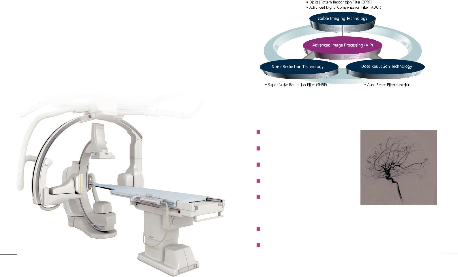
Advanced Image Processing (AIP) provides superb image quality for visualization of vessels and device.
Major improvements in image quality, patient access and ease of use
Comprehensive 12" x 12" biplane imaging without compromising patient access
High-resolution, flat panel images with uniform brightness and no distortion
Quiet instant-on, liquid-metal bearing X-ray tube for efficient exams
Unique lateral arm adjustment to quickly optimize imaging angles
Valuable dose-saving features:
-Various pulse rate controlled with grid
-X-ray beam filtration
-Variable frame rates in fluoroscopy and digital angiography
-Last image hold with virtual collimation
Major DICOM service classes included, which provide open access to patient information
New generation filter made it possible the reduction of noise with high spatial resolution
and less lag. The new filter enhances high-definition images of small devides and structures 3 (Super Noise Reduction Filter: SNRF).
Unparalleled patient access:
meeting the needs of all physicians
The VF-i/BP is designed to provide superior access to the patient — an
important point of distinction in the imaging landscape that now often requires
the attention of a wide range of specialists. In hybrid procedures that may require
a full complement of specialists including surgeons, neuroradiologists and
anesthesiologists, the VF-i/BP is at its best.
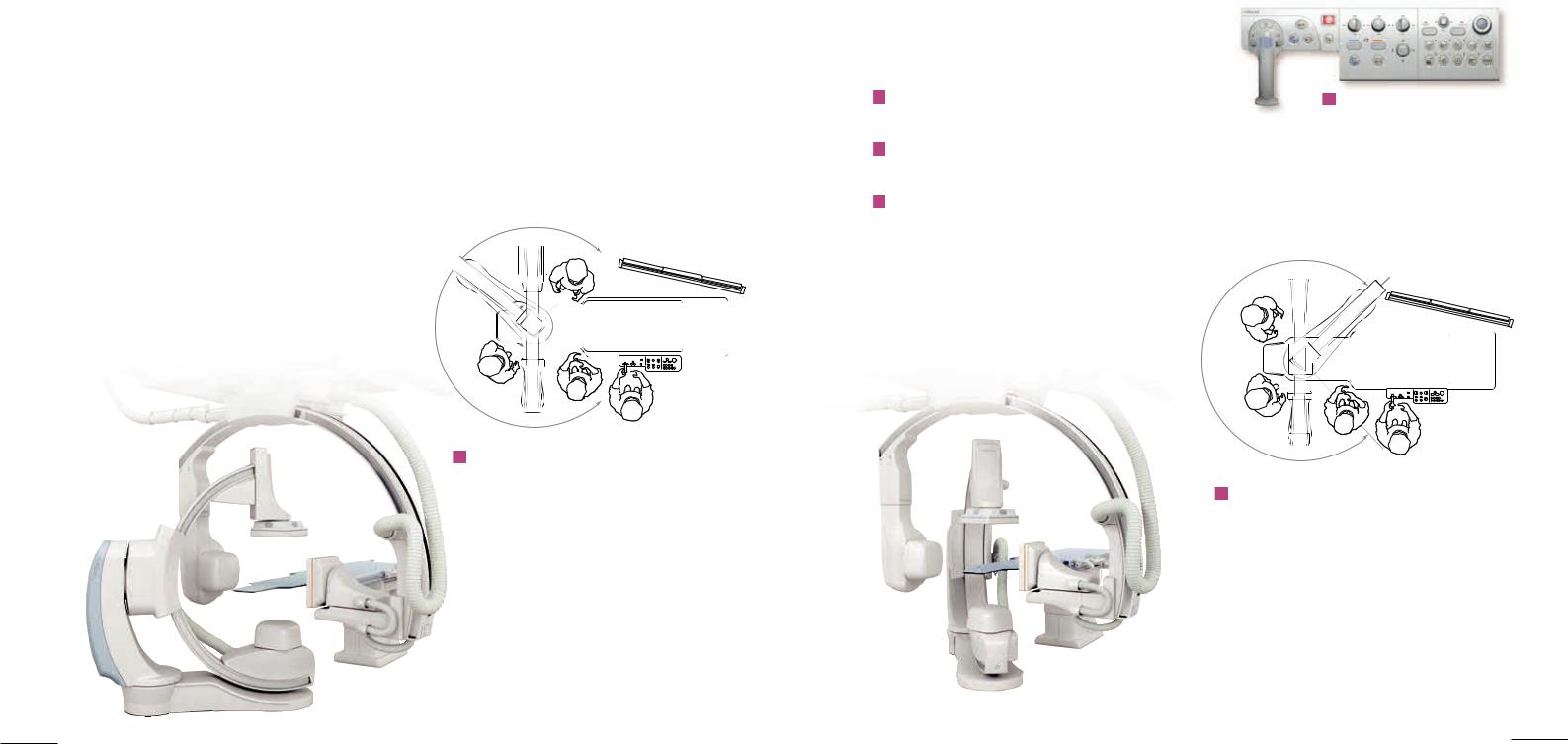
In this configuration, the head end of the table has 145 degrees of space allowing easy access for your anesthesiologist.
Efficient tableside control
The hyperhandle design and tableside console layout allow clinicians to more effectively concentrate on the patient and the image data providing a more patient focused examination.
Workflow is enhanced by tableside access to key functions through a specially designed graphical user interface
During image review, a single keystroke enables system setup from any selected image
Automatic archiving provides immediate recall of images at tableside without interruption
Compact tableside control provides ergonomic and tactile control of all exam functions for rapid component positioning, and control of digital processing functions.
The five-axis design provides a new level of access to the patient. The head end of the table has a full 180 degrees of space allowing the necessary physicians to conveniently access the patient and still provide biplane viewing.
4 |
5 |
|
 Loading...