


8000C
Digital Panoramic and Cephalometric System
8000C
User’s Guide

Notice
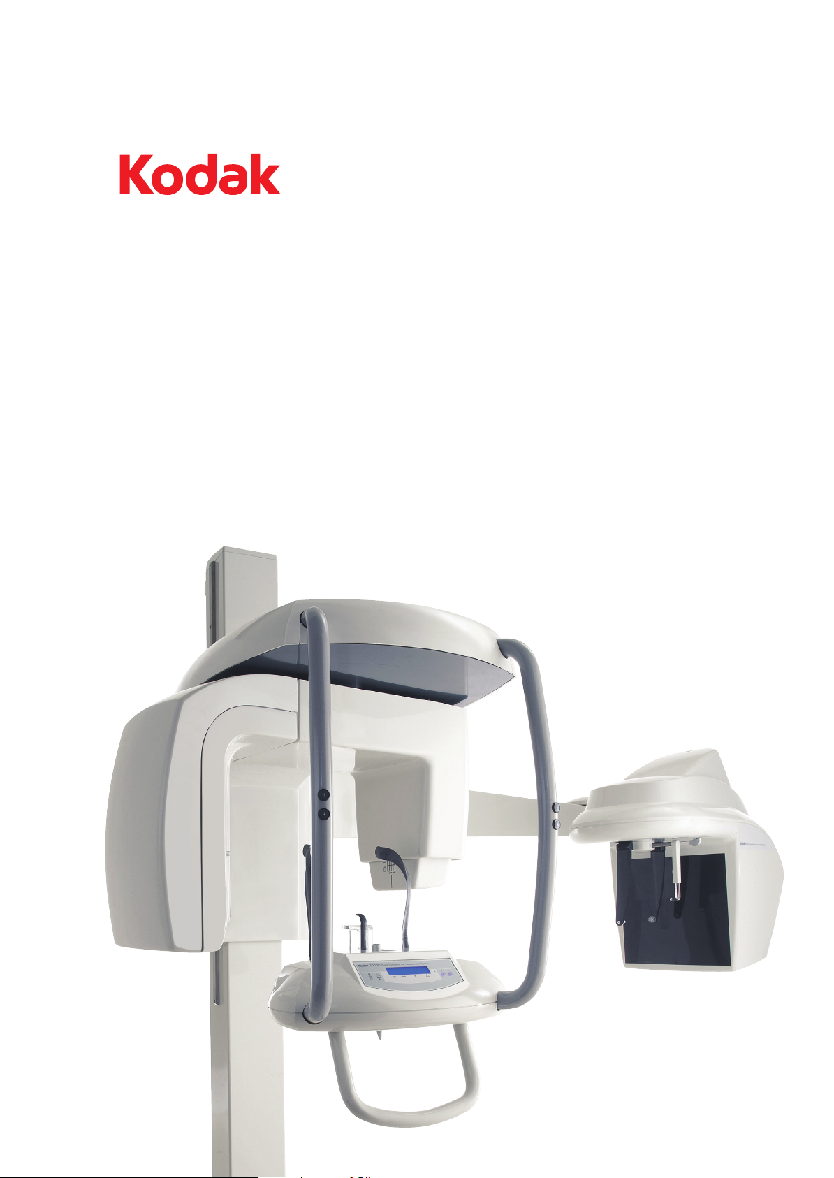
Congratulations on your purchase of the KODAK 8000C Digital Panoramic and Cephalometric Extraoral Imaging
System. The KODAK 8000C unit is the KODAK 8000 equipped with the cephalostat unit. Thank you for your
confidence in our products and we will do all in our power to ensure your complete satisfaction.
The User Guide for the KODAK 8000C Digital Panoramic and Cephalometric Extraoral Imaging System includes
information on the cephalometric features. For the panoramic features, see the KODAK 8000 Extraoral Imaging
System (SM722) User Guide. We recommend that you thoroughly familiarize yourself with this Guide in order to
make the most effective use of your system.
The information contained in this Guide may be subject to modification without notice, justification or notification
to the persons concerned.
No part of this Guide may be reproduced without the express permission of Carestream Health, Inc.
The US Federal law restricts this device to sale by or on the order of a physician.
This document is originally written in English.
Manual Name: KODAK 8000C Digital Panoramic and Cephalometric Extraoral Imaging System User Guide
Part Number: SM735
Revision Number: 02
Print Date: 03/2010
The brand names and logos reproduced in this Guide are copyright.
KODAK is a trademark of KODAK used under License.
KODAK 8000C Digital Panoramic and Cephalometric Extraoral Imaging System, complies with Directive
93/42/CEE relating to medical equipment.
.
Manufacturer
Authorized Representative in the European Community
TROPHY
4, Rue F. Pelloutier, Croissy-Beaubourg
77435 Marne la Vallée Cedex 2, France
WARNING: We recommend that you consult the “Safety, Regulatory and the Technical
Specification User Guide” before using the KODAK 8000C Extraoral Imaging Systems.
0086
Carestrea m Health, Inc.
150 Verona Street
Roche ster NY 14 608
EC REP

KODAK 8000C Digital Panoramic and Cephalometric Extraoral Imaging System User Guide (SM735)_Ed 02 iii
Contents
1—About This Guide
Conventions in this Guide. . . . . . . . . . . . . . . . . . . . . . . . . . . . . . . . . . . . . . . . . . . . . . . . . . . . . . . . . . . . . . . . . . . . . 1–1
2—KODAK 8000C UNIT OVERVIEW
General Overview . . . . . . . . . . . . . . . . . . . . . . . . . . . . . . . . . . . . . . . . . . . . . . . . . . . . . . . . . . . . . . . . . . . . . . . . . . . 2–1
Mobile Components . . . . . . . . . . . . . . . . . . . . . . . . . . . . . . . . . . . . . . . . . . . . . . . . . . . . . . . . . . . . . . . . . . . . . . 2–2
General Functional Components . . . . . . . . . . . . . . . . . . . . . . . . . . . . . . . . . . . . . . . . . . . . . . . . . . . . . . . . . . . . 2–3
Digital Sensor Locations. . . . . . . . . . . . . . . . . . . . . . . . . . . . . . . . . . . . . . . . . . . . . . . . . . . . . . . . . . . . . . . . . . . 2–4
Laser Locations . . . . . . . . . . . . . . . . . . . . . . . . . . . . . . . . . . . . . . . . . . . . . . . . . . . . . . . . . . . . . . . . . . . . . . . . . . 2–5
Control Panel . . . . . . . . . . . . . . . . . . . . . . . . . . . . . . . . . . . . . . . . . . . . . . . . . . . . . . . . . . . . . . . . . . . . . . . . . . . . . . . 2–6
X-Ray Remote Control Overview . . . . . . . . . . . . . . . . . . . . . . . . . . . . . . . . . . . . . . . . . . . . . . . . . . . . . . . . . . . . . . . 2–7
Positioning Accessories and Replacement Parts . . . . . . . . . . . . . . . . . . . . . . . . . . . . . . . . . . . . . . . . . . . . . . . . . . 2–8
3—IMAGING SOFTWARE OVERVIEW
Computer System Requirements . . . . . . . . . . . . . . . . . . . . . . . . . . . . . . . . . . . . . . . . . . . . . . . . . . . . . . . . . . . . . . . 3–1
General Software Overview . . . . . . . . . . . . . . . . . . . . . . . . . . . . . . . . . . . . . . . . . . . . . . . . . . . . . . . . . . . . . . . . . . . 3–2
KODAK Dental Imaging Software . . . . . . . . . . . . . . . . . . . . . . . . . . . . . . . . . . . . . . . . . . . . . . . . . . . . . . . . . . . 3–2
Cephalometric Acquisition Interface Module . . . . . . . . . . . . . . . . . . . . . . . . . . . . . . . . . . . . . . . . . . . . . . . . . . 3–2
Cephalometric Acquisition Interface Module . . . . . . . . . . . . . . . . . . . . . . . . . . . . . . . . . . . . . . . . . . . . . . . . . . . . . 3–3
Cephalometric Acquisition Window Overview. . . . . . . . . . . . . . . . . . . . . . . . . . . . . . . . . . . . . . . . . . . . . . . . . 3–3
Cephalometric Program Pane . . . . . . . . . . . . . . . . . . . . . . . . . . . . . . . . . . . . . . . . . . . . . . . . . . . . . . . . . . . 3–5
Cephalometric Patient Pane . . . . . . . . . . . . . . . . . . . . . . . . . . . . . . . . . . . . . . . . . . . . . . . . . . . . . . . . . . . . 3–6
Cephalometric Parameter Pane. . . . . . . . . . . . . . . . . . . . . . . . . . . . . . . . . . . . . . . . . . . . . . . . . . . . . . . . . . 3–7
4—GETTING STARTED
Switching on the Unit . . . . . . . . . . . . . . . . . . . . . . . . . . . . . . . . . . . . . . . . . . . . . . . . . . . . . . . . . . . . . . . . . . . . . . . . 4–1
Starting the Imaging Software . . . . . . . . . . . . . . . . . . . . . . . . . . . . . . . . . . . . . . . . . . . . . . . . . . . . . . . . . . . . . . . . . 4–2
Creating a Patient Record. . . . . . . . . . . . . . . . . . . . . . . . . . . . . . . . . . . . . . . . . . . . . . . . . . . . . . . . . . . . . . . . . . . . . 4–2
Accessing the Cephalometric Acquisition Window . . . . . . . . . . . . . . . . . . . . . . . . . . . . . . . . . . . . . . . . . . . . . . . . 4–3
5—ACQUIRING CEPHALOMETRIC IMAGES
Acquiring a Lateral Image . . . . . . . . . . . . . . . . . . . . . . . . . . . . . . . . . . . . . . . . . . . . . . . . . . . . . . . . . . . . . . . . . . . . . 5–1
Preparing the Unit and Setting the Acquisition Parameters . . . . . . . . . . . . . . . . . . . . . . . . . . . . . . . . . . . . . . 5–1
Acquiring a Frontal AP or PA Image. . . . . . . . . . . . . . . . . . . . . . . . . . . . . . . . . . . . . . . . . . . . . . . . . . . . . . . . . . . . . 5–5
Preparing the Unit and Setting the Acquisition Parameters . . . . . . . . . . . . . . . . . . . . . . . . . . . . . . . . . . . . . . 5–5
Preparing and Positioning the Patient . . . . . . . . . . . . . . . . . . . . . . . . . . . . . . . . . . . . . . . . . . . . . . . . . . . . . . . . 5–6
Acquiring an Oblique Image . . . . . . . . . . . . . . . . . . . . . . . . . . . . . . . . . . . . . . . . . . . . . . . . . . . . . . . . . . . . . . . . . . . 5–9
Preparing the Unit and Setting the Acquisition Parameters . . . . . . . . . . . . . . . . . . . . . . . . . . . . . . . . . . . . . . 5–9
Preparing and Positioning the Patient . . . . . . . . . . . . . . . . . . . . . . . . . . . . . . . . . . . . . . . . . . . . . . . . . . . . . . . 5–10
Acquiring a Submento-Vertex Image . . . . . . . . . . . . . . . . . . . . . . . . . . . . . . . . . . . . . . . . . . . . . . . . . . . . . . . . . . . 5–12
Preparing the Unit and Setting the Acquisition Parameters . . . . . . . . . . . . . . . . . . . . . . . . . . . . . . . . . . . . . 5–12
Preparing and Positioning the Patient . . . . . . . . . . . . . . . . . . . . . . . . . . . . . . . . . . . . . . . . . . . . . . . . . . . . . . . 5–13

iv
Contents
Acquiring a Carpus Image. . . . . . . . . . . . . . . . . . . . . . . . . . . . . . . . . . . . . . . . . . . . . . . . . . . . . . . . . . . . . . . . . . . . 5–16
Preparing the Unit and Setting the Acquisition Parameters . . . . . . . . . . . . . . . . . . . . . . . . . . . . . . . . . . . . . 5–16
Preparing and Positioning the Patient . . . . . . . . . . . . . . . . . . . . . . . . . . . . . . . . . . . . . . . . . . . . . . . . . . . . . . . 5–17
X-Ray Dose Emission Information . . . . . . . . . . . . . . . . . . . . . . . . . . . . . . . . . . . . . . . . . . . . . . . . . . . . . . . . . . . . . . 5–19
6—MAINTENANCE
Daily . . . . . . . . . . . . . . . . . . . . . . . . . . . . . . . . . . . . . . . . . . . . . . . . . . . . . . . . . . . . . . . . . . . . . . . . . . . . . . . . . . . . . . . 6–1
Monthly . . . . . . . . . . . . . . . . . . . . . . . . . . . . . . . . . . . . . . . . . . . . . . . . . . . . . . . . . . . . . . . . . . . . . . . . . . . . . . . . . . . . 6–1
Annually . . . . . . . . . . . . . . . . . . . . . . . . . . . . . . . . . . . . . . . . . . . . . . . . . . . . . . . . . . . . . . . . . . . . . . . . . . . . . . . . . . . . 6–1
7—TROUBLESHOOTING
Quick Troubleshooting. . . . . . . . . . . . . . . . . . . . . . . . . . . . . . . . . . . . . . . . . . . . . . . . . . . . . . . . . . . . . . . . . . . . . . . . 7–1

KODAK 8000C Digital Panoramic and Cephalometric Extraoral Imaging System User Guide (SM735)_Ed 02 1–1
Chapter 1
About This Guide
Conventions in this Guide
The following special messages emphasize information or indicate potential risk to
personnel or equipment:
WARN I NG
Warns you to avoid injury to yourself or others by following the
safety instructions
precisely.
CAUTION
Alerts you to a condition that might cause serious damage.
IMPORTANT
Alerts you to a condition that might cause problems.
NOTE
Emphasizes important information.
TIP
Provides extra information and hints.

Conventions in this Guide
1–2 About This Guide

KODAK 8000C Digital Panoramic and Cephalometric Extraoral Imaging System User Guide (SM735)_Ed 02 2–1
Chapter 2
KODAK 8000C UNIT OVERVIEW
The KODAK 8000C digital panoramic and cephalometric unit is designed to carry out the
following radiological examinations:
• Panoramic
• Maxillary Sinus
• Temporomandibular Joints (TMJ)
• Lateral cephalometric
• Frontal (PA or AP) cephalometric
• Oblique cephalometric
• Submento-vertex cephalometric
• Carpus cephalometric
General Overview
The KODAK 8000C digital panoramic and cephalometric unit is composed of the
following functional components:
• The unit head that contains all the electronic control
• The rotative arm
• The fixed arm with a control panel
• The panoramic digital sensor
• The x-ray source assembly
• The x-ray remote control
• The chin rest and bite block
• The chin rest base
• The panoramic chin rest and bite block
• The temple supports
• The hand grips
• The cephalostat arm
• The cephalostat head
• The head clamps and ear cones
• The nasion support
• The acquisition software (see “Imaging Software Overview”)
The following figures illustrate the general overview of the KODAK 8000C digital
panoramic and cephalometric units.

General Overview
2–2 KODAK 8000C UNIT OVERVIEW
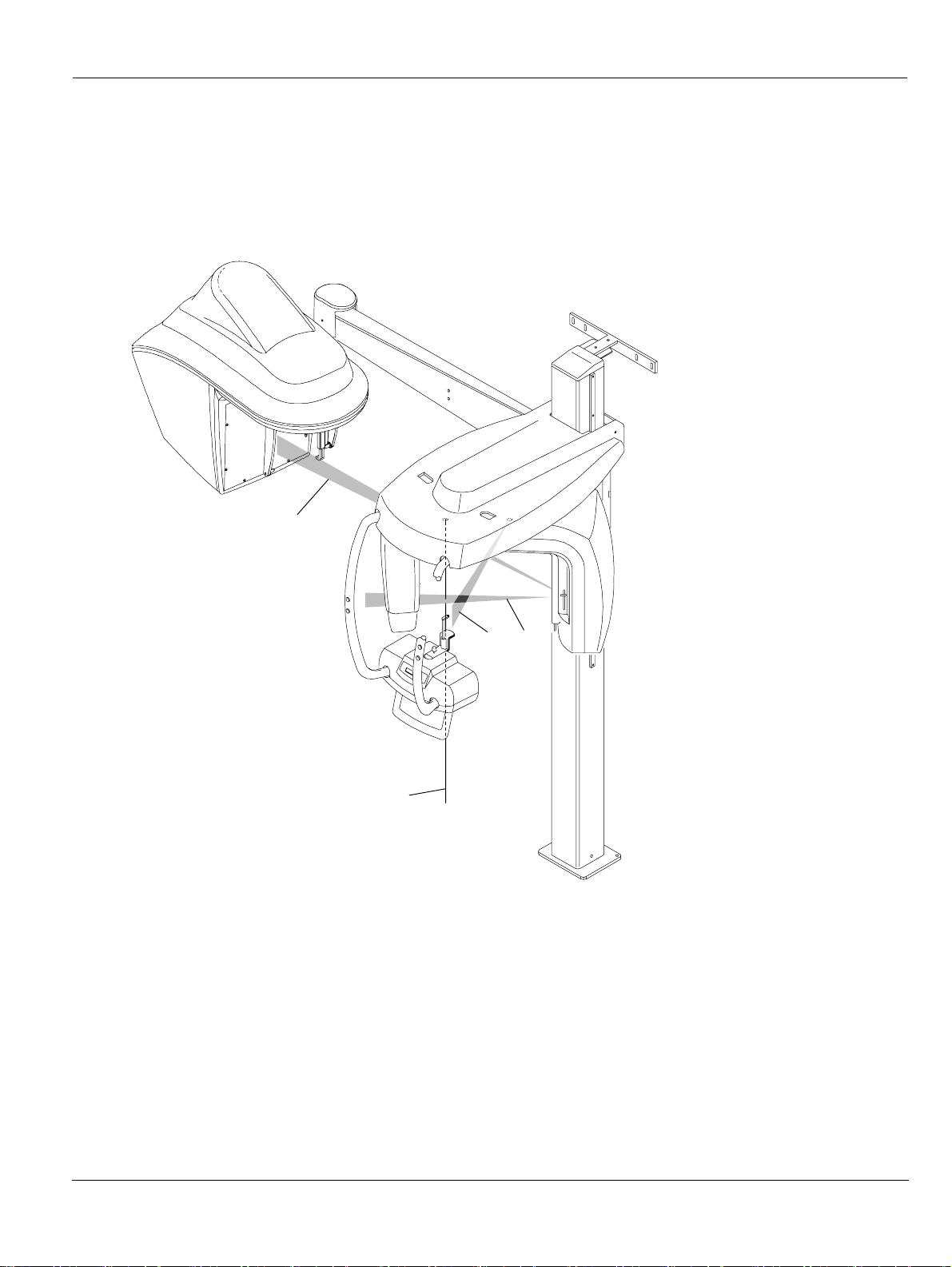
Mobile Components
Figure 2-1 illustrates the up and down movement of the KODAK 8000C digital
panoramic and cephalometric units mobile component and the rotation of the rotative
arm.
Figure 2–1 KODAK 8000 and KODAK 8000C Units Mobile Components
IMPORTANT
The Cephalostat can be positioned either on the right or the
left side of the KODAK 8000 unit
.

General Overview
KODAK 8000C Digital Panoramic and Cephalometric Extraoral Imaging System User Guide (SM735)_Ed 02 2–3
General Functional Components
Figure 2-2 illustrates the general functional components of the KODAK 8000C digital
panoramic and cephalometric units.
Figure 2–2 KODAK 8000 and KODAK 8000C Units Functional Components
1 ON/OFF button 10 Collimator selector
2 Chin rest and bite block 11 Unit rotative arm
3 Temple supports 12 X-Ray remote control
4 Temple supports control knob 13 PC hosting the imaging and the acquisition
software
5 Hand Grips 14 Cephalostat arm
6 Control panel 15 Cephalostat head
7 Height adjustment buttons 16 Head clamps and ear cones
8 Panoramic digital sensor 17 Nasion support
9 X-Ray source assembly
12
13
3
7
9
1
8
4
5
6
2
11
10
14
15
16
17
16

General Overview
2–4 KODAK 8000C UNIT OVERVIEW
Digital Sensor Locations
Figure 2-3 illustrates the locations of the digital panoramic and digital cephalometric
sensors of the KODAK 8000C digital panoramic and cephalometric units.
Figure 2–3 KODAK 8000 and KODAK 8000C Units Digital Sensor Locations
system
General Overview
KODAK 8000C Digital Panoramic and Cephalometric Extraoral Imaging System User Guide (SM735)_Ed 02 2–5
Laser Locations
Figure 2-4 illustrates the location of the lasers of the KODAK 8000C digital panoramic
and cephalometric units.
Figure 2–4 KODAK 8000 and KODAK 8000C Units Laser Beam Locations
1 Mid-sagittal plane positioning laser beam
2 Frankfort plane positioning laser beam
3 Canine plane positioning laser beam
4 Cephalometric Frankfort plane positioning laser beam
2
1
4
3
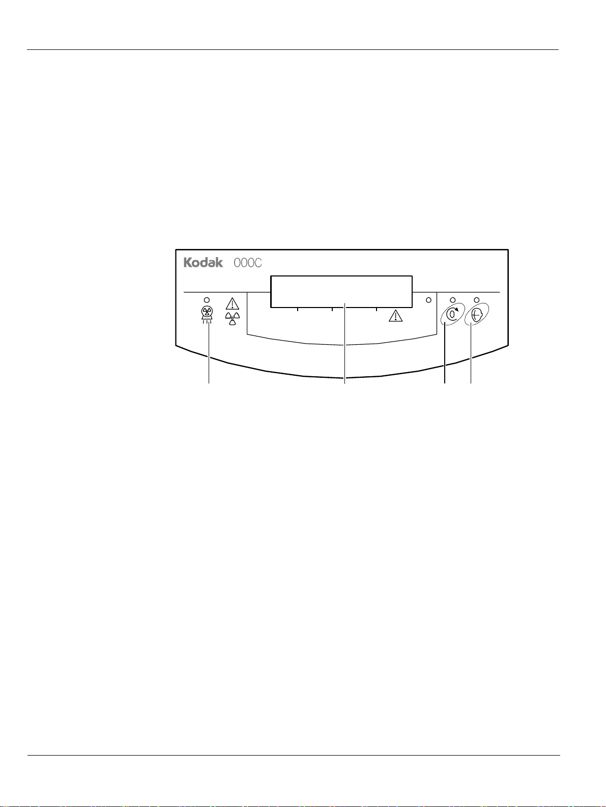
Control Panel
2–6 KODAK 8000C UNIT OVERVIEW
Control Panel
The control panel is an alphanumeric, digital soft touch console. It allows the operator to
control certain unit functions. It also displays the operating parameters and error
messages.
Figure 2–5 Unit Control Panel
8
Digital Panorami c a nd Cephalometric S ystem
kV
mA S
4321
1 X-Ray emis sion LE D: Yellow, indicates the x-rays are being emitted.
2 Display Screen: Displays the current acquisition parameters and the error messages.
3 Reset button: Resets the unit arm to the initial position to enable the patient to enter
and exit the unit.
4 Laser beam button: Activates the laser positioning beams to correctly position the
patient.
X-Ray Remote Control Overview
KODAK 8000C Digital Panoramic and Cephalometric Extraoral Imaging System User Guide (SM735)_Ed 02 2–7
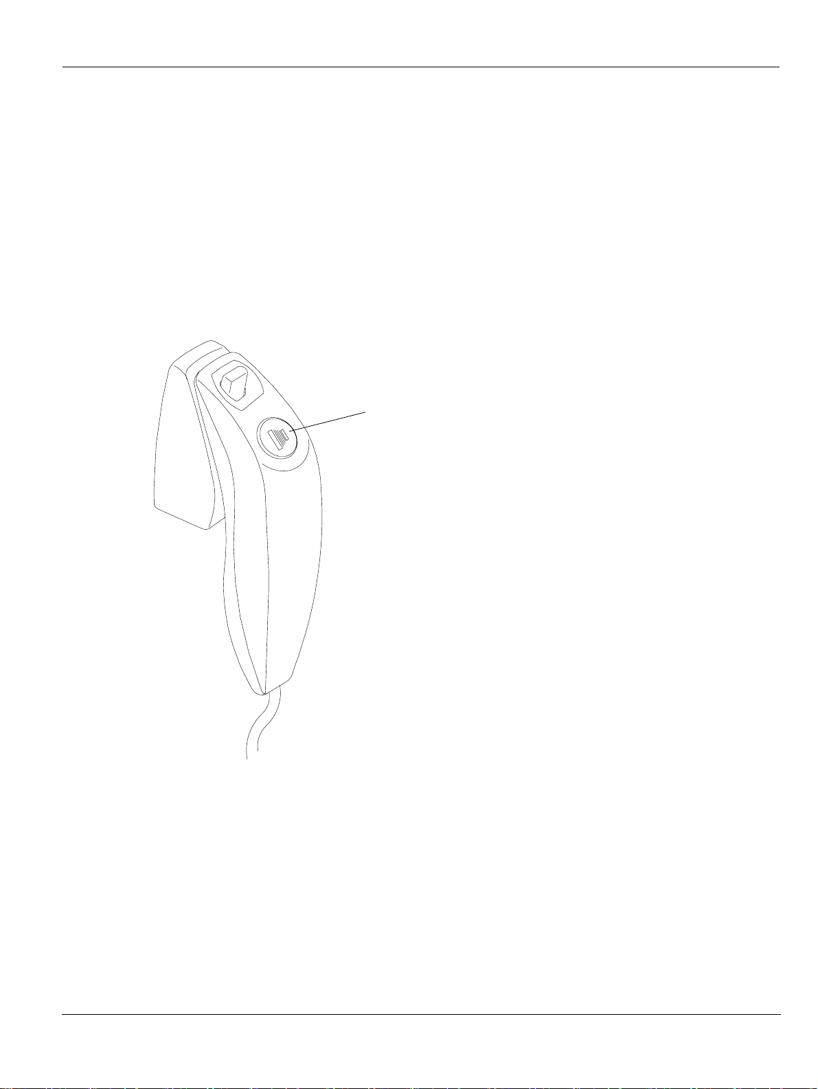
X-Ray Remote Control Overview
The x- ray remote control enables you to launch a radiological image acquisition via the
exposure button from outside the x-ray room. You must press and hold the exposure
button until the end of acquisition. Premature release of the exposure button interrupts
the acquisition.
Figure 2–6 X-R ay Re mote C ontrol
1 Exposure button: launches image acquisition.
1

Positioning Accessories and Replacement Parts
2–8 KODAK 8000C UNIT OVERVIEW
Positioning Accessories and Replacement Parts
The following accessories are used when positioning a patient. They are delivered with
the KODAK 8000 digital panoramic and KODAK 8000C digital panoramic and
cephalometric unit.
Table 2-1 and Table 2-2 list the panoramic and cephalometric positioning accessories.
Table 2–1 Panoramic Positioning Accessories and Replacement Parts
Accessory Description
Panoramic chin rest and TMJ x2
Maxillary sinus chin rest
TMJ x2 and TMJ x4 nose rest
Standard bite block
Bite block for edentulous patients
A set of right and left temple supports
Single use sheaths for bite blocks
(500 pcs box)
x2

Positioning Accessories and Replacement Parts
KODAK 8000C Digital Panoramic and Cephalometric Extraoral Imaging System User Guide (SM735)_Ed 02 2–9
Table 2–2 Cephalometric Positioning Accessories and Replacement Parts
Accessory Description
Head clamps with ear cones
Nasion support
x2

Positioning Accessories and Replacement Parts
2–10 KODAK 8000C UNIT OVERVIEW
 Loading...