

Page 1

Userguide
No 041 I Micromanipulation
Microinjection of cDNA into
Enrica Charbonnier, Devolopmental Biology, Biology I, University of Freiburg, 79104 Freiburg, Germany,
enrica.charbonnier@googlemail.com
Abstract
Germline transformation via microinjection of DNA into Drosophila melanogaster has been used to interfere with
gene expression and study their function for many years. Recently, Bischof and co-workers established a new system to circumvent typical limitations by optimizing a phiC31-based integration system [1]. This integration system
demonstrates to be extremely efficient in Drosophila embryo microinjection. Furthermore, in combination with the
ease-of-use plus versatility of this technique, a systematic high throughput screening of large cDNA sets and regulatory elements is now feasible.
In this Userguide, a step-by-step protocol using the Eppendorf programmable microinjector FemtoJet and ready to
use Femtotip II microcapillaries is presented for this application.
Introduction
Nowadays the scientific community invests great effort into
the identification and characterization of all genes which
are relevant to a specific signalling pathway or biological
process and their interaction between each other. Since
long the Drosophila genome has been sequenced and thus
makes it a powerful tool to study gene expression and gene
regulation even at the transcriptional level. P-elements are
a powerful tool to generate insertion mutagenesis thanks to
their random integration into the Drosophila genome. However, this random insertion causes difficulties to analyze the
expression pattern of the transgenic animal obtained with
this method. One of the biggest problems is the positional
effect, because the genomic environment can influence the
expression of the transgene.
Recently, the genome integration method based on the
site-specific phiC31 integrase [2], has been applied to
Drosophila [3].
Drosophila
The bacteriophage phiC31 encodes a serine integrase
that mediates sequence-directed recombination between
a bacterial attachment site (attB) and a phage attachment
site (attP) [4]. Bischof and co-workers generated a collection of Drosophila lines (available now at Bloomington
Drosophila Stock Center, http://flystocks.bio.indiana.edu/)
with precisely mapped attP sites that allow the insertion of
transgenes into many different predetermined intergenetic
locations throughout the fly genome. By using regulatory
elements of the nanos and vasa genes, they established
endogenous sources of the phiC31 integrase, eliminating
the difficulties of coinjecting integrase mRNA and raising
the transformation efficiency. Moreover, to discriminate
between specific and rare non-specific integration events, a
white gene-based reconstitution system was generated that
enables visual selection for precise attP targeting [1].
embryos
Page 2
Userguide No 041 | Page 2
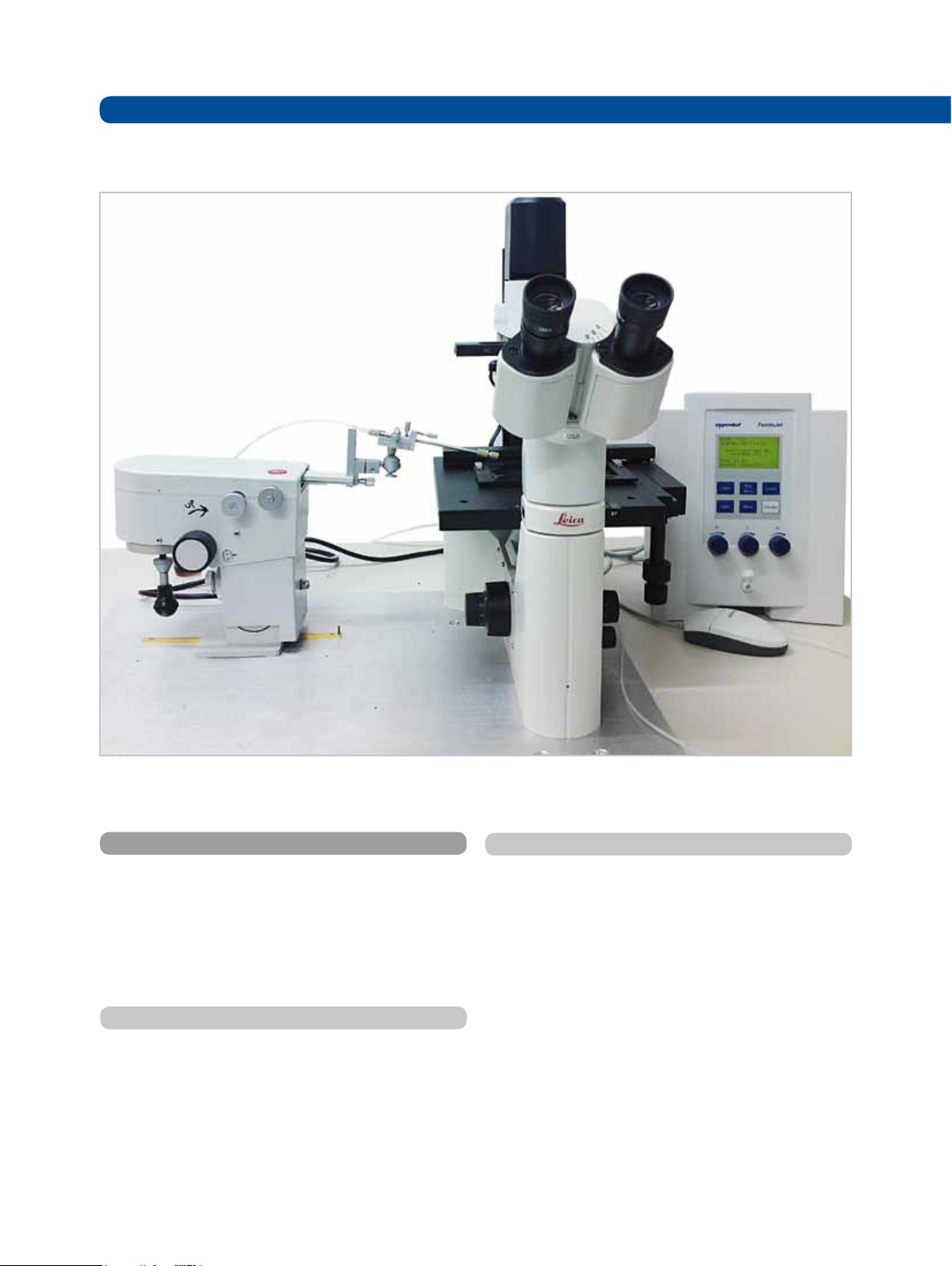
Fig. 1: Workstation for Drosophila injection, consisting of a Leica DM IL inverted microscope, a Leica manual micromanipulator and
an Eppendorf programmable microinjector FemtoJet.
Materials and Methods
DNA is introduced at 18 °C in Drosophila embryos by
microinjection, into the posterior end of the egg just before
pole cell formation using a manual micromanipulator (Leica
microsystems), Eppendorf programmable microinjector, the
FemtoJet, and ready to use, sterile Femtotip II microinjection
capillaries (see figure 1).
Instruments
‡ Inverted microscope (Leica DM IL, Leica Microsystems
CMS GmbH, Wetzlar, Germany)
‡ Manual Micromanipulator (Leica microsystems)
Programmable microinjector FemtoJet (Eppendorf AG,
Hamburg, Germany)
‡ Micropipette 0.5-10 µL
‡ Microcentrifuge Eppendorf 5424
Hamburg, Germany)
(Eppendorf AG,
Materials
‡
Femtotip II microinjection capillaries (Eppendorf AG,
Hamburg, Germany)
‡
Microloader (Eppendorf AG, Hamburg, Germany)
‡
Microscope slide 76x26 mm (Leica Microsystems)
Agar plates with grape juice
‡
Small mesh basket for collecting rinsing and dechorionat ing embryos
‡
Double sided tape (tesa SE, Hamburg, Germany)
‡
Embryo collection chambers
‡
Sodium hypochlorite (Sigma-Aldrich, Germany)
‡
Hair dryer
‡
Voltalef oil (VWR, Bruchsal, Germany)
‡
Plasmid purification kit (Qiagen, Hilden, Germany)
Page 3

DNA preparation
DNA used for injection is prepared with Qiagen Midiprep
kit, resuspended in injection buffer (5 mM KCI, 0.1 mM
2PO4
, pH 6.8).
NaH
The DNA to be injected is quantified and diluted at a
concentration of 0.3 µg/µL in a final volume of 20 µL. The
mixture is centrifuged at 20,000 x g for 15 min at RT (room
temperature) followed by additional 15 min at 4 °C to remove particles that may block the microinjection needle.
Preparation of the Drosophila embryos
Userguide No 041 | Page 3
Fig. 2:
Injection into the posterior of an embryo, injection is high-
lighted with red droplet
Freshly hatched y1w1118 flies with an integrase target site
th
(attP) and a phi-integrase source on the 4
chromosomes
were transferred to an egg laying cage 2-3 days prior to
injection in order to acclimatize them to the condition.
The microinjection preparation has the following scheme:
1. Let female lay for 30-40 min on freshly yeasted grape
juice agar plates in the dark at RT.
2. Change the plates and treat the laid embryos with sodium hypochlorite diluted in water in a 1:4 ratio for 2 min to
remove the chorion.
3. Collect the dechorionated embryos with a mesh basket
and wash thoroughly with water to remove traces of bleach.
4. Line up approximately 100 embryos at the edge of an
agar block with their anterior poles facing outwards.
5. Press carefully a microscope slide with a stripe of
double-sticky tape on the lined-up embryos and dry them
on the microscope slide with a hair-dryer with cool-shot
function.
Note: Adjust conditions (length of drying and distance to
the air stream) carefully in advance depending on the room
temperature and humidity.
6. Cover embryos with Voltalef 10S oil and inject them with
the DNA mixture at the posterior poles.
Injection procedure
by hand control (see figure 1), left mouse click induces injection pressure, right mouse click triggers the Clean function.
Another option is to trigger the injection by using a foot control. If the sample preparation was performed as described
above, the capillary is not blocked by particles and the
injection can be performed also with the continuous flow of
a high adjusted compensation pressure (Pc) of the FemtoJet
microinjector and constant moving the embryo against the
needle.
Note: Injection parameters need to be adjusted prior each
experiment, but we suggest as starting and optimal parameters an injection pressure (Pi) around 170 hPa and a compensation pressure around 200 hPa. The latter can be increased
up to 300 hPa if necessary.
3. The embryo is successfully injected when a droplet of
DNA is seen to diffuse in the embryo.
Trouble shooting:
In case the injection of some embryos is not successful
with the constant flow, use the injection button or hand control to trigger injection.
In case the needle gets clocked or stuck press the button
Clean. With the Clean function button or the right mouseclick of the hand control, a pressure of 6000 hPa is applied
as long as the button is pressed.
1. Focus on the embryo on the microscope slide at one end
of the line.
2. Fill the capillary bubble-free with the aid of Microloaders,
mount it in the holder and bring the tip of the needle as close
as possible to the first embryo in order to inject the embryo
(according to figure 2). In our laboratory, the injection ist triggered by using the respective buttons on the device or
After an injection session is finished carefully detach the
tape stripe with embryos and transfer it onto agar plates
with baker’s yeast in a humidified chamber.
After 2 days at 18 °C transfer the plates to a 25 °C incubator
until pupation occurred. Pupae were then carefully transferred into fresh fly tubes.
Page 4

Userguide No
041
References
[1] Bischof J, Maeda RK, Hediger M, Karch F, Basler K (2007) An optimized transgenesis system for Drosophila using
germ-line-specific phiC31 integrases. Proc Natl Acad Sci U S A 104(9):3312-7.
[2] Thorpe HM, Smith MC (1998) In vitro site-specific integration of bacteriophage DNA catalyzed by a recombinase of the
resolvase/invertase family. Proc Natl Acad Sci U S A 12;95(10):5505-10.
[3] Groth AC, Fish M, Nusse R, Calos MP (2004) Construction of transgenic Drosophila by using the site-specific integrase
from phage phiC31. Genetics 166(4):1775-82.
[4] Thorpe HM, Wilson SE, Smith MC (2000) Control of directionality in the site-specific recombination system of the
Streptomyces phage phiC31. Mol Microbiol 38(2):232-41.
[5] Spradling AC, Rubin GM (1982) Transposition of cloned P elements into Drosophila germ line chromosomes.
Science 218(4570):341-7.
Ordering information
Product Description Order no.
International
FemtoJet
Femtotip II™
®
Programmable microinjector with integrated pressure supply 5247 000.013 920010504
20 sterile glass capillaries for microinjection into adherent and
suspension cells
5242 957.000 930000043
Order no.
North America
TransferMan® NK 2 Micromanipulator for suspension cells 5188 000.012 920000011
InjectMan® NI 2 Dynamic micromanipulator for microinjection 5181 000.017 920000029
Microloader
Microcentrifuge
Eppendorf 5424
™
Capillaries for filling Femtotips, 2 racks of 96 5242 956.003 930001007
with rotary knobs, includes aerosol-tight 24 x 1.5/2.0 ml rotor
and lid
5424 000.410 022620401
Eppendorf AG · 22331 Hamburg · Germany · Tel: +49 40 53801-0 · Fax: +49 40 538 01-556 · E-mail: eppendorf@eppendorf.com
Your local distributor: www.eppendorf.com/worldwide
Eppendorf North America, Inc. · 102 Motor Parkway · Hauppauge, N.Y. 11788-5178 · USA
Tel: +1 516 334 7500 · Toll free phone: +1 800-645-3050 · Fax: +1 516 334 7506 · E-mail: info@eppendorf.com
Application Support Europe, International: Tel: +49 1803 666 789 (Preis je nach Tarif im Ausland;
9 ct/min aus dem dt. Festnetz; Mobilfunkhöchstpreis 42 ct/min) · E-mail: support@eppendorf.com
North America: Tel: +1 800 645 3050 · E-mail: techserv@eppendorf.com
Asia Pacific: Tel: +60 3 8023 6869 · E-mail: support_asiapacific@eppendorf.com
are registered trademarks of Eppendorf AG • Microloader™, TransferMan NK 2™, InjectMan NI 2™ and Femtotip II™ are trademarks of Eppendorf AG
is a registered trademark of Elf Atochem Deutschland GmbH.
®
®
and InjectMan
®
0T/1011/CREA • All rights reserved, including graphics and images • Copyright © 2011 by Eppendorf AG
TransferMan
®,
, FemtoJet
®
is a registered trademark of Tesa AG • Voltalef
®
Trademarks or registered trademarks of other manufacturers or distributors are acknowledged as the property of their respective owners. Trademarks as mentioned in this publication are included.
eppendorf
Tesa
Order-No. AU0 41WW 020/GB1/
 Loading...