

Eppendorf Cell Imagine Coverglasses User Manual
Instructions for use Eppendorf Cell Imaging Coverglassesseeon p.Fig.Tab.p.
English (EN)
Instructions for use
Eppendorf Cell Imaging Coverglasses
English (EN)
1 Product description
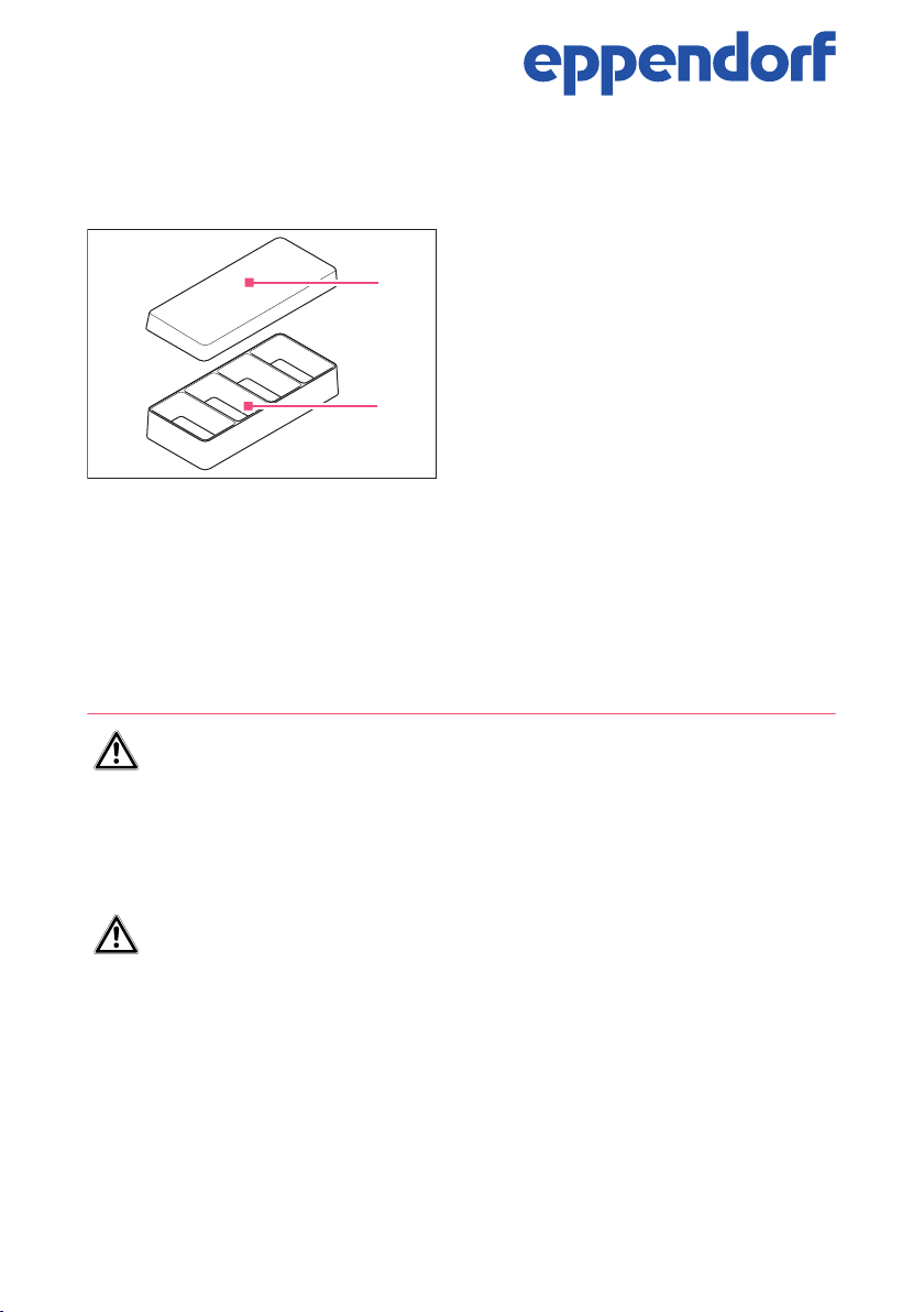
The Eppendorf Cell Imaging Coverglasses
are suitable for the growth and microscopy
1
of cells. There are chambers with 1, 2, 4 or
8 wells (2). The lid (1) prevents evaporation
and protects against contamination.
2
The bottom of the Eppendorf Cell Imaging Coverglasses is made of glass as thick as a
cover glass and is suitable for the microscopy of living cells.
The field of application of Eppendorf Cell Imaging Consumables is training, routine and
research laboratories in the areas of life science, industry, clinics or chemistry. The
Eppendorf Cell Imaging Consumables may only be operated by skilled personnel who
have been trained in the areas mentioned above.
2 Notes for use
WARNING! Risk of contamination
Consumables are only sterile in sealed packaging.
Please check that the packaging is undamaged.
Observe the expiry date printed on the packaging.
Only open the packaging immediately before use.
Only use visually perfect and undamaged items.
WARNING! Risk of contamination from multiple use.
Consumables are intended for single-use only.
After use, dispose of consumables in accordance with the substances with
which they have come into contact.
Instructions for use
2
Eppendorf Cell Imaging Coverglasses
English (EN)
WARNING! Damage to health due to infectious liquids and pathogenic
germs.
When handling infectious liquids and pathogenic germs, observe the national
regulations, the biological security level of your laboratory, the material safety
data sheets, and the manufacturer's application notes.
Wear personal protective equipment.
For full instructions regarding the handling of germs or biological material of
risk group II or higher, please refer to the "Laboratory Biosafety Manual"
(Source: World Health Organization, current edition of the Laboratory
Biosafety Manual).
CAUTION! Risk of injury from glass fragments!
Glass fragments can cause cuts.
Handle consumables made from glass with care. Avoid glass breakage.
Do not touch glass fragments with your hands. Use suitable tools to pick up
glass fragments.
Reagents, consumables contaminated by reagents and materials used for cleaning and
disinfecting must be disposed of in accordance with laboratory regulations.
The surface is tissue culture treated (TCT). The surface should be coated with an
appropriate biological coating (e.g. collagen) to support cell attachment and growth.
3 Application
1. Remove the lid from the Cell Imaging Coverglass.
2. Pipet the cell suspension into the single wells.
3. Incubate the Cell Imaging Coverglasses.
4. Microscope Cell Imaging Coverglass using an inverted microscope.
Eppendorf Cell Imaging Coverglasses are suitable for microscopy of living cells.
The chamber is tied to the cover glass by means of a silicone seal. The chamber
can be removed for further work steps or for archiving purposes.
Removing the chambers
1. Carefully press down the cover glass close to the inner wall of the chamber by means
of a flat device (e.g. spatula).
2. Lift the chamber upwards to remove it.
 Loading...