

Bio-Rad Bio-Plex Pro Human Angiogenesis Reagent and Diluent Kits User Manual
Bio-Plex
Manager™ software
recommended
Bio Plex ProTMAssays
-
Angiogenesis
Instruction Manual
For technical support, contact your local Bio-Rad office or
in the US, call 1-800-4BIORAD (1-800-424-6723).
For research use only. Not for diagnostic procedures.

Table of Contents
Section 1 Introduction . . . . . . . . . . . . . . . . . . . . . . . . . . . . . . 1
Section 2 Principle . . . . . . . . . . . . . . . . . . . . . . . . . . . . . . . . . 2
Section 3 Product Description . . . . . . . . . . . . . . . . . . . . . . .4
Section 4 Required Materials . . . . . . . . . . . . . . . . . . . . . . . . 5
Section 5 Recommended Materials . . . . . . . . . . . . . . . . . . 6
Section 6 Sample Preparation . . . . . . . . . . . . . . . . . . . . . . . 7
Section 7 Standard Preparation . . . . . . . . . . . . . . . . . . . . . 8
Section 8 Control Preparation (optional) . . . . . . . . . . . . . 10
Section 9 Assay Instructions . . . . . . . . . . . . . . . . . . . . . . . 12
Plan Experiment . . . . . . . . . . . . . . . . . . . . . . . . . . 12
Prepare Coupled Magnetic Beads . . . . . . . . . . . . 13
Calibrate Vacuum Apparatus . . . . . . . . . . . . . . . . 13
Assay Procedure . . . . . . . . . . . . . . . . . . . . . . . . . . 14
Section 10 Data Acquisition . . . . . . . . . . . . . . . . . . . . . . . . . 17
Section 11 Troubleshooting . . . . . . . . . . . . . . . . . . . . . . . . . 24
Section 12 Safety Considerations . . . . . . . . . . . . . . . . . . . .28

Section 1
Introduction
Angiogenesis is a critical component of various normal and pathological
processes. A regulated angiogenic process includes the growth of new
blood vessels for tissue repair, fetal development and female reproductive
cycle. An uncontrolled angiogenic growth contributes to a major
pathological component of diseases such as cancer, cardiovascular
disease, and rheumatoid arthritis. Many proteins involved in the
uncontrolled angiogenic growth are candidate drug targets relevant for the
development of cancer therapies. Developing these therapies involves
measurement of various angiogenesis biomarkers.
Bio-Plex Pro™ angiogenesis assays are magnetic bead-based multiplex
assays that allow the measurement of angiogenesis biomarkers in diverse
matrices including serum, plasma, and cell/tissue culture supernatant. The
multiplexing feature makes it possible to quantitate the level of multiple
angiogenesis targets in a single well of a 96-well microplate in just 3 hours,
using as little as 12.5 µl of serum or plasma, or 50 µl of culture
supernatant.
As one of the most recent additions to the Bio-Plex
system, these assays incorporate magnetic beads into their design. The
magnetic beads allow the use of an assay protocol similar to non-magnetic
Bio-Plex assays, with the option of using magnetic separation of wash
steps instead of vacuum filtration (and allows automation of many of the
steps).
®
suspension array
Bio-Plex Manager™ software is recommended for Bio-Plex Pro
angiogenesis assays. Instructions for Luminex xPONENT software are
also provided. For instructions using other xMAP system software
packages, contact Bio-Rad Technical support or your Bio-Rad field
application specialist.
For a current listing of Bio-Plex angiogenesis assays, visit us on the Web at
www.bio-rad.com/bio-plex/
1

Section 2
Principle
Technology
The Bio-Plex®suspension array system is built around three core
technologies. The first is a novel technology that uses up to 100 unique
fluorescently dyed beads (xMAP technology) that permit the simultaneous
detection of up to 100 different types of molecules in a single well of a
96-well microplate. The second is a flow cytometer with two lasers and
associated optics to measure the different molecules bound to the
surface of the beads. The third is a high-speed digital signal processor
that efficiently manages the fluorescent output.
Assay Format
The principle of these 96-well plate-formatted, bead-based assays is
similar to a capture sandwich immunoassay. An antibody directed
against the desired angiogenesis target is covalently coupled to internally
dyed beads. The coupled beads are allowed to react with a sample
containing the angiogenesis target. After a series of washes to remove
unbound protein, a biotinylated detection antibody specific for a different
epitope is added to the reaction. The result is the formation of a
sandwich of antibodies around the angiogenesis target. Streptavidinphycoerythrin (streptavidin-PE) is then added to bind to the biotinylated
detection antibodies on the bead surface.
Data Acquisition and Analysis
Data from the reaction are then acquired using the Bio-Plex suspension
array system (or Luminex system), a dual-laser, flow-based microplate
reader system. The contents of the well are drawn up into the reader.
The lasers and associated optics detect the internal fluorescence of the
individual dyed beads as well as the fluorescent signal on the bead
surface. This identifies each assay and reports the level of target protein
in the well. Intensity of fluorescence detected on the beads indicates the
relative quantity of targeted molecules. A high-speed digital processor
efficiently manages the data output, which is further analyzed and
presented as fluorescence intensity on Bio-Plex Manager™ software.
2
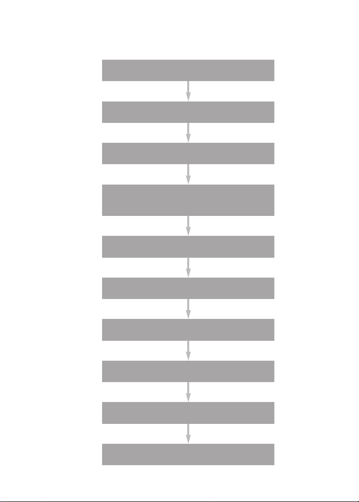
Assay Workflow
Prewet wells
Add beads
Wash
Add standards, controls,
and samples, 30 min
Wash
Add detection antibody, 30 min
Wash
Add streptavidin-PE, 10 min
Wash
Resuspend, acquire data
3

Section 3
Bio-Plex®Reagent Kit
Product Description
Bio-Plex Pro™ angiogenesis assays require the use of the Bio-Plex
angiogenesis reagent kit to run the multiplex panel.
Component of the
Bio-Plex Angiogenesis
Reagent Kit
Bio-Plex assay buffer
Store at 4ºC. Do not freeze.
Bio-Plex wash buffer
Store at 4ºC. Do not freeze.
Bio-Plex detection
antibody diluent
Store at 4ºC. Do not freeze.
Streptavidin-PE (100x)
Store at 4ºC. Do not freeze.
Sterile filter plate (96-well)
with cover and tray
Sealing tape 1 pack of 4 10 packs of 4 (40)
Angiogenesis Instruction
Manual
Storage and Stability
Kit components should be stored at 4ºC and never frozen. Coupled
magnetic beads and streptavidin-PE should be stored in the dark. All
components are guaranteed for up to 6 months from the date of
purchase when stored as specified in this manual.
171-304060
1x96-Well Format
1 x 75 ml 1 x 750 ml
1 x 150 ml 2 x 750 ml
1 x 15 ml 1 x 150 ml
1 vial 1 vial
1 plate 10 plates
1 1
10x96-Well Format
171-304061
®
4

Section 4
Required Materials
Bio-Plex Pro™ angiogenesis assays require the Bio-Plex Pro™
angiogenesis reagent kit and a multiplex angiogenesis assay panel. The
Bio-Plex Pro angiogenesis assay panels contain the following
components.
• Antibody-conjugated beads (25x concentration)
• Detection antibody (10x concentration)
• Angiogenesis standard (2 vials, lyophilized)
• Angiogenesis control (2 vials, lyophilized)
Visit us on the web at www.bio-rad.com/bio-plex/ for our latest list of
assays.
5

Section 5
Recommended Materials
Item Ordering Information
Bio-Plex human serum diluent kit
For tissue culture samples, dilute
samples and standards with appropriate
tissue culture media
Bio-Plex Pro human angiogenesis
standards
Additional standards sold separately
(optional)
Item Ordering Information
Bio-Plex®suspension array system
(or Luminex System)
Bio-Plex validation kit
Bio-Plex calibration kit
Microtiter plate shaker
IKA-Schuttler MTS-4 shaker for 4
microplates or Lab-Line Model 4625
Plate Shaker (or equivalent, capable of
300-1,100 rpm)
Filter plate vacuum apparatus
Bio-Rad Aurum™ vacuum manifold
IMPORTANT: The use of filter plate
manifolds other than the one specified
may result in diminished assay
performance; see section 8 for
instructions specific to this assay
Vortexer
VWR brand mini-vortexer
Scientific Instruments Vortex-Genie 2 mixer
Reagent reservoir
Corning, Inc. Costar 50 ml reagent
reservoir 4870
Other materials
Pipets and pipet tips, sterile distilled
water, aluminum foil, absorbent paper
towels, 1.5 ml microcentrifuge tubes,
15 ml culture tubes
Bio-Rad catalog #171-305000
(1x96)
Bio-Rad catalog #171-305001
(10x96)
Bio-Rad catalog #171-D40003
(2 vials, lyophilized)
Bio-Rad catalog #171-D40004
(50 vials, lyophilized)
Bio-Rad catalog #171-000205
Bio-Rad catalog #171-203001
Bio-Rad catalog #171-203060
IKA catalog #3208000
VWR catalog #57019-600
Bio-Rad catalog #732-6470
VWR catalog #58816-121
VWR catalog #58815-234
Bio-Rad catalog #224-4872
6

Section 6
Sample Preparation
This section provides instructions for preparing samples derived from serum,
plasma, and tissue culture supernatant. For sample preparations not
mentioned here, consult the publications listed in Bio-Rad bulletin 5297,
available for download at discover.bio-rad.com
Serum and Plasma Samples
Note that for plasma samples, both EDTA plasma and citrate plasma are
acceptable. Avoid using hemolyzed samples.
1. Collect and process the serum or plasma samples and assay immediately
or freeze at –20ºC. Avoid repeated freeze-thaw cycles.
2. Centrifuge the samples at 13,200 rpm for 10 min at 4ºC to clear the
samples of precipitate. Alternatively, carefully filter the samples with a
0.8/0.22 µm dual filter to prevent instrument clogging.
3. Immediately dilute 1 volume of sample with 3 volumes of sample diluent.
Keep the samples on ice until ready for use.
Tissue Culture Supernatant Samples
1. Collect and process the tissue culture supernatant samples and assay
immediately or freeze at –20ºC. Avoid repeated freeze thaw cycles.
2. If required, dilute the culture supernatant with culture medium.
Serum-free culture medium should contain carrier protein (such as BSA)
at a concentration of at least 0.5%. Keep the samples on ice until ready
for use.
7

Section 7
Standard Preparation
Two vials of Iyophilized angiogenesis standards are provided in each Bio-Plex
Pro™ angiogenesis assay. However, only one vial is required per 96-well plate.
The product insert provided with the assay lists the concentration of the
reconstituted standard. This procedure will prepare enough standard to run
each dilution in duplicate.
Reconstitute Standards
1. Gently tap the glass vial containing the lyophilized angiogenesis
standard on a solid surface to ensure the pellet is at the bottom.
2. Reconstitute 1 vial of lyophilized standard with 500 µl of the
standard diluent. Do not use assay buffer to dilute standards.
3. Gently vortex 1–3 sec and incubate on ice for 30 min.
4. Be consistent with the incubation time for optimal assay performance.
Prepare Standard Dilution Series
The angiogenesis concentrations specified for the 8-point standard
dilution set have been selected for optimized curve fitting using the 5parameter logistic (5PL) or 4-parameter logistic (4PL) regression in
Bio-Plex Manager™ software. Results generated using dilution points
other than those listed in this manual have not been optimized.
1. Label a set of 1.5 ml Eppendorf tubes as shown in the diagram on
the next page.
2. Pipet the appropriate volume of standard diluent into the tubes (see
diagram on next page). Use serum standard diluent for serum and
plasma samples and culture medium for culture samples.
8
 Loading...