

Page 1
Page 2

Thank you for purchasing the Welch Allyn RL-150™ Rhinolaryngoscope. The operating and maintenance instructions
found in this manual should be followed to ensure many years
of accurate and reliable service. Please read these instructions
thoroughly before attempting to use your new RL-150
Rhinolaryngoscope.
Warning
The user of the RL-150 Rhinolaryngoscope should be thoroughly trained in
the medical procedures appropriate to the equipment. Furthermore, time
should be taken to read and understand these instructions before performing any procedures. Failure to do so may result in injury to the patient
and/or damage to the instrument.
While this manual describes the recommended protocol for inspecting and
operating the equipment, it does not outline procedure techniques. Only
physicians trained and versed in the procedure of nasopharynlaryngoscopy
should use this equipment.
Table of Contents
Specifications............................................................................................1
Components.............................................................................................1
Nomenclature...........................................................................................2
Prior to Initial Use ...................................................................................3
System Set Up..........................................................................................3
System Inspection....................................................................................4
Operation .................................................................................................6
Cleaning....................................................................................................7
Disinfection ............................................................................................10
Sterilization and Aeration ......................................................................12
Storage....................................................................................................13
Service ....................................................................................................13
Page 3
Specifications
Welch Allyn
RL-100
LENS CLEANER
Angle of View .............................75°
Depth of Field ....................3-50 mm
Diopter ....................+2 to -8 diopter
Tip Deflection
Up ....................................130°
Down...............................130°
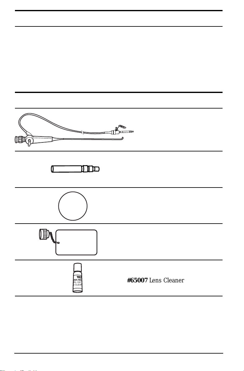
Components
Distal Rigid Portion
Diameter ....................3.4 mm
Insertion Tube Diameter .....3.5 mm
Insertion Tube
Working Length ...........30 cm
Total Length.........................53.5 cm
#65050 RL-150
Rhinolaryngoscope
#65015 LX-150 Adaptor
#65008 Eyepiece Cap
#65012 ETO Vent Cap
#65007 Lens Cleaner
1
Page 4
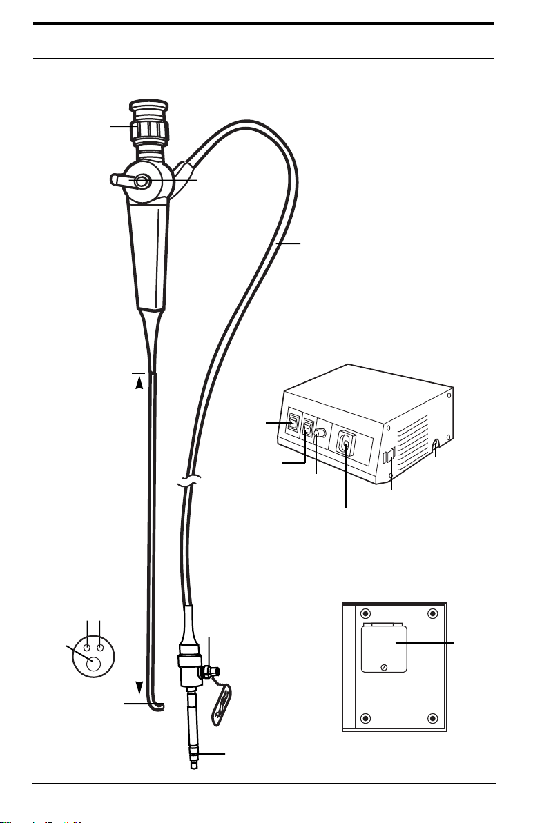
Nomenclature
RL-150 Rhinolaryngoscope
Diopter
Adjustment
Ring
Angulation
Lever
Power
Switch
Umbilical
Cord
LX-150 Light Source
Objective
Lens
Distal Tip
2
Light
Guides
Insertion Tube
Bending
Section
Air
Switch
ETO
Vent
Connector
Light Guide
Terminal
Illumination
Control
Endoscope
Connector Port
Air Output
Connector
Water Bottle
Bracket
Lamp
Access
Door
Bottom View
Page 5

Prior to Initial Use
Before set up and inspection of the equipment, check all components
received against the list of components (see Components section, pg. 1) to
verify complete set. If parts are missing, please notify Welch Allyn. Review
the Nomenclature, Set Up, Operation and Cleaning/Disinfection sections to
become familiar with the equipment.
Specifically Inspect:
Rhinolaryngoscope
Insertion Tube—For tears, cuts, dents, bubbles, bumps
Control Section—Test angulation lever/bending section deflection to
assure smooth articulation
Light Source
Cabinet—For any dents, scratches or other surface abnormalities
System Set Up
Rhinolaryngoscope
The No. 65015 adaptor is coupled to your RL-150 Rhinolaryngoscope
endoscope connector terminal when shipped from Welch Allyn. [In the
event that the adaptor should become uncoupled, Steps 1-2 (below)
should be undertaken. Otherwise, proceed to Step 3.]
1. Place the No. 65015 adaptor over the light guide terminal.
2. Thread the adaptor on until snug against endoscope connector terminal.
Endoscope
Connector
Terminal
RL-150 Umbilical
Cord
3. Insert the male end of the adaptor into the Light Source endoscope
connector port. Push until the ball detent in the light source “clicks”
into place to ensure a secure fit.
LX-150
Light Source
Adaptor
65015
Endoscope
Connector Port
3
Page 6

System Set Up (continued)
Light Source (See Light Source Manual for complete instructions.)
1. Plug power cord into the receptacle on the back of the cabinet.
2. Plug the remaining end into a properly grounded, hospital-grade
AC outlet.
3. Activate power and air switches to verify functionality of the lamp
and pump.
NOTE: The air pump that is built in to some light boxes is not used
for Rhinolaryngoscopy.
System Inspection
The following steps (in addition to leak testing, cleaning and disinfection)
should be repeated prior to every procedure to verify that the system is
working. If any problem is encountered, immediately contact Welch Allyn
Customer Service for assistance.
Insertion Tube
Check the entire surface of the
insertion tube for abnormalities
such as dents, wrinkles, or cuts.
Any indentation in a flexible
shaft of the endoscope can cause
damage to the internal mechanisms.
CAUTION: To avoid further damage to the endoscope or the possibility of malfunction during a procedure, do not use any instrument
with outward signs of damage.
NOTE: The distal end of the fiberscope must be
protected against damage from impact. Never
apply excessive force by twisting or severely
bending the insertion tube or bending section
of the instrument.
4
Page 7

System Inspection (continued)
Inspection of Light Guides and Optics
1. With the light source lamp on, hold distal tip
approximately 40 mm from any printed surface.
Verify light is being emitted from light guides.
2. Hold eyepiece to your eye and rotate diopter
ring until print is in focus. Verify focus from
10-40 mm from surface.
Inspection of Umbilical Cord
1. Check the umbilical cord for cracks,
dents, crushed and twisted areas.
2. Verify that the endoscope
connector terminal’s light
guide terminal fitting is tight
and does not move when
light pressure is applied.
NOTE: Instrument should be cleaned and disinfected or sterilized
prior to every use. (Follow steps starting on pages 7 and 10.)
Inspection of Light Source
1. Activate “power” switch. This will start the lamp and cooling fan.
CAUTION: Do not look directly into the endoscope connector port
when activating the light source.
2. The output of the light source is controlled by the illumination intensity
control. Clockwise rotation will
increase illumination output. Counterclockwise will decrease illumination.
NOTE: If your light source has an air pump built in, the air pump is
not used for Rhinopharynlaryngoscopy.
WARNING: The risk of thermal injury exists whenever fiber optic
instruments are used with high intensity light sources.
The risk of injury is greatest:
(A) During close stationary observation and/or prolonged close
contact with mucosa.
(B) When the fiberscope is advanced slowly through a narrow
lumen.
Close stationary viewing should be avoided and the level of illumination
should be limited to the level necessary for adequate visualization.
5
Page 8

Operation
Procedure
The methods and techniques of rhinopharynlaryngoscopy are well-defined
and documented. Endoscopy training seminars and programs are also in
existence.
No attempt is made in this manual to outline the medical procedure or
techniques of rhinopharynlaryngoscopy. The physician should always
take care to understand the clinical background of each patient and the
contraindications of the procedure.
Preparation Before Insertion
Note: The endoscope should be properly cleaned and disinfected
or sterilized prior to the first use and all subsequent uses. Only by
adhering strictly to the steps outlined elsewhere in this manual can
the user be assured of the effectiveness of the processes.
1. Gently clean the objective lens with a cotton-tip applicator moistened
with alcohol.
2. Gently wipe the insertion tube with gauze dampened with clean water.
3. The individual user should adjust the diopter adjustment ring to make
sure that a clear view can be obtained. No further adjustment should
be necessary during a procedure.
4. If desired, apply a water-soluble lubricant to the insertion tube. The
objective lens must be kept free of the lubricant which would obstruct
your view.
NOTE: Do not use lubricants that are petroleum based as these will
deteriorate the rubber covering the bending section.
Insertion and Operation
1. Always insert the instrument slowly under direct vision.
2. Once lumen is visible, adjust the light intensity control on the light
source to desired setting.
Note: The distal tip of the rhinolaryngoscope may become warm if
in continuous operation with the illumination set too high; this can
damage the mucosa. Use the lowest light setting possible.
3. Tip deflection must be performed under direct observation. Deflect the
bending section gently using minimal force.
4. When withdrawing the instrument, pull back slowly under direct obser-
vation. Do not remove the instrument “blindly”.
6
Page 9

Cleaning
T
E
S
T
D
A
N
G
E
R
0.1
0.2
0.3
0.4
0
65009
Endoscopic instruments should be cleaned immediately after each use.
Endoscopes are delicate and will degrade if not cleaned promptly, due to
the effects of blood, mucous, etc. The steps outlined on the following pages
have been tested and verified to have no damaging effects. Therefore, these
procedures should be adhered to.
Supplies Needed For Cleaning
Large basin of water Disposable gloves
Cleaning solution (soap solution) Leakage tester
Gauze pads Soft scrub brush
Immediately After Procedure…
1. Immediately after removing the instrument from the patient, gently
wipe all blood, mucous and debris from the insertion tube with a
gauze pad moistened in clean water.
2. Turn of f the “power”
switch on the light
source.
3. Disconnect endoscope
from light source by
grasping endoscope connector terminal/adaptor
and pulling back gently.
7
Page 10

T
E
S
T
D
A
N
G
E
R
0.1
0.2
0.3
0.4
0
Cleaning (continued)
T
E
S
T
D
A
N
G
E
R
0.1
0.2
0.3
0.4
0
Perform Leak Test Procedure
The leakage test allows for a simple two stage test of the water tight
integrity of the instrument. Air pressure is introduced into the interior
of the instrument by means of a hand pump. The instrument is then
checked for pressure decay via a gauge and then visually.
Green Zone
Red Zone
Air Bladder
Scope Connector
Pressure
Release
Hand Bulb
Leakage Tester
Stage-One Test
BEFORE IMMERSION, the instrument should be tested for any major loss of
integrity in its watertight construction (example: major tear in the bending
section).
1. Secure the Leakage Tester to
the ETO vent connector on the
instrument’s endoscope connector
terminal. The Leakage Tester
connector and the ETO vent
connector MUST be dry before
securing. Proper connection will
require alignment of the air vent
pin and clockwise rotation.
2. Turn the gauge faceplate to “zero” on
the pressure indicator.
8
Page 11

Cleaning (continued)
T
E
S
T
D
A
N
G
E
R
0.1
0.2
0.3
0.4
0
T
E
S
T
D
A
N
G
E
R
0.1
0.2
0.3
0.4
0
T
E
S
T
D
A
N
G
E
R
0.1
0.2
0.3
0.4
0
3. Pressurize the scope by pumping the hand bulb
until the indicator on the gauge is in the green
zone. DO NOT pressurize into the red zone; it
may cause serious damage to the scope.
4. Observe the gauge pressure to determine if the
indicator remains in the green zone. If the
indicator drops from the green zone rapidly,
a major leak may be indicated.
NOTE: Be certain that the pressure release valve on the handle of
the leakage tester has been tightened. DO NOT IMMERSE the entire
instrument if the gauge indicator does not remain in the green zone.
Instead, contact Welch Allyn Customer Service.
Stage-Two Test
After determining the absence of any major leak in stage-one testing, the
instrument may be immersed in clean water to test for loss of integrity in
the watertight construction.
1. With the leakage tester securely attached to the instrument and the
scope pressurized with the gauge indicator in the green zone, the
entire scope may be immersed in clean water.
NOTE: Only the leakage tester
connector and a small portion of
its tubing should be immersed.
Never immerse the entire
leakage tester.
2. Observe the instrument carefully. A few bubbles may initially rise
from recessed areas of the scope. This is normal. If a continuous stream
of bubbles is observed from
the same spot, a leak is indicated. Immediately remove the
scope from the water. DO
NOT use the scope. Contact
Welch Allyn Customer Service
immediately.
3. After removing the instrument from water, release the air pressure by
opening the pressure release valve on the handle of the leakage tester.
After the gauge indicates “zero”, disconnect the leakage tester from
the scope.
9
Page 12

Cleaning (continued)
NOTE: Never connect or disconnect the leakage tester under water.
This will allow water into the instrument and leakage tester.
4. If leakage was discovered during stage two, thoroughly dry the instrument and contact Welch Allyn Customer Service.
5. If no leakage was discovered during stage two, you may proceed with
cleaning and disinfection of the instrument as outlined in the following
pages.
Cleaning Procedure
1. Prepare a basin with warm water and a mild detergent. Immerse the
instrument in the solution.
2. Thoroughly (but gently) wash the entire outer surface of the instrument
with a soft brush or guaze pad. Remove the instrument from the basin.
NOTE: Do not squeeze or severely bend the insertion tube. Do not
use any abrasive materials. Be careful to avoid damage to the ocular
and distal lenses.
3. Place in basin of clean water. Thoroughly rinse the outside of the
instrument.
4. Gently dry all external surfaces of the fiberscope with soft gauze or a
towel. DO NOT put tension on the insertion tube of the endoscope
while drying since the outer cover of the bending section may be
excessively stretched. Dry the ocular lens with a cotton-tip swab.
Disinfection
Before any attempt is made to disinfect the fiberscope, the complete cleaning procedure, as previously described in this manual, must be completed.
Complete Immersion
Before complete immersion in any disinfecting solution, the instrument
should be LEAK TESTED as described elsewhere in this manual. Before
immersion, the Red ETO vent cap MUST be removed.
1. Immerse the instrument in a basin filled with a compatible disinfection
solution.
WARNING: Using a disinfectant other than those tested for compatibility with the instrument may result in severe damage to the instrument. Contact Welch Allyn Customer Service with any questions
regarding solution compatibility.
10
Page 13

Disinfection (continued)
2. The instrument should remain in contact with the disinfecting solution
for the time period recommended by the manufacturer of the solution
and accepted by the user as appropriate.
3. After the instrument has been in contact with the disinfecting solution
for an appropriate time, remove the instrument from the solution.
4. Place in a basin filled with clean water to rinse all residual disinfecting
solution from the instrument. Remove instrument from water.
5. Gently dry all external surfaces of the instrument with soft gauze or
towel. Do not put tension on the insertion tube while drying since the
outer cover of the bending section may be excessively stretched. Dry
the ocular lens with a cotton-tip swab.
Using the Disinfection Tray
1. Fill the clear tube of the disinfection tray with
disinfectant up to the black indicator dot.
2. Slide insertion tube into the disinfection solution and
position the instrument’s control section securely in
the space provided on the disinfection center.
3. Carefully wind the umbilical
cord and place next to the
tray as illustrated.
4. After soaking for the disinfection solution manufacturer’s
recommended time, remove
the instrument from disinfection tray.
5. Immerse the insertion tube in
clean water to rinse.
6. Remove instrument from water
and dry surfaces of the instrument
thoroughly.
7. Hang scope with insertion tube
straight to dry.
11
Page 14

Disinfection (continued)
Disinfecting Solutions
Specific reference to brand names is not an endorsement of their efficacy
as a disinfecting solution. Tests have shown these solutions to be compatible with the RL-150 Rhinolaryngoscope, providing the manufacturer’s directions are followed.
Solution Brand Name Source Usage
Glutaraldehyde Cidex (14 Day) J&J Follow manufacturer’s
(2%) Medical instructions
Glutaraldehyde Wavicide-01 Wave Energy Follow
(2%) Systems manufacturer’s
instructions
Glutaraldehyde Metricide Metrex Follow manufacturer’s
(14 Day) (2%) Research Corp. instructions
Sterilization and Aeration
Before any attempt is made to sterilize the fiberscope, the complete
cleaning procedure as described elsewhere in this manual must be
completed.
Only the user can assure that sterilization techniques, like those described
here, will accomplish the desired clinical effect.
IMPORTANT WARNING:
Never place the fiberscope in a steam autoclave! Never subject the
fiberscope to ultrasonic cleaning methods!
Ethylene Oxide (ETO) Gas Sterilization can be performed on the RL-150
Rhinolaryngoscope provided the following special instructions, which differ
from other endoscopes, are followed to ensure proper performance of the
instrument.
1. The endoscope must first have been properly cleaned and thoroughly
dried according to the instructions in this manual.
a) The Red ETO Gas Sterilization Venting Cap MUST be on securely.
b) Temperature must not exceed 55°C (131°F).
c) Pressure must not exceed 24 PSI.
d) Humidity must not exceed 50%.
e) Sterilization procedure must not exceed 4 hours.
12
Page 15

Sterilization And Aeration (continued)
2. Following ETO Gas Sterilization, aeration time of 72 hours at room
temperature is required.
3. Aeration Chamber: To shorten the aeration time to 12 hours, an
aeration chamber may be used, provided the temperature does not
exceed 55°C (131°F).
NOTE: Prior to placing the RL-150 Rhinolaryngoscope in an
aeration chamber, the Red ETO Gas Sterilization Vent Cap MUST
be on securely.
Cold Sterilization
For those situations where Ethylene Oxide (ETO) Gas Sterilization capability is not available, Welch Allyn endoscopes have been designed to
withstand immersion in Glutaraldehyde solution for a maximum of 10
hours to achieve “cold sterilization”. Never fully immerse the instrument
without first conducting the leakage test procedure, as described elsewhere in this manual, to verify the watertight integrity of the scope.
WARNING: Never exceed 10-hour maximum immersion.
After 10-hour immersion, the instrument must be thoroughly rinsed to
remove all residual Glutaraldehyde solution.
After being thoroughly rinsed, the instrument must be allowed to
“breathe”, balancing internal and external humidity, by attaching the
Red ETO Venting Cap.
Storage
Thoroughly dry the instrument and accessories. Place in the wall hanger.
1. During storage, the insertion tube of the scope and the light guide
should be kept as straight as possible.
2. The storage area should be dry and airy. Avoid high humidity, high
temperature and areas exposed to direct sunlight.
3. The Red ETO Venting Cap should be placed on the scope to allow
the scope to “breathe”, balancing internal and external humidity, while
it is in storage.
Service
Customer Service Telephone Assistance:
(315) 685-4560
For repairs on the
RL-150 Rhinolaryngoscope, ship to:
Welch Allyn Technical Service
4341 State Street Road
Skaneateles, NY 13153
13
Page 16

4341 State Street Road
Skaneateles Falls, NY 13153
315-685-4560
800-535-6663
Printed in U.S.A.
Part No. 650033 Rev. C
 Loading...