

Olympus CF-HQ190L, CF-HQ190I Specification

EVIS EXERA III COLONOVIDEOSCOPE
CF-HQ190L /I
Amazing image quality and handling for colonoscopy
EVIS EXERA III COLONOVIDEOSCOPE
OLYMPUS CF-HQ190L /I
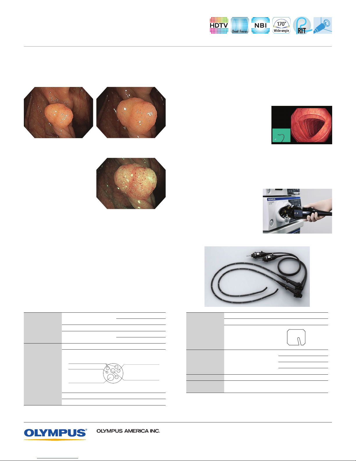
Dual Focus
Dual Focus, a unique Olympus optical innovation, allows the
user to select between two focus settings. With the simple push
of a scope button, the desired depth of eld for observation can
be optimized to either the near eld or normal eld.
Normal-focus mode Near-focus mode
(Narrow Band Imaging)
NBI
NBI in EVIS EXERA III 190
Series scopes provides twice
the viewable distance of EVIS
EXERA II 180 Series scopes.
(Responsive
RIT
Insertion Technology)
RIT combines three proprietary
insertion tube technologies: HFT (High Force Transmission),
PB (Passive Bending), and variable stiffness. These technologies are
designed to facilitate complete colonoscopies by improving scope
handling, insertability, and ergonomics. PB helps EVIS EXERA III 190
Series scopes move through acute bends in the colon. HFT provides
improved operator control for both pushing and twisting maneuvers.
Variable stiffness allows the physician to adjust the rigidity of Olympus
scopes as needed by simply turning an adjustment ring on the
scope’s control section.
Enhanced Image Quality
An enhanced level of resolving power is achieved owing to a
new optical system. Signicantly less halation and noise is
visible on screen. Improved optics, a brighter image, increased
contrast, reduced noise, and reduced halation are the result of
a new CCD available in the EVIS EXERA III, providing superior
image quality even during electronic zoom.
ScopeGuide
ScopeGuide is an integrated
technology in EVIS EXERA III 190
Series HQ colonoscopes.
ScopeGuide provides real-time 3D
visualization of scope position and
conguration. This new level of
visualization provides physicians with the ability to recognize loops
as they form, potentially leading to shorter insertion time and less
patient discomfort.
Waterproof One-touch Connector
A new connector design
minimizes the effort required for
setup prior to and in between
cases. In addition, it is fully
submersible and eliminates the
need for a water-resistant cap
and the associated risk of an
expensive repair due to
accidental immersion.
Field of view
Optical System
Insertion Section
Direction of view Forward viewing
Depth of eld
Distal end outer diameter 13.2 mm
Distal end enlarged
Light-guide Lens
Auxiliary Water Channel
Right Left
Instrument Channel Outlet
Insertion tube outer diameter 12.8 mm
Working length L: 1680 mm I: 1330 mm
3500 Corporate Parkway, PO Box 610, Center Valley, PA 18034
Normal 170º
Near 160º
Normal 5-100 mm
Near 2-6 mm
Up
Down
Channel inner diameter 3.7 mm
Minimum visible distance
Instrument Channel
Air/Water Nozzle
Objective Lens
Specifications, design, and accessories are subject to change without any notice or obligation on the part of the manufacturer.
Bending Section Angulation range
Total Length L: 2005 mm I: 1655 mm
Compatible EVIS
EXERA System
Olympus is a registered trademark of Olympus Corporation, Olympus America Inc., and/or their affiliates.
Direction from which
endotherapy accessories
enter and exit the
endoscopic
image
Video System Center OLYMPUS CV-190
Xenon Light Source OLYMPUS CLV-190
For more information, contact your local Olympus
©2012 Olympus America Inc. All rights reserved.
4.0 mm (Normal) from the distal end
Up 180º
Down 180º
Right 160º
Left 160º
Image courtesy of Roy Soetikno, MD.
sales representative, or call 800-848-9024.
www.olympusamerica.com
Printed in USA OAIGI0312BRO8796
 Loading...