

Page 1
Leica DM IL LED
Brilliant Optics Combined with Innovative Illumination
The New Inverted Microscope for Routine and Laboratory Microscopy
in Cell Biology and Medicine
Page 2
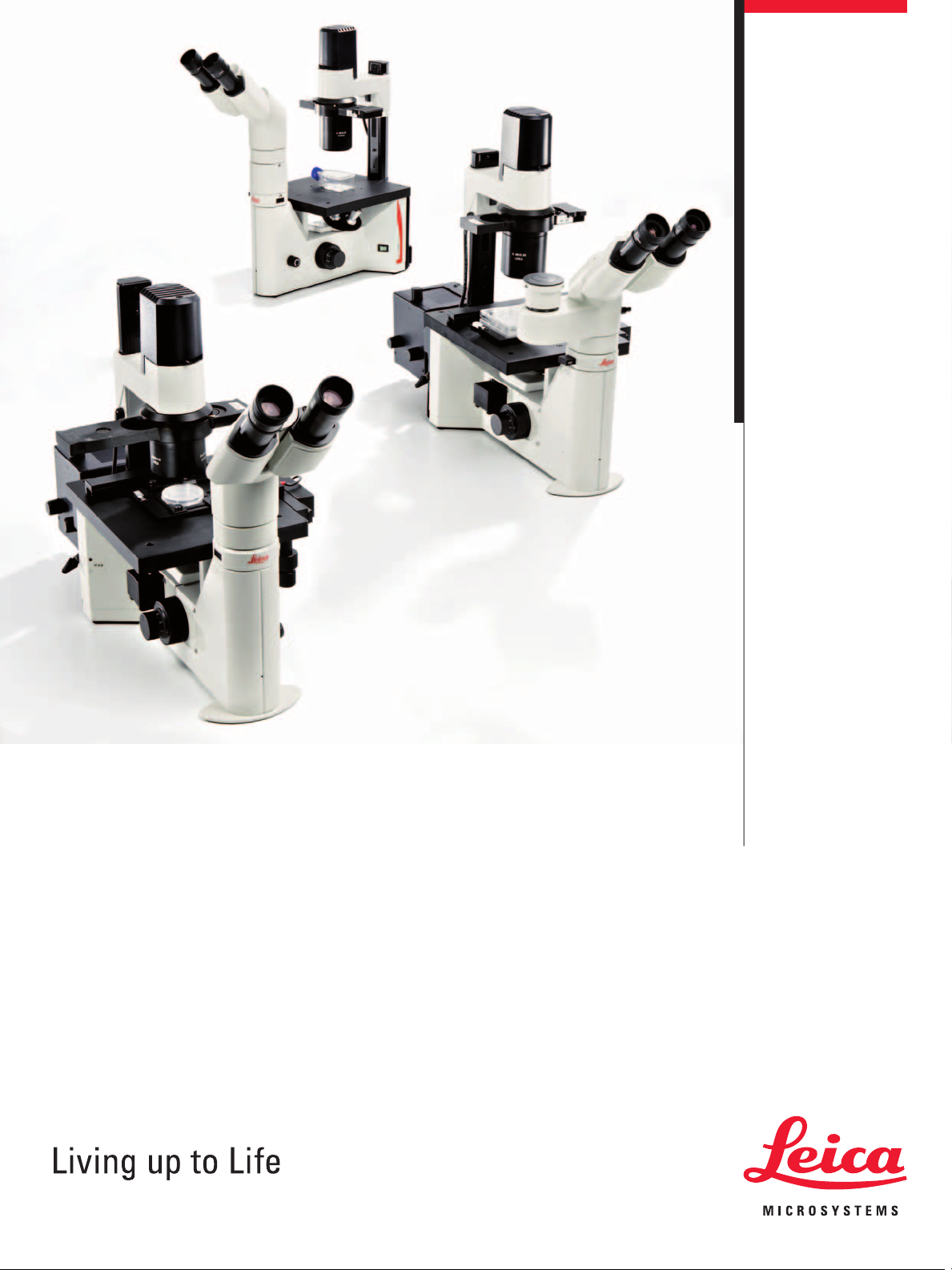
Inverted Routine
Microscopy in a New Light
Compact and stable
Lean and sturdy design•
Plenty of space for operation•
Low stage height•
Large dimensions and low•
center of gravity of microscope
Large working distances •
Wide variety of possible applications
Cell biology and medicine•
Micromanipulation (injection, IVF, ICSI)•
Medicine•
Biotechnology•
Developmental biology•
Transgenics•
Optical performance and illumination are key elements in microscopy. Both characteristics are unifi ed in the new design of the
Leica DM IL LED. As the fi rst inverted routine microscope, the
Leica DM IL LED is not only equipped with outstanding Leica HC
optics, but also features innovative LED illumination. The transmitted-light illuminator including optimized condensers and improved
contrast methods are adapted specifi cally for cell biology applications. High stability, plenty of space for operation, large working
distances, illumination without heat development and the separately accommodated electronics provide optimum conditions for
microscopy. The Leica DM IL LED is exceptionally well-suited for
uses ranging from various cell and tissue culture examinations in
life sciences, developmental biology studies or micromanipulation
in cell biology to living cell examinations in transgenics or electrophysiology.
Molecular biology•
Fluorescence applications •
2
Page 3
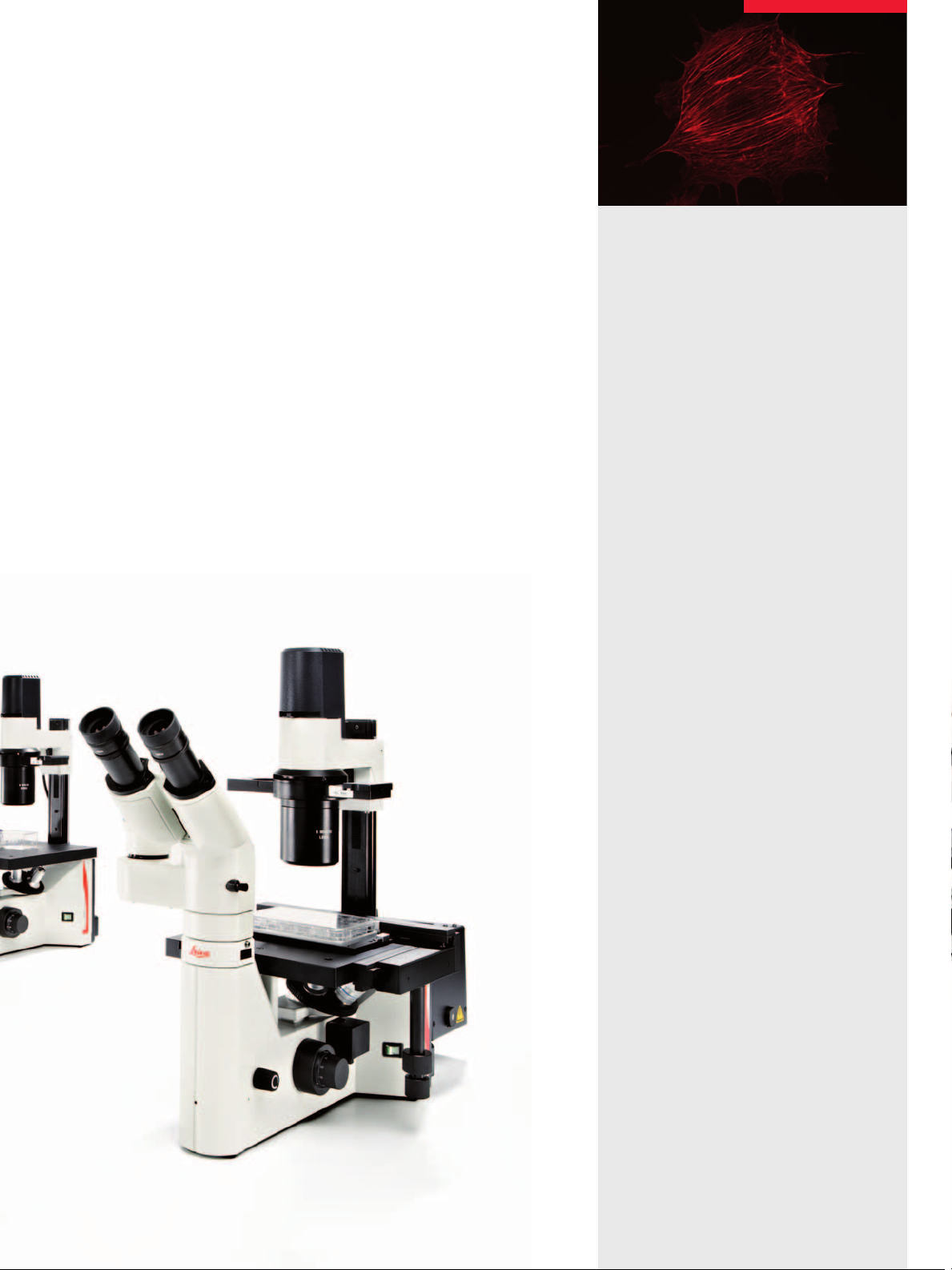
The fl uorescence version, the Leica DM IL LED Fluo, also offers a
variety of possible applications. Optionally, it is also available with
the new LED illumination.
Integrated fl uorescence
Manual fl uorescence with three fi lter cubes•
Heatable microscope stages and 3-plate cross-stages provide
great fl exibility for experiments on living cells under physiological
conditions.
The Leica DM IL LED has a further advantage that distinguishes
it from other microscopes in its class: The stand is highly compatible with components of the Leica research microscopes.
Objectives, eyepieces, tubes, camera ports, contrast methods.
Additionally, special tubes and condensers have been developed
for the Leica DM IL LED.
Integrated shutter•
Optionally LED, classic mercury •
illumination or fi ber optic coupling
Flexible and modular
A full range of optical components•
Compatible with research stands •
Unheated and heated stages•
Large selection of tubes •
Comprehensive range of accessories•
for special applications
3
Page 4
The Most Comprehensive Array
of Contrast Methods
All available contrast methods can be adapted to individual
applications easily and quickly. Two condensers have been developed specifi cally for the Leica DM IL LED, which can be used for
the entire magnifi cation range of the respective contrast method.
The high-resolution S40/0.45 condenser makes even tiny details of
a specimen visible. Both condensers, the S40/0.45 and S80/0.30,
allow for use of phase contrast up to the 63x objective as well as
Integrated Modulation Contrast (IMC) up to the 40x objective.
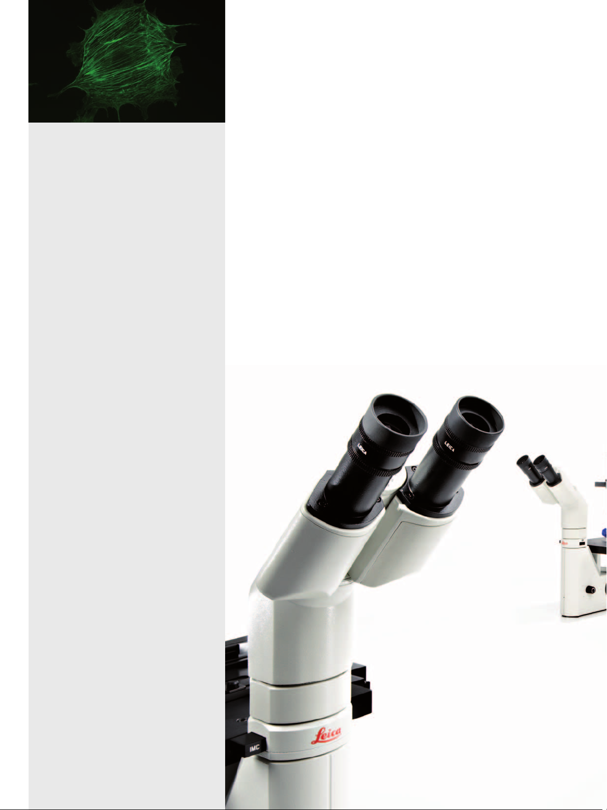
Fatigue-free operation
The ergonomic arrangement of all controls such as the focus dial,
brightness controller, condenser height adjustment, objective
nosepiece and XY stage adjustment allow users to be relaxed
while working with the microscope – even for hours. The heightadjustable stages, Ergo tubes with variable tube height, fl exible
viewing height, and the interpupillary distance and diopter setting
enable each user to confi gure his or her personal Leica DM IL LED.
The large working distances provide suffi cient room for large culture fl asks, and the unobstructed view of the specimen area facilitates handling more diffi cult specimens.
4
Page 5

Brightfi eld
All Leica brightfi eld and phase objectives from 2.5x to 100x can
be used for stained specimens. The bright fi eld method can be
used even for low magnifi cation levels without condenser. A
working distance of 200 mm is realized simply by unscrewing the
condenser head.
Phase contrast
Phase contrast is used primarily in live cell microscopy to make
structures in unstained specimens visible. Three preadjusted light
rings on a slider allow phase contrast for all objectives from 5x to
63x. No readjustment is necessary when changing the objectives.
The intelligent LED illumination adjusts the brightness automatically when switching between the phase contrast and brightfi eld
method.
PAP smear, Brightfi eld
Integrated Modulation Contrast (IMC)
IMC creates relief-type images and has proven to be an alternative to Differential Interference Contrast (DIC), particularly in
micromanipulation. The IMC developed by Leica Microsystems
does not require special objectives because the IMC modulator is
not integrated into the objective, but operated via a separate slider.
The IMC illumination slider is encoded and controls the LED illumination. IMC is available for both condensers and for standard 10x,
20x, 32x and 40x objectives.
Fluorescence
Incident-light fl uorescence is an integral part of the Leica DM IL
LED
Fluo microscope variant. The fl uorescence slider holds three
fi lter blocks. The transmitted light method and fl uorescence can
be used simultaneously. This way, object structures can be clearly
assigned. Each fi lter block comprises an optimally matched combination of excitation, refl ection and barrier fi lters. Illumination can
be generated via the Leica SFL100 LED illumination, the classic
mercury illumination or the Leica EL6000 fi ber optic coupling.
Section taste buds rabbit, Phase Contrast
C. elegans, Integrated Modulation Contrast (IMC)
Convallaria lilly of the valley,10x, Fluorescence
5
Page 6

Perfectly Illuminated
High-intensity and high-contrast
5 watt LED illumination•
Constant color temperature•
Automatic brightness adjust-•
ment to the contrast method
Phase contrast from 5x to 63x•
Modulation contrast for •
10x, 20x, 32x and 40x
Integrated modulation contrast•
Without special objectives; •
for all condensers
Cost-effective and effi cient
Low energy consumption•
No heat buildup•
LED with a service life of 50,000 hours•
The Leica DM IL LED is the fi rst inverted routine microscope with
LED illumination for the transmitted-light method. The compact
illumination unit includes a precentered light emitting diode that
has a service life of 50,000 hours.
The LED, with a service life at least 250 times longer than that of
conventional halogen lamps, is easy to maintain and very costeffective. The 5 watt power of the LED is completely converted
into light while maintaining uniform color temperature. Nearly no
unwanted heat is generated. Optionally, users can activate the
integrated automatic shut off – an additional contribution to energy
savings.
In particular the phase contrast and IMC are optimized by the
warm hue of the LED. With the help of the attachable fi lter, the
illumination impression can be individually adapted in both directions of the color spectrum.
The integrated collector attains optimum light utilization and the
integrated aperture diaphragm creates optimum contrast and
resolution for every specimen and every objective.
"Auto-off" function for illumination •
Rat testes, Integrated Modulation Contrast (IMC)
6
Page 7

Anything Goes
For the fi rst time, a condenser concept has been realized in the
Leica DM IL LED that allows all contrast methods with all condensers. With a working distance of at least 40 mm and a numerical aperture of 0.45, the S40 condenser is the perfect tool for
applications for which optimum resolution is the most important
parameter. Phase contrast and IMC ensure optimum contrasting.
The working distance of at least 80 mm and 0.30 aperture of the
S80 condenser are the ideal prerequisites for achieving maximum
possible free room around the specimen and optimum contrasting
at the same time. The continuous adjustment of the condenser
height depending on specimen vessel and liquid layer is a one-ofa-kind feature. It ensures maximum fl exibility when using peripheral microtools.
Whether you work with thin sections or thick specimens, phase
and modulation contrast produce a brilliant microscopic image for
all specimens and applications.
Perfect for your applications –
the S40 and S80 condensers
High-intensity and high-contrast –
the 10 W LED illumination
7
Page 8

Innovative LED illumination for fl uorescence applications
with the Leica SFL100.
Flexible Fluorescence
Fluorescence applications, in particular GFP labeling, play an ever
more important role in clinical diagnostics and routine microscopy.
The Leica DM IL LED Fluo has been designed in consideration of
this trend. The microscope has been equipped with a fl uorescence
axis and a 3-position slider to ensure fast and easy switching to
different fl uorochromes. The slider glides smoothly in an elaborately constructed dovetail guide. An extensive, constantly growing range of fi lters allows a wide variety of fl uorescence examinations. The fi lter blocks are optimized for minimizing stray light.
Excitation, refl ection and barrier fi lters adapt to your application.
Transmitted-light methods can be used simultaneously or as an
alternative so that fl uorescent and nonfl uorescent structures can
be clearly assigned. An integrated shutter protects the specimen
against bleaching.
The Leica DM IL LED Fluo is the fi rst routine fl uorescence microscope
that allows users to choose between classic illumination (halogen,
mercury or high-pressure xenon lamps), the ”cold” light guide coupling of the Leica EL6000 and the new LED illumination, Leica SFL100.
This gives users the ability to excite and examine fl uorochromes
to see more detail under the microscope. A dark background and
the bright emitted fl uorescence produce brilliant color images.
The fi eld of applications ranges from DAPI (UV) for nuclear stain-
ing to CY5 (IR) for immuno-
histochemical arrays. The
Leica DM IL LED Fluo thus is a
high-performance instrument for
use in immunology, cytology, virolo-
gy – and in any fi eld that requires fl uo-
rescence techniques for living specimens.
Leica DM IL LED with fl uorescence axis and 3-position slider
8
Page 9

Capturing Every Detail
A large selection of tubes is available for the Leica DM IL LED. All
tubes can be rotated individually by 360° and are equipped with a
1x tube lens and an eyepiece holder for HC optics.
In addition, two special tubes have been developed for the
Leica DM IL LED:
– ILB binocular tube with a viewing angle of 45°
– ILT trinocular tube with a viewing angle of 45° and vertical cam-
era port with selectable light path (100% photo or 100% visual).
The port is positioned 88 mm to the side and allows an unobstructed view of the specimen at all times. It is also possible to
center the camera port.
Nine other tubes from the range of accessories for upright Leica
microscopes are also available. These include different tubes
with fi xed viewing angles and Ergo tubes with variable viewing
angles, Ergo tubes with camera port and different splittings of the
light path.
Leica DM IL LED with trinocular tube
for transmitted-light applications
Apart from the Ergo Modules for variable height adjustment, Leica
Microsystems offers a drawing attachment for special examinations and a discussion attachment for two observers.
A large selection of TV adapters is available for a wide variety of
camera types. Leica digital cameras offer many advantages for
live cell microscopy. The product range includes everything from
color cameras for various applications to monochrome camera
systems for fl uorescence applications. Leica digital cameras offer
variable resolutions for live imaging; Resolutions range between
1.3 and 12 megapixels at a color depth of up to 14 bits per color
channel.
Discussion attachment for two observers
on the Leica DM IL LED
9
Page 10

Heating insert for Petri dishes
TempControl 37 for Leica DM IL LED stands
with heating stage
Properly Cultured
In live cell microscopy, the right microscope stage and the corresponding accessories are important prerequisites for the best
results. In addition to the fi xed stages with or without mechanical
stage, Leica Microsystems offers 3-plate cross-stages with different inserts for a wide variety of culture fl asks. All microscopes are
also available with heating stages or heating inserts. The sturdy
mechanical system and the compact stand ensure best possible
stability.
Leica DM IL LED
Optics Infi nity corrected (HCS), tube factor 1x
Field of view 20 mm
Lamp power supply External power supply
AC input: 100-240 V 0.33-0.19 A
DC output: 5 V 2 A
Illumination 5 watt LED
Focusing Coarse and fi ne adjustment, nosepiece
focusing, vertical travel 7 mm
Objective Nosepiece
Stage Fixed work stage with 3-point support
Transmitted-light
illuminator arm
Additionally for fl uorescence versions:
Lamp housing Interchangeable lamp housings for
Fluorescence Integrated lamp mount in a massive, stable
4-position, M25x0.75 objective thread
248 x 212 x 20 mm
or
Heating stage 248 x 212 x 20 mm
incl. TempControl 37
or
3-plate stage, 150 x 150 mm insert plate,
adjustment range 60 x 40 mm
with illumination unit, with precentered LED
illumination incl. collector, diffusion fi lter, iris
aperture diaphragm, condenser holder
fl uorescence
back panel, integrated fl uorescence axis,
3-position fl uorescence slide for three
different fi lter blocks, dark stop
10
Page 11

Overview of the Leica DM IL LED
Optics Leica HC optics (infi nity corrected)
HC objectives: 2.5x–100x
Objective Nosepiece Four positions
Focus Coaxial coarse and fi ne adjustment, travel path 7 mm, nosepiece focusing
Transmitted-light
illuminator
Condenser Interchangeable condenser heads: S40/0.45: available working distance 40 mm,
Contrasting Precentered insert with four positions
Contrast methods Brightfi eld, Phase Contrast, Integrated Modulation Contrast
Fluorescence Fluorescence slider with three positions for fi lter cubes
Fluorescence
illumination
Stages Fixed stage, fi xed heating stage, 3-plate stage, attachable mechanical stage for both
Documentation Camera port for all Leica digital cameras and common camera models
Tubes – Binocular tube 45°, interpupillary distance 55–75 mm, fi eld of view 20 mm
5 watt LED, external power supply (in 100-240, out 5 V/2 A)
Filter holder for TL fi lter Ø 32 mm, collector, scattering fi lter
aperture 0.45 S80/0.30: available working distance 80 mm, aperture 0.30
(Brightfi eld, 5x–63 x Phase Contrast)
Insert for IMC illumination
Manual light stop
Fluorescence LED Leica SFL100,
50 W Hg, 100 W Hg, Leica EL6000 fi ber optic coupling
fi xed stages
– Trinocular phototube 45°, interpupillary distance 55–75 mm, fi eld of view 20 mm,
with camera port positioned 88 mm to the side, selectable 100% photo or 100% visual
Additional options in the Leica DM product line:
– Standard binocular tube 30°, Ergo binocular tube 15°
– Ergo Vario binocular tube 7.5–15°, Ergo Vario binocular tube 0–55°
– Ergo Vario binocular tube 5°–32° and eyepiece extension 0–30 mm
– Standard trinocular phototube 30°, Ergo Vario trinocular phototube 0°–35°
(Brightfi eld, 10x, 20x, 32x, 60x IMC)
DM IL LED
Fluo
•
•
•
•
•
•
•
•
•
•
•
•
DM IL LED
•
•
•
•
•
•
•
–
–
•
•
•
415 mm
210 mm
430 mm
187 mm
370 mm 190 mm
530 mm
187 mm
210 mm
11
Page 12

“With the user, for the user”
Leica Microsystems
Leica Microsystems operates globally in four divi sions,
where we rank with the market leaders.
Life Science Division
•
The Leica Microsystems Life Science Division supports the
imaging needs of the scientifi c community with advanced
innovation and technical expertise for the visualization,
measurement, and analysis of microstructures. Our strong
focus on understanding scientifi c applications puts Leica
Microsystems’ customers at the leading edge of science.
Industry Division
•
The Leica Microsystems Industry Division’s focus is to
support customers’ pursuit of the highest quality end result.
Leica Microsystems provide the best and most innovative
imaging systems to see, measure, and analyze the microstructures in routine and research industrial applications,
materials science, quality control, forensic science investigation, and educational applications.
Biosystems Division
•
The Leica Microsystems Biosystems Division brings histopathology labs and researchers the highest-quality,
most comprehensive product range. From patient to pathologist, the range includes the ideal product for each
histology step and high-productivity workfl ow solutions
for the entire lab. With complete histology systems featuring innovative automation and Novocastra™ reagents,
Leica Microsystems creates better patient care through
rapid turnaround, diagnostic confi dence, and close customer collaboration.
Surgical Division
•
The Leica Microsystems Surgical Division’s focus is to
partner with and support surgeons and their care of patients with the highest-quality, most innovative surgi cal
microscope technology today and into the future.
The statement by Ernst Leitz in 1907, “with the user, for the user,” describes the fruitful collaboration
with end users and driving force of innovation at Leica Microsystems. We have developed fi ve
brand values to live up to this tradition: Pioneering, High-end Quality, Team Spirit, Dedication to
Science, and Continuous Improvement. For us, living up to these values means: Living up to Life.
Active worldwide
Australia: North Ryde Tel. +61 2 8870 3500 Fax +61 2 9878 1055
Austria: Vienna Tel. +43 1 486 80 50 0 Fax +43 1 486 80 50 30
Belgium: Groot Bijgaarden Tel. +32 2 790 98 50 Fax +32 2 790 98 68
Canada: Richmond Hill/Ontario Tel. +1 905 762 2000 Fax +1 905 762 8937
Denmark: Herlev Tel. +45 4454 0101 Fax +45 4454 0111
France: Nanterre Cedex Tel. +33 811 000 664 Fax +33 1 56 05 23 23
Germany: Wetzlar Tel. +49 64 41 29 40 00 Fax +49 64 41 29 41 55
Italy: Milan Tel. +39 02 574 861 Fax +39 02 574 03392
Japan: Tokyo Tel. +81 3 5421 2800 Fax +81 3 5421 2896
Korea: Seoul Tel. +82 2 514 65 43 Fax +82 2 514 65 48
Netherlands: Rijswijk Tel. +31 70 4132 100 Fax +31 70 4132 109
People’s Rep. of China: Hong Kong Tel. +852 2564 6699 Fax +852 2564 4163
Portugal: Lisbon Tel. +351 21 388 9112 Fax +351 21 385 4668
Singapore Tel. +65 6779 7823 Fax +65 6773 0628
Spain: Barcelona Tel. +34 93 494 95 30 Fax +34 93 494 95 32
Sweden: Kista Tel. +46 8 625 45 45 Fax +46 8 625 45 10
Switzerland: Heerbrugg Tel. +41 71 726 34 34 Fax +41 71 726 34 44
United Kingdom: Milton Keynes Tel. +44 1908 246 246 Fax +44 1908 609 992
USA: Bannockburn/lllinois Tel. +1 847 405 0123 Fax +1 847 405 0164
and representatives in more than 100 countries
LEICA and the Leica Logo are registered trademarks of Leica Microsystems IR GmbH.
Order no.: English 914 675 • VII/09/DX/N.H.
www.leica-microsystems.com
 Loading...