

Page 1
RESEARCH STEREOMICROSCOPE SYSTEM
Page 2
Reaching a new dimension of microscopy —
Reaching a new dimension of microscopy —
beyond the imagination.
beyond the imagination.
Olympus expertise raises stereomicroscopes to a new level with the world’s highest
zoom ratio* and maximum numerical aperture (NA). Unmatched image clarity and
a highly flexible optical system assure operational ease. The SZX2 Research
Stereomicroscope System reveals a new world, far beyond expectations.
Cutting-edge biological and medical laboratories are environments that require the most
effective imaging and observation of a vast quantity of live specimens. By meeting these
needs, the research stereomicroscope system has advanced. Pursuant to its mission of
“looking into the world of the unknown”, the SZX2 Stereomicroscope Series has been
refined to a higher level of quality and performance that redefines the possible for
stereomicroscopes.
Superb optical performance and maximum comfort have therefore been realized. Image
clarity exceeding that of conventional stereomicroscopes derives from unprecedented high
resolution achieved by the highest available NA and a depth of field perceived as deeper
from multi-wave length astigmatism-free design that absolutely minimizes astigmatism.
SZX2 is ergonomically redesigned especially to reduce fatigue and to provide ultimate
ease of use for long-time observation and other tasks. SZX2 opens the door for you to a
new world, untouched and unseen.
*Olympus finding, as of March 31, 2006.
Page 3
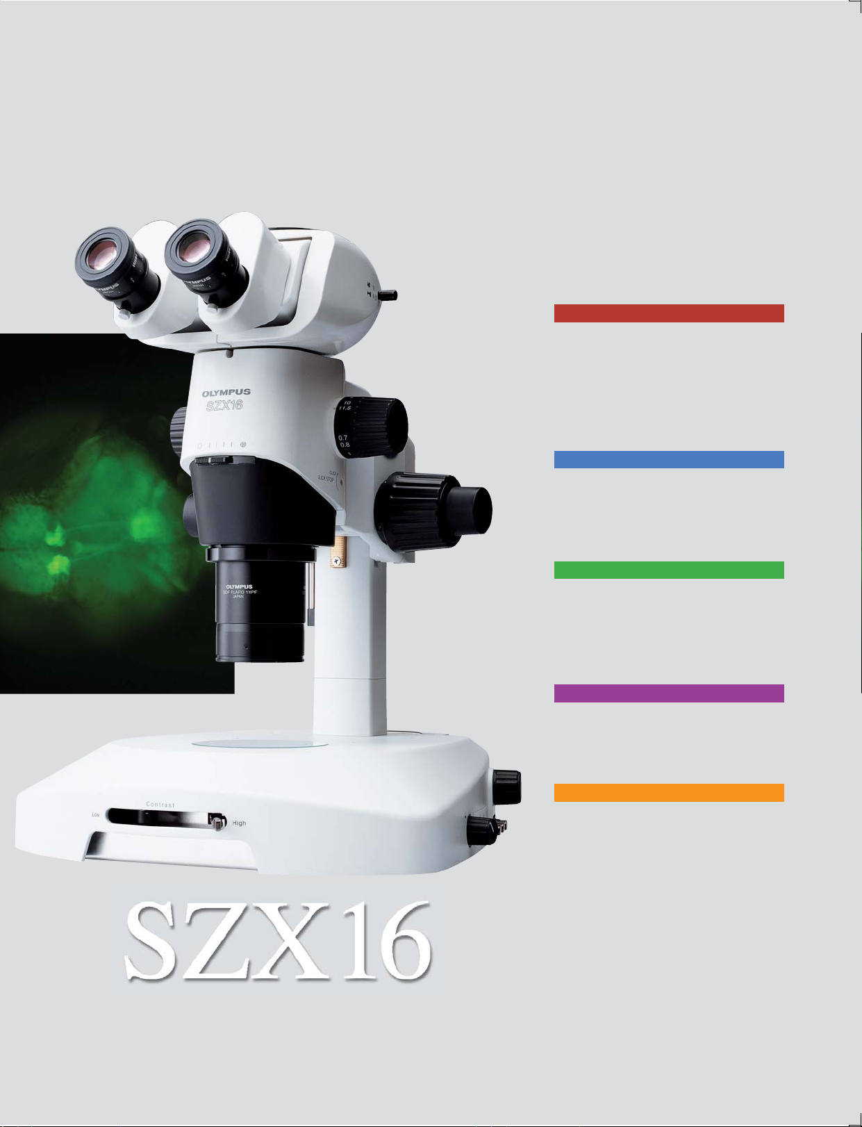
P03-P08
A new dimension in image clarity
Unparalleled sharp viewing is the result of
a multi-wavelength, astigmatism- free
design that absolutely minimizes
aberration and the world's highest NA.
From low to high magnification,
unprecedented bright and even
fluorescence observation is achieved.
P09-P10
Comfortable operability
Long working distance (WD) and high NA
are featured. Overall work efficiency is
assured, including the illumination base
design.
P11-P12
Ergonomic design for working ease
Tilting trinocular tube, with an optimum
convergence angle and new slim
illumination base, effectively eliminates
fatigue resulting from long-time
observation and other tasks.
P13-P14
Digital imaging
From brightfield to fluorescence
observation, image acquisition of various
specimens is possible at high resolution.
P15-P16
Equipment answering various needs
Accessories for maximizing optical
performance and operability include a
variety of illumination bases, light guides,
and stage plates.
Page 4
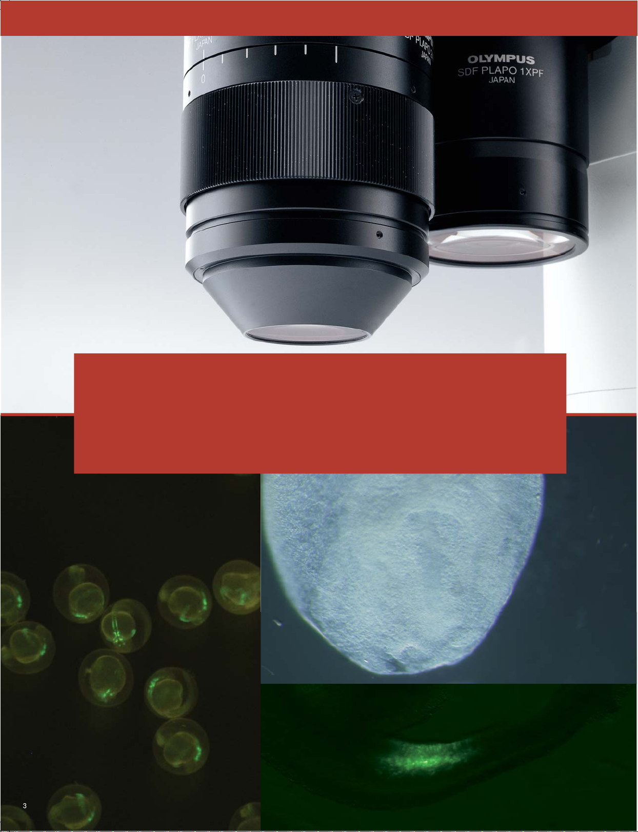
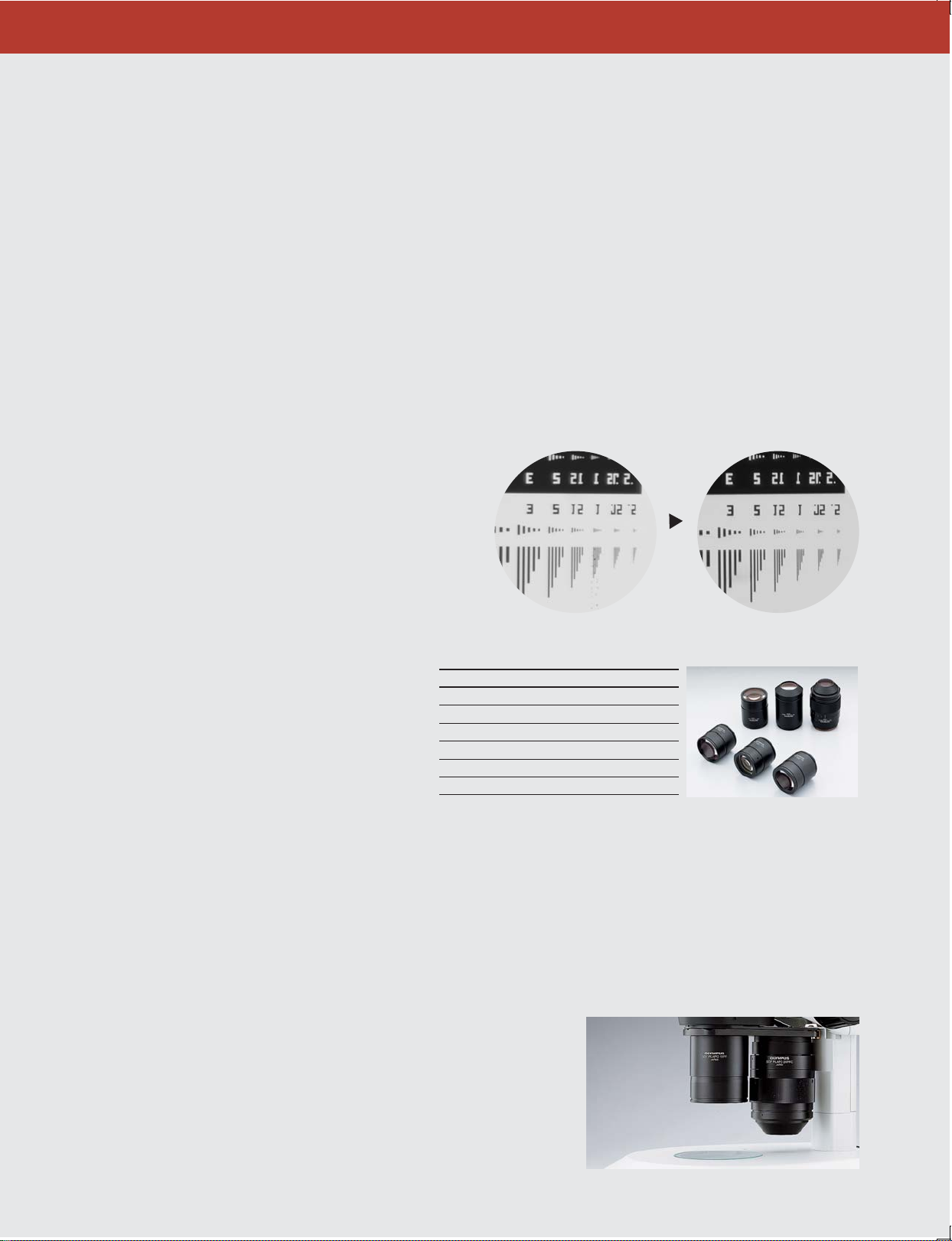
A new dimension in image clarity
Olympus’ new SDF objective lenses provide the highest
NA with 900lp/mm resolution. Optimum specimen viewing
from large field overview to microstructure, along with
instant zoom function to select observation points, is assured.
Page 5
A new dimension in image clarity
The world’s highest zoom ratio of 1:16.4 is achieved.
From low to high magnification, several different observation tasks
are available on one microscope.
The SZX16 offers optical performance at the highest global standard. SDF objective lenses with the
highest available NA enable microstructural observation that could not be achieved before. And with
the world’s highest zoom range of 7.0x-115x, this all-in-one microscope answers a wide range of needs
related to operation at low magnifications to detailed observation at high magnifications. These
outstanding features meet the need for clear observation of transparent specimens of minimal
contrast to microstructural viewing. Manipulation of live specimens has never been easier.
■ Highest available NA
The SZX16 realizes the highest NA (0.3) among
stereomicroscopes with 2x objectives. Optical performance
is 30% better than that of comparable products and allows
for significantly more image information.
■
SDF lineup: six objectives for various uses
The broad lineup of the SZX16 PLAN APO objective
series covers several needs with features ranging from
long WD objectives for operations requiring wide
working space (such as sample acquisition or selection)
to high magnification objectives with the world's
highest NA for microstructural observation.
■
World’s highest zoom ratio for several observation tasks
With a zoom range of 7.0x-115x (using SDFPLAPO 1X and
WHN10X-H), the SZX16 has the world’s highest zoom ratio of
1:16.4. From sample verification and selection at low
magnification to microstructure verification at high
magnification, seamless observation is available.
Model WD (mm) Magnification*
SDFPLFL0.3X 141 2.1x-34.5x
SDFPLAPO0.5XPF 70.5 3.5x-57.5x
SDFPLAPO0.8X 81 5.6x-92x
SDFPLAPO1XPF 60 7x-115x
SDFPLAPO1.6XPF 30 11.2x-184x
SDFPLAPO2XPFC 20 14x-230x
*Using WHN 10X-H
Conventional model
SZX16(withSDFPLAPO2×PFC)
SDF Objective Lens Series
■
Two objectives combine with revolving nosepiece for
3.5x – 230x zoom
The Parfocal (PF) Series of 0.5x, 1x, 1.6x, and 2x comprises
four PF objectives. The revolving nosepiece allows easy
switching between two objective lenses and smooth
zooming between 3.5x and 230x (using WHN10X-H).
4
Page 6
A new dimension in image clarity
Unprecedented depth and sharp images
dramatically enhance efficiency for a variety
of tasks including specimen manipulation.
5
Page 7
A new dimension in image clarity
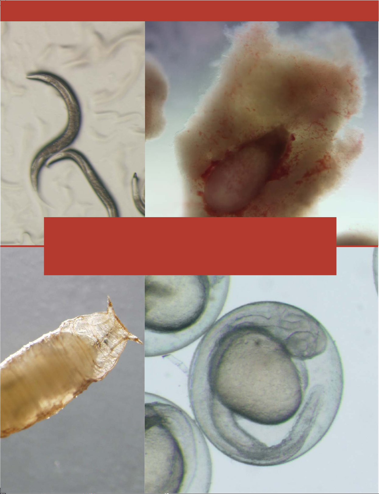
Multi-wave length, astigmatism-free design for sharp 3D observation.
New standards in image clarity begin with this design.
“Multi-wave length astigmatism-free”: a new design effectively eliminating image-deforming
aberration and enabling remarkably sharp 3D imaging and dramatically enhanced specimen
manipulation. Also, with an apochromatic lens system that effectively reduces chromatic aberration,
the latest proprietary SZX 16 optical system provides sharp 3D observation images of various
specimens.
■ Sharp, detailed observation of specimens
Newly designed SDF objective lenses keep astigmatism to a
minimum. This effectively eliminates image deforming at
pre- and post-focal plane and thus the depth of field is
perceived as deeper than before. These design features
enable stress-free use of forceps in the field of view during
live sample selection and acquisition. SZX2 puts power into
action for long-time observation. When these objectives are
combined with the newly developed transmitted light
illumination base, clear observation is even possible for
transparent specimens where contrast is low. Oversights are
thus minimized for specimen selection, dissection, and
manipulation.
■
Integration of apochromatic system
The apochromatic system — integrated into observation tubes,
zoom body, and objectives — eliminates chromatic aberration
throughout the zoom range and ensure excellent image quality
without chromatic blur.
Non astigmatismfree design
Astigmatism effects greater
deforming of the image at
pre- and post-focal plane.
•
Depth of field seen in focal plane will vary according to individual differences in users’ vision.
SDF objective
lenses
As deforming of the image at
pre- and post-focal plane is
kept to a minimum, the eye can
focus around the point the user
wants to observe. Thus depth
of field is perceived as deeper.
Pre-focal
plane
Focal plane
Post-focal
plane
■Optical performance with less fatigue
A 360º view of balanced images is made possible by
accommodating vertical and horizontal parameters.
Discomfort in the eyes and body, as well as stress from
prolonged observation or operation, is effectively eliminated.
Previous model
■
SZX16: new optics for equally easy handling of thick specimens
Obtaining a lucid visual perspective, such as a clear feel for dimension, is essential for manipulation of thick specimens like
eggs or embryos. The latest proprietary SZX16 delivers the images needed for observation of such specimens. 3D observation
images from surface to interior are also particularly effective in the dissection and manipulation of live specimens.
6
Page 8
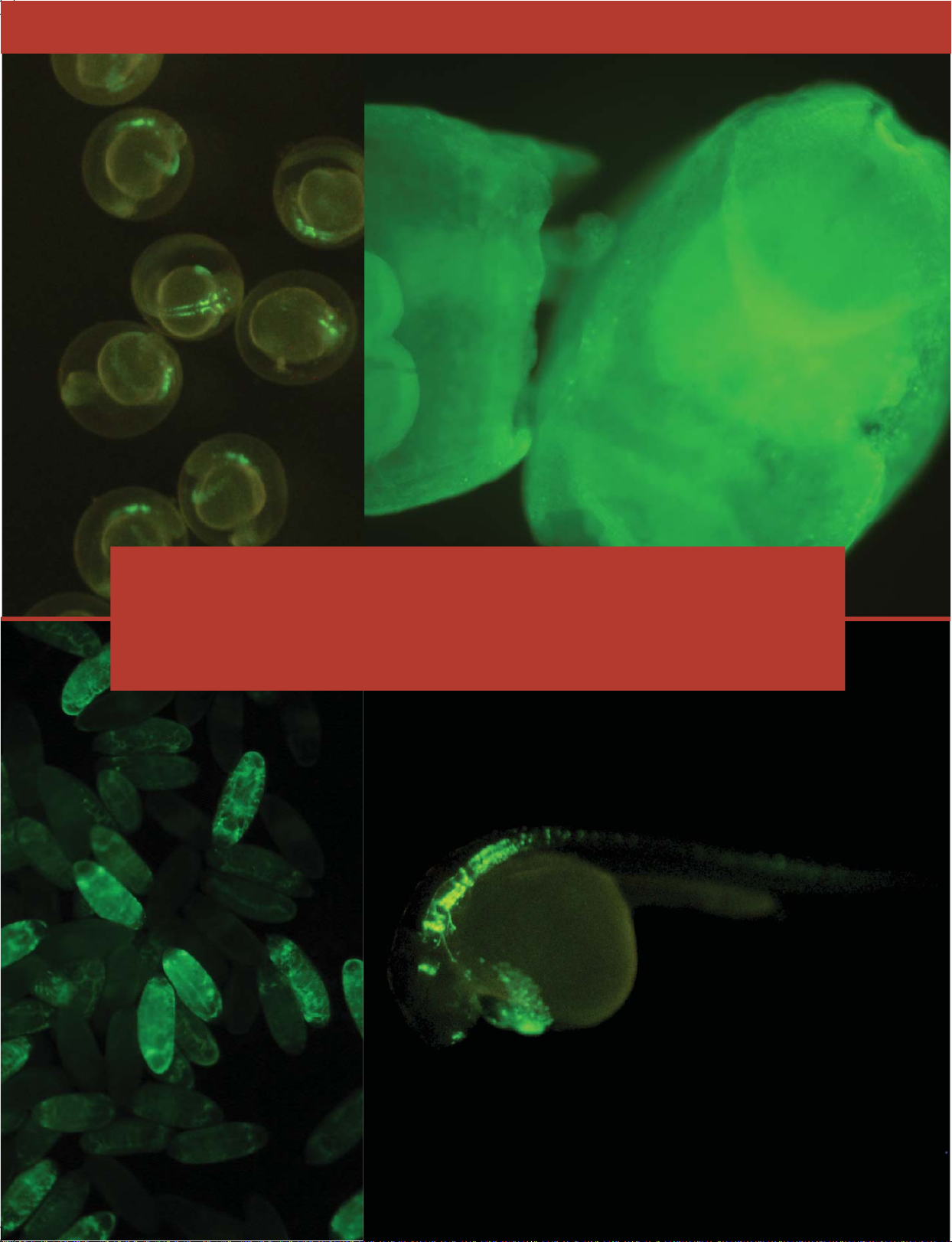
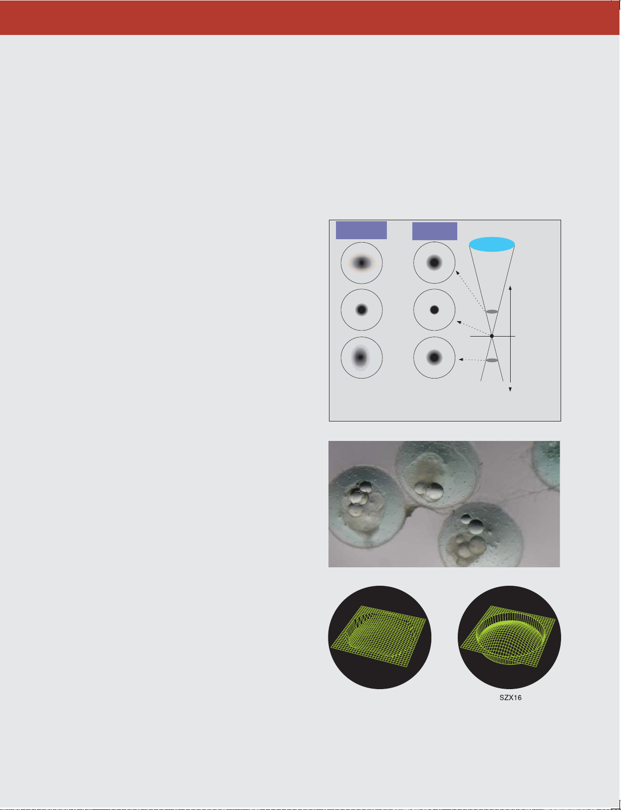
A new dimension in image clarity
From low to high magnification, exceptionally
bright and even fluorescence is assured for
efficient screening.
7
Page 9

A new dimension in image clarity
Newly designed SDF objective lenses of the highest NA and Olympus’
newly developed near-vertical reflected light illuminator will
significantly improve signal intensity and support bright fluorescence
observation.
Bright fluorescence observation is essential in recent biological and medical research. Observation of
weak fluorescence at low magnification under stereomicroscope has especially been a problem.
However, the SZX16 enables even and overwhelmingly bright fluorescence observation from low to
high magnifications.
■
Highest NA among stereomicroscopes provides
extraordinarily bright fluorescence observation
SDF objective lenses of the highest NA dramatically
improve fluorescence detection sensitivity. Furthermore,
the newly designed near-vertical reflected light illuminator’s
excitation light paths are independent from the observation
paths, allowing for substantially improved excitation light
efficiency. These features provide far brighter fluorescence
observation than conventional stereomicroscopes at all
magnifications. Transmitted light observation for verification
of specimen outline is possible even under reflected light
fluorescence observation.
Fluorescence and transmitted light illuminationFluorescence illumination only
■
Even and seamless fluorescence observation from
low to high magnification
In correspondence with zoom function, the reflected light
illuminator provides even illumination from low to high
magnification. This will eliminate oversights along the periphery
of the field of view during live specimen screening and
dramatically improve work efficiency.
■Five-position turret with nine-filter selection
Nine filter units, ranging from UV excitation to RFP, respond to
applications using various fluorescent dyes and protein. Olympus
High Quality (HQ) filters have an edge steepness and high
transmission that efficiently detect the light of fluorescence to
enhance and capture brighter fluorescence images in precise
detail.
Filter unit Model Remarks
For UV excitation SZX2-FUV Ex330-385/Em420-
For BV excitation SZX2-FBV Ex400-440/Em475High performance for CFP SZX2-FCFPHQ Ex425-445/Em460-510
For GFP SZX2-FGFP Ex460-490/Em510-
For GFP separation SZX2-FGFPA Ex460-495/Em510-550
High performance for GFP SZX2-FGFPHQ Ex460-480/Em495-540
High performance for YFP SZX2-FYFPHQ Ex490-500/Em510-560
For RFP 1 SZX2-FRFP1 Ex530-550/Em575-
For RFP 2 SZX2-FRFP2 Ex540-580/Em610-
SZX16 reflected light fluorescence illumination stand
SZX16 Fluorescent filter unit
8
Page 10

Comfortable operability
Effective combination of long WD and high NA.
Development of an ergonomic design.
Design focus on creating a wide working space and comfort.
9
Page 11

Comfortable operability
Operability configured as a whole-system characteristic. Design is focused
not only on WD and NA, but also on a stage that is slim.
SZX2 responds well to a variety of specimens and operations ― from large specimens like mice to small ones like zebra
fish, nematode or drosophilia eggs ― because of the effective combination of high numerical aperture and wide working
space. Moreover, the transmitted light illumination base is newly designed thin (only 41mm) to provide a wide working
space and allow various users to work comfortably.
■ Wide working space and high NA
NA
0.15
Field of view
31.4mm
WD
60mm
WD60mm and NA0.15 from the 1x objective
The 1x objective has a WD (60mm) that gives the user room to move
and an NA (0.15) that meets the needs of advanced research. Also
available are 0.8x objectives that have a longer WD of 81mm, which
provides not only a larger working space between objective lenses and
sample but also a total magnification of 5.6x-92x (using WHN10x-H).
2x objectives with ease of access and correction collar
A highly flexible design assures a wide access angle to objectives with the
highest NA (0.3) for specimen selection. An additional correction collar can
adjust image quality independently of the specimen — a first in
stereomicroscopes.
■ Ergonomic design for user-friendly base
Offering a wide working space in which users can place several Petri dishes, these illumination bases have an ergonomic,
beveled design for users to work comfortably and naturally.
High-level transmitted light
illumination base (SZX2-ILLB)
This unit provides effective contrast from
oblique illumination and easily selected
“High” and “low” contrast settings. Light
volume and color temperature are adjusted by
means of built-in filters (LBD/ND). It also has a
cooling fan to prevent overheating of the base
surface.
Brightfield/darkfield transmitted light illumination
base (SZX2-ILLD)
This base enables darkfield observation under illumination
twice as bright as conventional models. Flat and thin
specimens like brain tissue slices are vividly displayed on a
black background. A cooling fan prevents the illumination base
surface from overheating.
Transmitted light illumination base (SZX2-ILLK)
Offering outstanding cost-efficient performance, this
illumination base uses oblique illumination to provide highcontrast images of transparent specimens.
■ Slim 41mm LED illumination base
Slim LED transmitted light illumination base (SZX2-ILLT)
With a slim design of 41mm — approximately half that of conventional models
— the SZX16 transmitted light illumination base has a lower height to enable
a low eyepoint and easy access to base-mounted samples during observation
and operation. The world’s first LED 4-position turret enables contrast
adjustment between brightfield, oblique, and darkfield illumination with a
simple turn. This makes the SZX16 the all-in-one microscope for various
samples and observation tasks. Another advantage of LED illumination is
keeping down the temperature of the base surface, which is suitable for longtime manipulation of live specimens. Power consumption is about half that of
a conventional 30W Halogen light source. A life cycle of over 10,000 hours
significantly reduces operation costs.
10
Page 12

Ergonomic design for working ease
Users can work at ease, regardless of height.
Ergonomic design reduces fatigue for
maximum comfort.
11
Page 13

Ergonomic design for working ease
Optimal convergence angle plus tilting trinocular tube
reduces fatigue.
The SZX2 brings greater working comfort with an observation tube featuring a convergence angle that
relieves eyestrain. Moreover, the tilting trinocular tube and slim transmitted light illumination base
enable natural posture for increased work efficiency during observation and manipulation tasks of
long duration.
Convergence
■ Observation tube with convergence angle
relieves eyestrain
In cooperation with opthalmologists, a correlation between
stereomicroscope optical systems and eyestrain has been
confirmed. Specifically, the angle between right and left lines
of vision (convergence angle) is directly related to it. The
SZX2 series provides an optimum convergence angle that is
designed to allow users to observe in a natural position
suited to the eye. This solution effectively eliminates eyestrain
during long-time observation.
Observation tube with convergence angle
angle
■ Tilting trinocular tube allows for natural
posture, reduces fatigue
The tilting trinocular tube easily adjusts to the exact angle
desired (5º – 45º). Regardless of desk height, the tilting
trinocular tube assures a natural posture during long-time
observation. As fatigue and stress are greatly relieved,
oversights are avoided and work efficiency is increased.
■
Designed for efficiency: zoom and focus handles
Close positioning of zoom handle and focus handle enable
stress-free, blind operation. Fine adjustment of the
focusing handle offers increased sensitivity for easy
focusing at high magnification. In addition, the rigid body
provides stable observation.
■
Slim design minimizes fatigue and lowers eyepoint
Illumination bases are designed not only to be easy to use
but fatigue-free. The Slim LED transmitted light illumination
base, at 41mm, lowers the eyepoint and makes access to
specimens easier than ever. The wide stage surface easily
accommodates Petri dishes and other specimen containers
during observation and manipulation.
Tilting trinocular tube
Slim LED transmitted light Illumination base
12
Page 14

Digital imaging
From brightfield to fluorescence observation,
several different specimens can be viewed at
a high resolution recognized as leading the
digital imaging world.
13
Page 15

Digital imaging
Microscope digital camera that reproduces true-to-life images
Each microscope digital camera in the SZX2 lineup captures images at high resolution. Olympus
stereomicroscopes and digital cameras contribute to cutting-edge research in biology and medicine.
■ The high-performance Digital Camera (DP71)
provides accurate and detailed image capture
Digital camera (DP71)
With the DP71, live images are displayed with 1360 x 1024 pixel
resolution at a rate of 15 fps. Thanks to this smooth and fast display
of live images, users now can perform observation without stress.
By shifting the 1.45 mega pixel CCD, the camera delivers extremely
high-resolution images equivalent to 12.5 million pixels. The highspeed image processing hardware captures these quality images in
3 seconds. Furthermore, when acquiring monochrome fluorescence
images in gray scale, the custom gray-scale mode maintains a dynamic
range with full RGB intensity.
Digital camera (DP20)
DP20 is a stand-alone type without the need of a PC. Featuring highresolution imaging displayed at the high rate of 15fps for smooth,
easy-to-view display of live specimen images. Digital zooming
function wastes no time in focusing on selected areas. The RGB 24-bit
(16.7 million colors) configuration will bring about rich, high-contrast
streaming and video images that offer true-to-life colors. From
various settings to the scales display, operations are done on the
compact control box.
■ Motorized zoom and focus unit (SZX-FOA2)
Handling is made easier than ever, even when mounting a heavy unit
like a large camera (maximum load of 14kg). Another advantage of this
unit is that the user can stand away from the microscope for monitor
observation. Manual coarse focusing and fine focusing are both possible
when motorized control is engaged. A foot switch controls motorized
zooming and focusing, which frees the user’s hands for higher
concentration on operation, improving overall work efficiency during
observation and image capture.
■ Vertical observation
The revolving nosepiece
matches the objective lens
center to the zoom lens optical
path for images with reduced
aberration. Image shifting from
focus change is eliminated for
effective 3D rendering by
software.
SZX2-2RE16
Coaxial light axis
Coaxial
light axis
Ordinary image (9x zoom)
Coaxial optical path
image (9x zoom)
14
Page 16

Equipment answering various needs
A wide array of accessories to observe
various specimens
■Stands and optional units
Standard stand (SZX2-ST) Universal stand type2 (SZ2-STU2)
This standard reflected light illumination stand
supports observation conditions where no transmitted
light is needed.
Smooth horizontal movement and rotation enable
specimen observation from various angles.
CO2 incubator* (MI-IBCSZXF)
This CO2 incubator is developed especially for
the SZX2. Transparent glass heaters
positioned above and below samples will
create a stable environment within,
equivalent to a CO
observation of specimens during cultivation
is therefore possible. The glass heater above
the chamber dries off condensation for a vivid
imaging environment.
incubator. Long-time
2
Large stand (SZX2-STL)
This stand provides a large working space to
accommodate large specimens.
Thermo plate*
(MATS-55SZX2A/
MATS-55SZX2B)
Compatible with the SZX2 only,
this thermo plate has a
temperature range that may be set
from room temperature to 50˚ to
keep the specimen warm.
Large stage plate (SZX-CL)
This stage plate attaches to the top
of a transmitted light illumination
base and can be detached for easy
sterilization and other antiseptic
procedures.
15
Page 17

■Transmitted/Reflected light illumination base
Equipment answering various needs
Transmitted light guide adaptor (SZX-TLGAD)/
Light guide (LG-SF)
As the light guide LG-SF power source is mounted away
from the transmitted light illumination base, increases in
illuminator surface temperature are prevented.
Dual combination light guide (LG-DFI)
The SZX2 light guide can be mounted directly onto the
focus drive, keeping the observation position properly
illuminated even when focus is adjusted or when the
specimen is exchanged.
Dual inter-lock light guide (LG-DI) Ring light guide (LG-R66)
This light guide can be positioned as the observer likes
for bright, even illumination - especially effective when
high-contrast images are required. The spot lens HLL301*
can be mounted.
With its ø66mm diameter mount, this ring light
illuminator has been specially developed for
stereomicroscope compatibility. When mounted with ring
light adapter SZX-LGR66*/LGR66, it provides bright,
uniformly lit images especially avoiding glaring
reflections or obscuring shadows.
Coaxial illuminator
(SZX2-ILLC16/SZX2-ILLC10*)
Used with the dual flexible light guide LG-DF*, this
illuminator provides bright, even illumination without the
need for centering adjustments to the lamp.
* Compatible with the SZX10 only.* Due to be launched in the summer of 2006.
■Accessories
Light beam splitter (SZX2-LBS)
The adapter allows a digital camera or
other imaging unit to be attached on
both sides of the SZX2-LBS body. The
light path to the camera port is switched
between 100% and 50% light. The 100%
light path to the camera port enables
image capturing of dark specimens.
Simple polarizer (SZX-PO)
and analyzer (SZX2-AN)
This simple polarizer should be used
with a transmitted light illumination
base. It provides double-refractile
image observation of such specimens
as sea urchin larvae. Analyzer should
be attached on the tip of objectives.
16
Page 18

A zoom ratio of 1:10 is suitable for operations like
specimen selection or dissection. SZX10 provides
wide viewing and assures fewer oversights while
relieving fatigue. Choose from a wide range of
accessories to suit your sample needs.
17
Page 19

SZX10: the highly versatile research stereomicroscope
This outstanding model assures cost-effective performance
and faithful reproduction of images.
■ Distortion-free design provides accurate observation of images
Distortion-free design that has been constantly improved by Olympus over the
years minimizes embossment of image plane and provides accurate images.
■ Maximum depth of field from the finest built-in AS zoom body
Closing the aperture increases the depth of field.
■A wide array of accessories upgrades the system for various observation and documentation methods
The SZX10 responds to a wide range of accessories and achieves high performance during image
capture and monitor observation. This versatile system can be used for a variety of applications.
Eyepoint adjuster (SZX-EPA)
Allows users to assume a natural
posture during observation.
Binocular tubes (SZX-B130/TBI/B145)
These binocular tubes allow for
variable eye points. Users will find
observation can be done in a natural
posture, thanks to the tilting head
with an incline angle varying between
5º and 45º.
Side-by-side discussion tube (SZX-SDO)
Suitable for teaching because primary
and secondary observers are seated
beside one another.
Drawing attachment (SZX-DA)
Enables users to accurately draw the
specimen for scientific study or
illustration - a traditional alternative
to photomicrography. The accessory
can be mounted on either side of the
microscope, depending on preference.
Discussion tube (SZX-DO)
Face-to-face, discussion-style
intermediate tube allows primary and
secondary observers to sit opposite
one another during specimen
observation. The secondary observer
can support primary observer more
effectively in their tasks.
Coaxial fluorescence illumination
stand (SZX-RFA)
This fluorescence unit allows
observation of fluorescent proteins
introduced into living cells.
18
Page 20

SZX16/SZX10 specifications
Item
Zoom ratio: 16.4 (0.7x-11.5x)
Zoom
microscope body
Objective
Eyepiece
Observation tube
Focusing assembly:
Stands
Magnification indication: 0.7/0.8/1/1.25/1.6/2/2.5/3.2/4/5/6.3/8/10/11.5
Objectives
SDFPLFL0.3X
SDFPLAPO0.5XPF
SDFPLAPO0.8X
SDFPLAPO1XPF
SDFPLAPO1.6XPF
SDFPLAPO2XPFC
SZX2-TTR/SZX2-TTRPT: Tilting trinocular tube
Convergence angle, Tilting angle:5˚-45˚, 2 steps optical path selectable (TTR observation: straight port = 100:0, 50:50) (TTRPT observation: straight port = 100:0, 0:100)
Interpupillary distance adjustment: 52-76mm
SZX2-TR30/SZX2-TR30PT: 30 degree trinocular tube
Convergence angle, Tilting angle:30˚, 2 steps optical path selectable (TR30 observation: straight port = 100:0, 50:50) (TR30PT observation: straight port = 100:0, 0:100)
Interpupillary distance adjustment: 52-76mm
SZX2-FO: Focusing unit / focus: rack and pinion with roller guide (with torque adjustment ring for coarse focusing), optional counter balance,
coarse handle stroke: 80mm, coarse handle stroke per rotation: 21mm, Load capacity: 0-10.0kg
SZX2-FOF: Fine focusing unit / focus: rack and pinion with roller guide (with torque adjustment ring for coarse focusing), coarse and
fine coaxial handle, built-in counter balance, coarse handle stroke: 80mm, coarse handle stroke per rotation: 36.8mm, fine handle
stroke: 80mm, fine handle stroke per rotation: 0.77mm, load capacity: 2.7-15.0kg
SZX2-FOFH: Fine focusing unit for heavy loading / focus: rack and pinion with roller guide (with torque adjustment ring for coarse focusing),
coarse and fine coaxial handle, built-in gas spring counter balance, coarse handle stroke: 80mm, coarse handle stroke per rotation: 36.8mm,
fine handle stroke: 80mm, fine handle stroke per rotation: 0.77mm, load capacity: 8.0-25.0kg
SZX-FOA2: Motorized focus unit / focus: rack and pinion with roller guide (with torque adjustment ring for coarse focusing),
focusing stroke: 75mm, motorized focusing speed coarse:1.5mm/sec fine:0.3mm/sec load capacity: 2.7-15.0kg
SZX-ST: Standard stand / Pillar height: 270mm, base dimension: 284(W)x335(D)x31(H)mm, Stage clips are mountable, with stage adapter fixing screw holes
SZX2-STL: Large stand / Pillar height: 400mm, base dimension: 400(W)x350(D)x28(H)mm, Stage clips are mountable, with stage adapter fixing screw holes
Specifications
SZX10SZX16
Zoom ratio: 10 (0.63x-6.3x)
Magnification indication: 0.63/0.8/1/1.25/1.6/2/2.5/3.2/4/5/6.3
Zoom drive system: Horizontal handle Click-stop for various zoom positions incorporated
For SZX-ZB16 For SZX-ZB10
WHN10X-H F.N. 22
WHSZ15X-H F.N. 16
WHSZ20X-H F.N. 12.5
WHSZ30X-H F.N. 7
Zoom variable magnification system with parallel optical axis
Built-in AS zoom body
Objective mounting: screw mount
N.A.
0.045
0.075
0.12
0.15
0.24
0.3
W.D.(mm)
141
70.5
81
60
30
20
Objectives
DFPL0.5X-4
DFPL0.75X-4
DFPLAPO1X-4
SZX-ACH1X
DFPLAPO1.25X
SZX-ACH1.25X-2
DFPL1.5X-4
DFPL2X-4
SZX-BI30: 30˚ binocular tube Tilting angle:30˚ Interpupillary distance adjustment: 51-76
SZX-BI45: 45˚ binocular tube Tilting angle:45˚ Interpupillary distance adjustment: 52-76
SZX-TBI: tilting binocular tube Tilting angle:5-45˚
Interpupillary distance adjustment: 51-76
N.A.
0.05
0.075
0.1
0.1
0.125
0.125
0.15
0.2
WHSZ10X-H F.N. 22
WHSZ15X-H F.N. 16
WHSZ20X-H F.N. 12.5
WHSZ30X-H F.N. 7
W.D.(mm)
171
116
81
90
60
68
45.5
33.5
Transmitted illumination base specifications
Item
Light source
Light intensity adjustment
Effective illuminated area
Built-in filter
Add-on filter
Illumination mode
Contrast selection
Cooling fan
The height of stage
(from desk surface)
Pillar height
Weight
Power source
19
LED (Average service life: Over 10,000 hrs by rated use.)
Brightfield: ø63mm Darkfield / Oblique: ø35mm
SZX2-ILLT SZX2-ILLB SZX2-ILLK SZX2-ILLD
6V30W Halogen 6V30WHAL PHILIPS 5761 (average lamp service life: approx. 100 hours by rate use.)
LBD, ND6, ND25 one for each
Brightfield illumination
Oblique illumination
Darkfield illumination
41mm
Approx. 3.7kg
AC adaptor
Brightfield illumination
Oblique illumination
2-step selection of High and Low
Approx. 5.0kg Approx. 4.6kg
Specifications
Continuously variable system
ø40mm
ø45LBD filter
Brightfield illumination
Oblique illumination
270mm
Built-in trance power unit
Brightfield: ø40mm Darkfield: ø35mm
LBD (bright field only)
Brightfield illumination
Darkfield illumination
Built-in
82mm
Approx. 5.4kg
Page 21

Reflected light illuminators specifications
Type
Features
Illumination
specification
Light source
specifications
Ring light guide LG-R66 Dual ring light guide LG-DFI/DI Coaxial illuminator SZX2-ILLC16/10
Bright, uniformly lit images without
glaring reflections or obscuring
shadows
Minimum WD: 30mm
Mount diameter: 66mm
Flexible part: 1000mm
Attachment adapter*: SZX-LGR66
*No adapter required for SZX16-LGR66
*Unable to attach to SDFPLAPO2XPFC/SDFPLAPO1.6XPF
Type: LG-PS2
Functions: Light intensity control and lamp ON/OFF control by external signal (DC0-5V), mechanical adjustment function
Power consumption: 150W (350VA)
Rated voltage: 100-120V/220-240V 50/60Hz
Dimensions: 120(H)x120(H)x235(D)mm Weight: approx. 1.5kg
Flexible illumination for any angle and
position
LG-DFI: Flexible part 900mm
Inter-lock part 500mm
LG-DI: Inter-lock part 500mm
Bright high contrast coaxial illumination.
Effective for observing structure, such as imperfections
on metal surfaces, patterns on IC or LCD
Magnification factor: 1.5x
Light guide: LG-DF
Flexible part 1000mm
1/4 wave plate included
Option
LG-R66PL: Polarizer/analyzer
set for LG-R66
HILL301: spot lens
LG-FAD: ø25 filter adapter
Reflected light fluorescence illuminator
Type Reflected light fluorescence illuminator/Fine focusing unit SZX2-RFA16
Illumination method
Near vertical reflected light fluorescence illumination which is corresporded to microscope zoom function
Zooming of illuminator independent to zoom function of microscope body is possible.
Five-position turret
Filter turret
Maximum 5 sets of excitation/emission filter sliders are attachable.
Comes with shutter that prevents flash-light caused by switching.
Filter holder slider
Filter slider
Three-step switch by shutter and two holes. ND filter can be attached at the holes.
One excitation balancer can be attached.
Built-in
Fine focusing unit / focus: rack and pinion with roller guide (with torque adjustment
Focusing assembly
ring for coarse focusing), coarse and fine coaxial handle, built-in counter balance,
coarse handle stroke: 69mm, coarse handle stroke per rotation: 36.8mm, fine handle
stroke: 69mm, fine handle stroke per rotation: 0.77mm, load capacity: 2.7-15.0kg
Light source specifications
100W mercury lamp
Total magnifications and actual field diameters of SZX2-ZB16
Eyepiece
Objective
SDFPLFL0.3X
SDFPLFL0.5XPF
SDFPLAPO0.8X
SDFPLAPO1XPF
SDFPLAPO1.6XPF
SDFPLAPO2XPFC
WHN10X-H WHSZ15X-H WHSZ20X-H WHSZ30X-H
total mag.
2.1x-34.5x
3.5x-57.5x
5.6x-92x
7x-115x
11.2x-184x
14x-230x
ø104.8-ø6.4
ø62.9-ø3.8
ø39.3-ø2.4
ø31.4-ø1.9
ø19.6-ø1.2*
ø15.7-ø1*
total mag. total mag. total mag.
3.2x-51.8x
5.3x-86.3x
8.4x-138x
10.5x-172.5x
16.8x-276x
21x-345x
ø76.2-ø4.6
ø45.7-ø2.8
ø28.6-ø1.7
ø22.9-ø1.4
ø14.3-ø0.9
ø11.4-ø0.7*
Some vignetting may occur from optical characteristics. This occurs in observations at low magnification.
4.2x-69x
7x-115x
11.2x-184x
14x-230x
22.4x-368x
28x-460x
Total magnifications and actual field diameters of SZX2-ZB10
Eyepiece
Objective
DFPL0.5X-4
DFPL0.75X-4
DFPLAPO1X-4
SZX-ACH1X
WHSZ10X-H WHSZ15X-H WHSZ20X-H WHSZ30X-H
total mag.
3.2x-31.5x
4.7x-47.3x
6.3x-63x
field diameter (mm) field diameter (mm) field diameter (mm) field diameter (mm)
ø69.8-ø7.0
ø46.6-ø4.7
ø34.9-ø3.5
total mag. total mag. total mag.
4.7x-47.3x
7.1x-70.9x
9.5x-94.5x
ø50.8-ø5.1
ø33.9-ø3.4
ø25.4-ø2.5
6.3x-63x
9.4x-94.5x
12.6x-126x
Reflected light fluorescence illuminator SZX-RFA
Coaxial illumination
Four-step slide switch
Maximum 3 mirror units are attachable.
Comes with shutter that prevents flash-light
caused by switching.
field diameter (mm)field diameter (mm)field diameter (mm)
ø59.5-ø3.6
ø35.7-ø2.2
ø22.3-ø1.4
ø17.9-ø1.1
ø11.2-ø0.7
ø8.9-ø0.5
ø39.7-ø4
ø26.5-ø2.6
ø19.8-ø2
6.3x-103.5x
10.5x-172.5x
16.8x-276x
21x-345x
33.6x-552x
42x-690x
9.5x-94.5x
14.2x-141.8x
18.9x-189x
field diameter (mm)
ø33.3-ø2.0
ø20.0-ø1.2
ø12.5-ø0.8
ø10.0-ø0.6
ø6.3-ø0.4
ø5.0-ø0.3
ø22.2-ø2.2
ø14.8-ø1.5
ø11.1-ø1.1
DFPLAPO1.25X
SZX-ACH1.25X-2
DFPL1.5X-4
DFPL2X-4
7.9x-78.9x
9.5x-94.5x
12.6x-126x
ø27.9-ø2.8
ø23.3-ø2.3
ø17.5-ø1.7
11.8x-118.1x
14.2x-141.8x
18.9x-189x
ø20.3-ø2
ø16.9-ø1.7
ø12.7-ø1.3
15.8x-157.5x
18.9x-189x
25.2x-252x
ø15.9-ø1.6
ø13.2-ø1.3
ø9.9-ø1
23.6x-236.3x
28.4x-283.5x
37.8x-378x
ø8.9-ø0.9
ø0.7
ø7.4-
ø5.6-ø0.6
20
Page 22

Eyepieces
30˚ trinocular tube Tilting trinocular tube
Filter sets
100W mercury lamp housing
100W mercury apo lamp housing
Power unit
Reflected light fluorescence illuminator
1/4 wavelength
Ring light guide
retardation plate*
1
Rotatable
analyzer
SZX16 zoom body
Light beam splitter
Photo adapter
Coaxial reflected light illuminator
Filter
adapter
Revolving nosepiece*
Spot lens
Light protective shield
Dual flexible light guide
Dual inter-lock
light guide
2
Light source
Dual combination
light guide
*1 Incorporated in SZX2-ILLC16 *2 Cannot be attached to SZX2-FO
Accessories Focusing units
Black/White
stage plate
Focusing unit
Stage plate
for fluorescence
Cup stage Gliding stage
Circular
rotatable stage
Simple
polarizer
Manipulator
adapter
Circular
rotatable stage
BX stage adapter
type 1
MATS-55SZX2A/
MATS-55SZX2B
Thermo plate
Large stage plate
Square mechanical stage
BH stage adapter
type 1
MI-IBCSZXF
CO2 incubator
Illuminators
400mm pillar
600mm pillar
Fine focusing for heavy loading
Slim LED transmitted
light illumination base
AC adapter
Page 23

Tilting binocular tube
Eyepieces
Mirror units
Coaxial reflected
light fluorescence
illuminator
Light beam
splitter
100W mercury lamp housing
100W mercury apo lamp housing
Light protective shield
Ring light guide adapter for SZX
Ring light guide
*1 Incorporated in SZX2-ILLC10
30 degree
trinocular
tube
Photo adapter Eyepoint
adjuster
Power unit
1/4 wavelength
retardation plate*
1
Analyser
Polarizer
LG-R66PL
Analyzer and polarizer set
for LG-R66
Analyzer
Tilting trinocular
tube
Drawing attachment
SZX10
Zoom body
Binocular
tube
Discussion tube
Coaxial illuminator
Filter
adapter
Revolving nosepiece
30 degree
binocular
tube
Dual flexible light guide
Spot lens
Side-by-side
discussion tube
Dual inter-lock
light guide
Power unit
Large stand for SDO
Light source
Dual combination
light guide
unit
Drop prevention collar
High-level transmitted
light illumination base
Transmitted light
illumination base
Dumper for SZX2 illumination base
6V30W lamp socket
Transmitted light guide adapter unit
Focusing unitFine focusing
BF/DF transmitted
light illumination base
Light guide
Motorized focusing unit
Standard
stand
Control unit
AC adapter
BX stage
adapter type2
Large stand
Foot Switch
Hand Switch
Universal stand type 2
Mechanical stage
Page 24

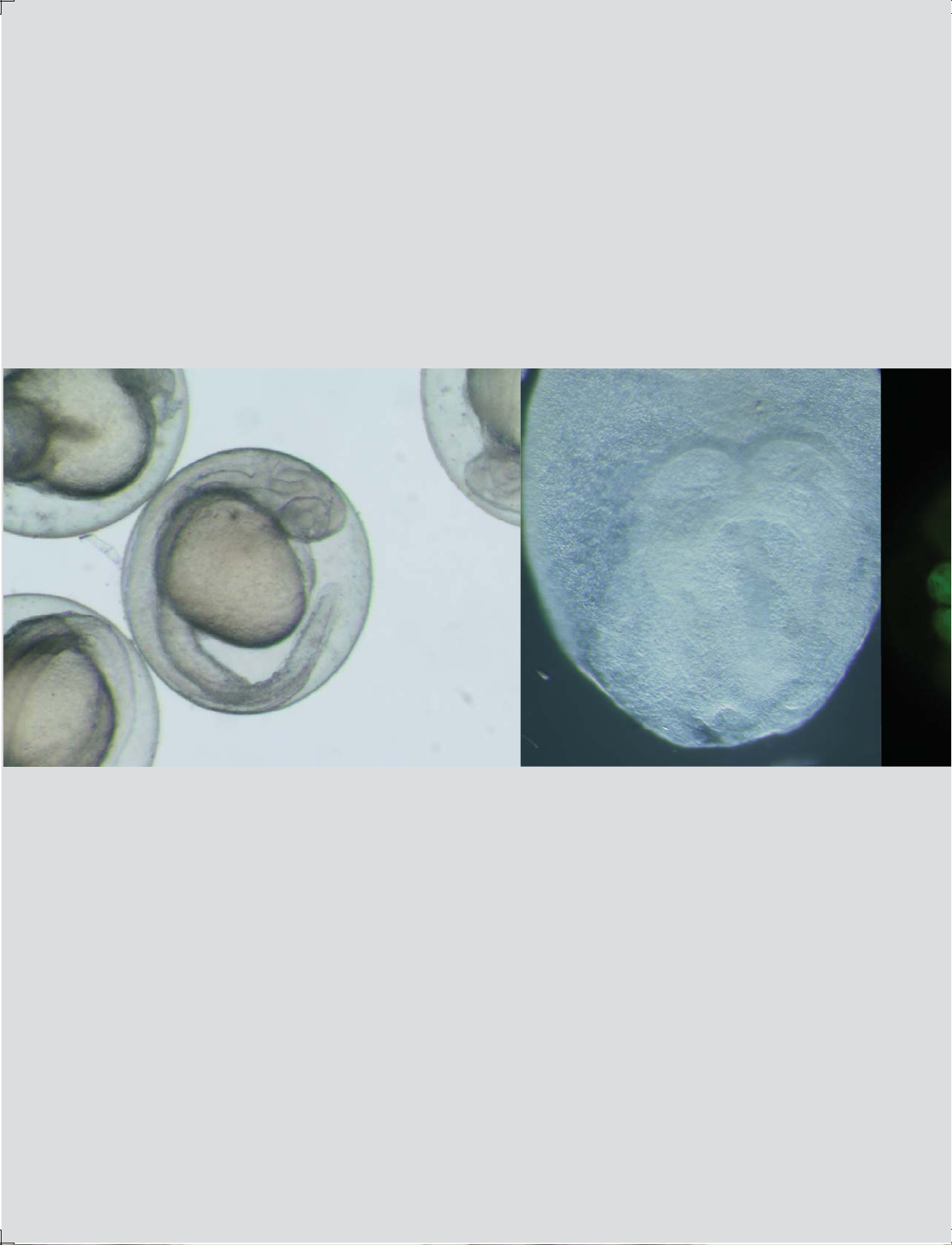
Images are courtesy of the following institutions:
RIKEN Brain Science Institute, Laboratory for Developmental Gene Regulation (page 1, left; page 3,
lower left; pages 5, 7, & 8, lower right). RIKEN Center for Developmental Biology, Laboratory for Cell Asymmetry,
Dr. Ayano Kawaguchi (page 3, lower right). Graduate School of Medicine and Faculty of Medicine,
the University of Tokyo, Department of Cell Biology and Anatomy, Dr. Yasushi Okamoto (page 1, right; page 3,
top; page 5, top right; page 7, top right). National Institute of Advanced Industrial Science and Technology,
Research Institute for Cell Engineering, Neuronics Research Group (page 1, right).
ISO 9001
Certification
Design and production
adheres to ISO9001
international quality standard.
Certified ISO 14001 by
ENVIRONMENTAL
UKAS
MANAGEMENT
008
ISO14001
Certification
Design and production at the OLYMPUS
CORPORATION Ina Plant conforms with
ISO14001 specifications for
environmental management systems.
Olympus has acquired ISO9001/ISO14001 certification for its quality/environmental
management systems. In line with this qualification, newly developed SZX16/SZX10
products are manufactured without lead, hexavalent chromium, or cadmium.
Olympus products contribute to society through their environmentally friendly designs.
Specifications are subject to change without any obligation on the part of the manufacturer.
 Loading...