

Page 1
R05
Page 2

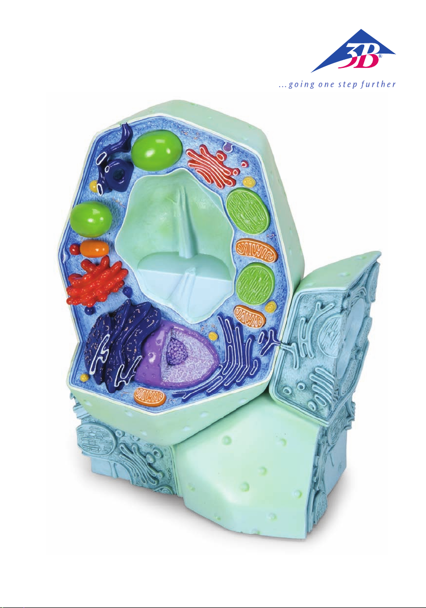
EnglishModel of a Plant Cell
(Magnification approx. 500,000-1,000,000)
The history of cytology
Cytology is an independent science in botany and deals with the structure and function of plant cells. The
term cell (Latin: cellula = chamber, compartment, cell) was coined in 1665 by Robert Hooke, after he had
discovered and recorded the cells within the tissue of a bottle cork with the help of one of the earliest light
microscopes. At the beginning of the 19th Century, Franz Meyen (1804-1840) recognised cells as the elementary units of plant organs. In 1838/1839, Matthias Jacob Schleiden and Theodor Schwann establish cell
theory: “Cells are the basis for all plants and animals.” In 1845, Karl Theodor Ernst von Siebold, based on
his observations on protozoa (unicellular organisms), wrote that cells can exist independently and represent
the smallest unit of life. At the same time, Louis Pasteur and other scientists refuted the prevailing theory
of the time which stated that cells can originate spontaneously out of dead organic matter (generatio
spontanea). In 1855, Rudolf Virchow confirmed Meyen’s theory which stated that every cell originates from
another cell (“omnis cellula ex cellula”). In 1879, Eduard Strasburger discovered the division of the nucleus
in plants. An important breakthrough in understanding the structure and function of cells was achieved by
E. Ruska and H. Mahl in 1940, thanks to the development of the transmission electron microscope.
As in the animal world, plant cells, too, are characterised by the following:
• They have a more complex structure than their environment
• They react to inner and outer stimuli
• They have the ability to reproduce
Differences between animal and plant cells
In spite of the consistency regarding the cellular structure of animal and plant cells – which had been
detected by Schleiden and Schwann in 1838 – there still are important differences in their basic structural
plan. The following three features characterise the differences between most plant cells and animal cells:
1. Plant cells are enclosed by a cell wall which is responsible for resisting the inner osmotic pressure of the
cell (turgor pressure), thereby giving it rigidity and increased stability.
2. As organelles, only plant cells possess plastids. These include, for example, the green chloroplasts, the
scene of photosynthesis.
3. They possess the sap vacuoles characteristic of plants, in which dissolved substances are stored and
macromolecules broken down.
®
Plant cells have an average size of 10-100 µm and can be observed by using a simple light microscope. A
tree is made up of 10
in part strongly differentiated and specialised cells (= tissues).
Structure and function of a plant cell
(For numbering see diagram)
Note: Unlike the model presented, all components of a living cell are in a state of constant motion!!!
The individual cell components have differences in their composition, e.g. proteins/enzymes, ionic milieu,
etc., and can best be classified according to their functions. An important term in plant cytology is proto-
plast, which refers to a cell surrounded by a plasma membrane in which the cell wall has been removed.
Cytoplasm with cytoskeleton (1)
In the course of evolution, a kind of division of work originated in a cell. This division of work is called
compartmentation. It is achieved when special reaction complexes, the organelles (Greek: organon =
tool), are surrounded and defined by membranes. These organelles can be detected, with the help of a
light microscope, in the fluid and colourless cytoplasm of protoplasts (60 to 90% water, proteins, lipids,
nucleic acids). The cell membrane (2) forms the boundary of the cell, marking it off from its exterior surroundings. The cell membrane consists of monomolecular layers of phospholipids and proteins which can
move in the lipid matrix (“fluid mosaic” – model). Incidentally, all plant and animal membranes are based
on the same elementary principle (unit membrane).
13
(= 10 trillion) cells! In multicellular organisms, they form groups of homogenous,
Page 3

English
Model of a Plant Cell
(Magnification approx. 500,000-1,000,000)
This membrane is responsible for controlling the selective transport into and out of the cell. It has the
same function with regard to the organelles, too. The cytoskeleton in the cytoplasm, which is made up
of proteins, guarantees not just the stability of the cell but also the most diverse intracellular movements
(e.g. visible plasma streams).
Nucleus (3a) with nucleolus (3b)
The nucleus (approx. 5-25 µm) is the information centre of the cell. It is enclosed within a double membrane lining with defined channels (= nuclear pores for controlling the metabolic flow between the nucleus and the cytoplasm) and contains the main part of the cell’s genetic information, present in the form of
chromatin. Only during cell division is the chromatin (normally not visible under a light microscope) transformed into its more compact form, viz. chromosomes. In the process, the DNA, which is bound by proteins, is strongly reduced by condensation and spiralisation. The nucleoli occur exclusively in the interior of
the nucleus and are the site for the synthesis of preliminary stages of cytoplasmic ribosomes (5).
Endoplasmic Reticulum (smooth ER (4a) and rough ER (4b)) Ribosomes (5)
All proteins of the cell are built at the “sewing machine” of proteins, the ribosomes. These extremely small
organelles (approx. 20-30 nm) can float freely in the cytoplasm or get attached to the sacciform or tubular
membrane system of the endoplasmic reticulum (rough ER). Inside the ER´s compartment, proteins are in
part transformed into helper proteins, commonly known as molecular chaperones, and are transported
to their biophase. The smooth ER, without ribosomes attached, is mainly responsible for the synthesis of
lipids. The structure of the ER is extremely dynamic and always subject to constant reorganisation. The ER
is also connected to the membrane coating of the nucleus. That is to say, the membrane and the lumens of
both of the organelles blend directly with one another.
Plasmodesmata (6)
Plasmodesmata constitute contact structures between neighbouring plant cells. In the process they form
connections, in the form of fine channels, between the living protoplasts through the cell wall and the
middle lamella. The coupling is built through tubular ER cisternae of both of the cells and its function is to
transfer low-molecular substances between cells.
®
Plastids
Plastids are compartments typical to plant cells. They are always surrounded by a double membrane. The
inner membrane is formed for the purpose of enlarging the reactive surface into the interior of the plastids. Plastids emerge on their own from the division of young proplastids and spread themselves during
mitosis into daughter cells. Chloroplasts possess their own genetic information (ring-shaped, extrachromosomal genome; plastids DNA).
The green chloroplasts (7) are the site of photosynthesis and the synthesis of numerous plant constituents
(e.g. fatty acids). The colourless and fluid matrix is denoted as stroma; the enlarged lamellar/sacciform
inner membranes are called thylakoids. The stacked membrane areas are called grana thylakoids. The
protein-bound pigments responsible for photosynthesis are found in these membranes (chiefly chlorophyll
and carotenoids). These photosynthesis pigments are also responsible for the Hill reaction. The CalvinBenson cycle or the photosynthetic carbon reduction cycle (PCR) CO2 fixation as well as the formation of
carbohydrates and starch take place in the stroma region.
Other plastids:
Chromoplasts: are inactive photosynthesis plastids responsible for the colouration of plant organs
Leucoplasts: are responsible for storing starch (amyloplasts), proteins (proteinoplasts), oils (elaioplasts)
Etioplasts: are the preliminary stage of chloroplasts and originate in the dark
Gerontoplasts: are all plastids at a very mature age
Mitochondria (8)
Mitochondria are the organelles responsible for cell respiration and energy conversion. They are therefore the “power plant” of every cell. Only mitochondria can give rise to mitochondria. Just like plastids,
Page 4

Model of a Plant Cell
English
(Magnification approx. 500,000-1,000,000)
mitochondria, too, are enclosed in a double membrane coating and possess their own genetic information.
The components/proteins responsible for the respiratory chain (ATP synthesis) are located on the inner side
of the membrane. The citrate cycle and the fatty acid oxidation cycle take place inside the mitochondrial
matrix.
Endosymbiont theory
The endosymbiont theory attempts to explain the origin of mitochondria and plastids. According to this
theory, mitochondria and plastids go back to intercellular protozoan (bacterial) symbiosis. In other words:
plastids have developed from cyanobacteria, and mitochondria have developed from purple bacteria. At
some point in the course of evolution, a “prototype“ cell with a nucleus (urocyte) incorporated prokaryotes
and integrated them into its cellular functions. A strong indication of this is the fact that mitochondria and
plastids have the following in common:
• A double membrane coating (inner and outer membranes are quite different in their chemical composition; the inner membrane resembles bacterial membranes)
• Inherent ring-shaped genome
• Inherent ribosomes (correspond to bacterial ribosomes, differ from cytoplasmic ribosomes)
Dictyosomes/Golgi apparatus (9)
Dictyosomes are disc-shaped, membranous hollow cavities (cisternae). The sum of all dictyosomes in a cell
are termed the Golgi apparatus. They are closely connected to the ER and are responsible for the conversion, storage and transfer of the products of the ER. Consequently, a distinction can be drawn between a
generation side (facing the ER, regeneration from the ER) and a secretion side (facing away from the ER)
which forms a significant cellular transport system responsible for exocytosis (elimination of substances
from the cell), the construction of biomembranes and is also involved in cell-wall formation.
Vacuole (10)
The vacuole is an organelle only to be found in plant cells. It is a space filled with fluid and is surrounded
by a simple membrane (= tonoplast). In mature plant cells, the volume of the central vacuole can constitute up to 80% of the total volume of the cell. In the cell, vacuoles serve as reaction, storage (e.g. of ions,
organic acids, saccharides, proteins, pigments), transport and deposit compartments (for substances that
can be harmful to the cell, e.g. toxins, tanning agents). The breakdown of macromolecules (lytic compartment) is also carried out in the vacuoles.
®
Microsomes/Microbodies (11)
Microsomes are organelles with a homogenous structure (simple membrane, spherical, size: 1 µm, granular
matrix) on the one hand, and strong biochemical and functional differences on the other.
Different functions:
− Lysosomes: are responsible for the break-up of proteins, polysaccharides and nucleic acids
− Glyoxysomes: play an important part in converting depot fats to carbohydrates
− Oleosomes (oil globules): are responsible for the break-up of fats and oils
− Peroxisomes: play an important part in photorespiration. Peroxisomes also break up the glycollate
which is inevitably created during CO2 fixation. Carbon is fed back into the photosynthesis cycle, and two
amino acids are produced for protein synthesis.
Cell wall (12)
Possessing a rigid cell wall is an additional feature which distinguishes plant cells from animal cells. The
cell wall gives the plant cell rigidity and form (exoskeleton) by resisting the interior osmotic pressure
(= turgor pressure) of the cell. It is a product secreted by the protoplasts (apoplast). From a chemical point
of view, the cell wall is made up of polysaccharides and proteins.
The cell wall is made up of up to three layers.
Middle lamella: a gelatinous layer, only a few nm in thickness, made up of pectin compounds with a low
quantity of proteins. It has no fibril structure and is therefore elastic.
Page 5

English Model of a Plant Cell
(Magnification approx. 500,000-1,000,000)
Primary cell wall: a gelatinous base substance (matrix) made up of pectin compounds, hemicellulose com-
ponents and proteins. In the matrix, fibril structures can be detected (10-25%) which are arranged in an
irregular, scattered texture (elasticity still present).
Secondary wall: is chiefly composed of 90% cellulose fibrils. The arrangement of the fibrils is primarily in
a parallel texture. There are often deposits of lignin, tanning agents, CaCO3, SiO2 or colouring agents. Cells
having a marked secondary wall are no longer capable of growth.
Author: Dr. Gerd Vogg, University of Würzburg, Germany
Numbering:
1 Cytoplasm with cytoskeleton
2 Cell membrane
3 a Nucleus
3 b Nucleolus
4 a Smooth Endoplasmic Reticulum (smooth ER)
4 b Rough Endoplasmic Reticulum (rough ER)
5 Ribosomes
6 Plasmodesmata
7 Chloroplasts
8 Mitochondria
9 Dictyosomes/Golgi apparatus
10 Vacuole
11 Microsomes/Microbodies
12 Cell wall (layered structure)
®
Page 6

Modell der Pflanzenzelle
Deutsch
(Vergrößerung etwa 500.000 - 1.000.000-fach)
Historisches zur pflanzlichen Zelllehre (Zytologie)
Die Zytologie ist eine eigenständige Wissenschaft innerhalb der Botanik, die sich mit der Struktur und
den Funktionen der pflanzlichen Zelle beschäftigt. Den Begriff Zelle (lat. cellula = Kämmerchen) prägte
im Jahre 1665 Robert Hooke, nachdem er diese im Gewebe des Flaschenkorks mit Hilfe eines der ersten
Lichtmikroskope entdeckte und detailliert aufzeichnete. Zu Beginn des 19. Jahrhunderts wurde die Zelle
von Franz Meyen (1804 – 1840) als Elementareinheit der Pflanzenorgane erkannt. 1838/1839 begründen
Matthias Jacob Schleiden und Theodor Schwann die Zellentheorie: „Pflanzen und Tiere sind gleichermaßen stets von Zellen aufgebaut“. 1845 veröffentlichte Karl Theodor Ernst von Siebold aufgrund der
Beobachtung an Protozoen (Einzeller), dass Zellen unabhängig voneinander leben können und die kleinste lebensfähige Einheit darstellen. Zur gleichen Zeit widerlegten Louis Pasteur und andere die damals
geltende Theorie, dass Zellen spontan aus toter organischer Materie (generatio spontanea) entstehen
können. 1855 bestätigte Rudolf Virchow die Theorie Meyens, dass jede Zelle aus einer anderen entsteht
(„omnis cellula ex cellula“). 1879 entdeckte Eduard Strasburger die Kernteilung bei Pflanzen. Ein wichtiger
Schritt im Verständnis von Bau und Funktion der Zelle wurde mit der Entwicklung des TransmissionsElektronenmikroskopes im Jahr 1940 durch E. Ruska und H. Mahl erzielt.
Wie im tierischen System zeichnen sich auch die pflanzlichen Zellen dadurch aus,
• dass sie komplexer organisiert sind als ihre Umgebung
• dass sie auf Reize aus ihrem Inneren und aus ihrer Umgebung reagieren
• dass sie die Fähigkeit haben, sich zu vermehren.
Unterschiede im Grundbauplan von pflanzlichen und tierischen Zellen
Trotz der 1838 von Schleiden und Schwann gefundenen Übereinstimmung im zellulären Aufbau von
Pflanzen und Tieren gibt es wichtige Unterschiede in deren Grundbauplan. So unterscheidet sich die
Mehrzahl der Pflanzenzellen von den tierischen Zellen in folgenden drei Merkmalen:
1. Die pflanzlichen Zellen sind von einer Zellwand umhüllt, die dem osmotischen Innendruck der Zelle
(= Turgor) entgegensteht und ihr dadurch eine hohe Festigkeit verleiht.
2. Nur die pflanzlichen Zellen besitzen als Organellen die Plastiden, dazu gehören z.B. die grünen
Chloroplasten, die Orte der Photosynthese.
3. Sie besitzen die pflanzentypischen Zellsaftvakuolen, in denen gelöste Stoffe gespeichert und
Makromoleküle abgebaut werden.
®
Die pflanzliche Zelle besitzt eine durchschnittliche Größe von 10 - 100 µm und kann mit einfachen
Lichtmikroskopen beobachtet werden. Ein Baum besteht aus bis zu 10
zelligen Organismen bilden sie Verbände aus gleichartigen, zum Teil stark differenzierten und dadurch
spezialisierten Zellen (= Gewebe).
Bau und Funktion der pflanzlichen Zelle
(Nummerierung siehe Abbildung)
Wichtig: Im Gegensatz zum vorliegenden Modell sind in einer lebenden Zelle alle Bestandteile ständig
in Bewegung und im Fluss !!!
Die einzelnen Zellbestandteile, besitzen unterschiedliche Ausstattungen z.B. an Proteinen/Enzymen,
Ionenmilieu etc. und lassen sich am sinnvollsten entsprechend ihrer Funktionen einteilen. Ein wichtiger Begriff in der pflanzlichen Zytologie ist der Protoplast, darunter versteht man eine von einer
Zytoplasmamembran umgebenen Zelle bei der die Zellwand entfernt wurde.
Zytoplasma mit Zytoskelett (1)
Im Laufe der Evolution wurde eine Art Arbeitsteilung innerhalb einer Zelle eingeführt, die man
Kompartimentierung nennt. Dies wird erreicht indem spezielle Reaktionsbereiche, die Organellen (griechisch: Organon = Werkzeug), durch Membranen umhüllt und abgegrenzt werden. Diese Organellen
kann man im flüssigen und farblosen Zytoplasma des Protoplasten (60 bis 90 % Wasser, Proteine,
13
(= 10 Billionen) Zellen! In mehr-
Page 7

Deutsch
Modell der Pflanzenzelle
(Vergrößerung etwa 500.000 - 1.000.000-fach)
Lipide, Nukleinsäuren) bereits mit dem Lichtmikroskop erkennen. Nach außen wird die Zelle durch die
Zellmembran (2) abgegrenzt. Diese besteht aus zwei monomolekularen Schichten von Phospholipiden und
Proteinen, welche sich in der Lipidmatrix bewegen können (’fluid mosaic’ - Modell). Im Übrigen basieren
alle pflanzlichen und tierischen Membranen auf diesem gleichen Grundbauprinzip (= Einheitsmembran).
Durch die Membranen wird der selektive Transport in und aus der Zelle sowie in und aus den Organellen
kontrolliert. Das aus Proteinen aufgebaute Zytoskelett im Zytoplasma gewährleistet die Stabilität der Zelle
aber auch vielfältigste intrazelluläre Bewegungen (z. B. sichtbare Plasmaströmungen).
Zellkern (Nucleus) (3a) mit Kernkörperchen (Nucleolus) (3b)
Der Zellkern (ca. 5-25 µm) ist das Informationszentrum für die Zelle. Er wird von einer doppelten
Membranhülle mit definierten Kanälen (= Kernporen zur Steuerung des Stoffflusses zwischen Kern und
Zytoplasma) umgeben und enthält den Hauptanteil der genetischen Information der Zelle in Form des
Chromatins. Nur für eine Kernteilung wird das ansonsten lichtmikroskopisch nicht sichtbare Chromatin in
die kompakte Transportform, die Chromosomen, umgewandelt. Dabei wird die an Proteine gebundene
DNA durch Kondensation und Spiralisierung stark verkürzt. Die Kernkörperchen (Nucleoli) treten ausschließlich im Innern des Kerns auf und sind der Ort der Synthese von Vorstufen der cytoplasmatischen
Ribosomen (5).
Endoplasmatisches Retikulum (glattes ER (4a) und raues ER (4b)) und Ribosomen (5)
Alle Proteine (Eiweiße) der Zelle werden an den „Nähmaschinen“ der Proteine, den Ribosomen, gebildet. Diese sehr kleinen Organellen (ca. 20 x 30 nm) können frei im Zytoplasma liegen oder an das
sack- oder röhrenförmige Membransystem des Endoplasmatischen Retikulums gebunden sein (raues ER).
Im Kompartiment des ER werden die Proteine zum Teil durch Helferproteine verändert und an ihren
Wirkort transportiert. Das glatte ER, ohne aufgelagerte Ribosomen, ist vor allem für die Synthese von
Lipiden zuständig. Die Struktur des ER ist sehr dynamisch und einer ständigen Reorganisation unterworfen. Weiterhin steht das ER mit der Membranhülle des Zellkerns in Verbindung. Das heißt, sowohl die
Membranen als auch das Lumen beider Organellen gehen direkt ineinander über.
Plasmodesmata (6)
Die Plasmodesmata stellen Kontaktstrukturen zwischen benachbarten pflanzlichen Zellen dar. Sie verbinden dabei als feine Kanäle die lebenden Protoplasten durch die Zellwand und Mittellamelle hindurch.
Die Verbindung wird durch schlauchförmige ER–Zisternen beider Zellen gebildet. Die Funktion ist der
Stofftransport niedermolekularer Substanzen zwischen den Zellen.
®
Plastiden
Die Plastiden sind für die Pflanzen typische Zellkompartimente, die stets von einer Doppelmembran umgeben sind. Die innere Membran ist zur Vergrößerung der reaktiven Oberfläche ins Innere der Plastiden ausgeformt. Plastiden gehen durch Teilung der jugendlichen Proplastiden aus sich selbst hervor und verteilen
sich bei der Mitose auf die Tochterzellen. Die Chloroplasten besitzen ihre eigene genetische Information
(= ringförmiges, extrachromosomales Genom; Plastiden DNA).
Die grünen Chloroplasten (7) sind die Orte der Photosynthese und der Synthese vieler pflanzlicher
Inhaltstoffe (z. B. Fettsäuren). Die farblose flüssige Matrix wird als Stroma, die lamellen- bis sackartig
vergrößerten Innenmembranen als Thylakoide bezeichnet. Die gestapelten Membranbereiche werden
dabei Granathylakoide genannt. In diesen Membranen sind die Photosynthesepigmente proteingebunden
lokalisiert (v.a. Chlorophylle, Carotinoide) und verantwortlich für die Lichtreaktion der Photosynthese. Im
Stromabereich finden die Dunkelreaktion der CO2-Fixierung und die Bildung von Kohlenhydraten und
Stärke statt.
Weitere Plastiden:
Chromoplasten: photosynthetisch inaktive Plastiden zur Färbung der Pflanzenorgane
Leukoplasten: Speicherung von Stärke (Amyloplasten), Proteinen (Proteinoplasten), Ölen (Elaioplasten)
Etioplasten: im Dunkeln entstandene Vorstufen der Chloroplasten
Gerontoplasten: Alterstadien aller Plastiden
Page 8

Modell der Pflanzenzelle
Deutsch
(Vergrößerung etwa 500.000 - 1.000.000-fach)
Mitochondrien (8)
Die Mitochondrien sind die Organellen der Zellatmung und Energieumwandlung. Sie stellen dadurch die
„Kraftwerke“ der Zelle dar. Mitochondrien können nur aus sich selbst gebildet werden. Wie die Plastiden
sind sie von einer doppelten Membranhülle umgeben und besitzen ihre eigene genetische Information.
An der inneren Membran sind die Bestandteile/Proteine der Atmungskette lokalisiert (Synthese von ATP). In
der Mitochondrienmatrix laufen der Zitratzyklus und die Fettsäureoxidation ab.
Endosymbiontentheorie
Die Endosymbiontentheorie versucht die Herkunft der Mitochondrien und Plastiden zu erklären. Nach
dieser Theorie gehen die Mitochondrien und Plastiden auf protocytische (bakterielle) intrazelluläre
Symbiosen zurück. Das heißt: Plastiden sind demnach aus Cyanobakterien, Mitochondrien aus atmenden
Purpurbakterien entstanden. Eine „Urzelle“ mit Zellkern (Ureuzyt) hat sich im Laufe der Evolution
Prokaryoten einverleibt und in ihr zelluläres Funktionsgefüge integriert. Dafür sprechen die folgenden
Gemeinsamkeiten von Mitochondrien und Plastiden:
• doppelte Membranhülle (innere und äußere Membran sind in ihrer chemischen Zusammensetzung sehr
verschieden; die innere ähnelt bakteriellen Membranen)
• eigenes ringförmiges Genom
• eigene Ribosomen (entsprechen den bakteriellen Ribosomen, unterscheiden sich von den zytoplasmatischen Ribosomen)
Dictyosomen/Golgi-Apparat (9)
Die Dictyosomen sind scheibenförmige, membranumgebene Hohlräume (Zisternen). Alle Dictyosomen
einer Zelle werden als Golgi-Apparat bezeichnet. Sie stehen in engem Kontakt zum ER und sind
für die Umwandlung, Speicherung und Weiterleitung der Produkte des ER zuständig. Folglich kann
eine Bildungsseite (dem ER zugewandt, Neubildung aus dem ER) und eine Sekretionsseite (dem ER
abgewandt) unterschieden werden. Sie sind ein wichtiges zelluläres Transportsystem, zuständig für
Exocytose (Ausscheidung von Stoffen aus der Zelle), dem Aufbau von Biomembranen und beteiligt an der
Zellwandbildung.
®
Vakuole (10)
Die Vakuole ist ein rein pflanzliches Organell. Sie ist ein flüssigkeitsgefüllter Raum, der von einer
einfachen Membran (= Tonoplast) umgeben ist. In ausgewachsenen Pflanzenzellen kann das
Volumen der Zentralvakuole bis über 80 % des Zellvolumens ausmachen. Sie dienen in der Zelle als
Reaktions–, Speicherungs- (z. B. Ionen, organische Säuren, Zucker, Proteine, Pigmente), Transport- und
Abladekompartimente (für zellschädigende Substanzen z.B. Toxine, Gerbstoffe). Auch der Abbau von
Makromolekülen (lytisches Kompartiment) erfolgt in der Vakuole.
Mikrosomen/Microbodies (11)
Mikrosomen sind Organellen mit einheitlichem Aufbau (einfache Membran, kugelig, Größe: 1 µm, granulöse Matrix), aber starken biochemischen und damit funktionellen Unterschieden.
Verschiedene Funktionen:
− Lysosomen: zuständig für den Abbau von Proteinen, Polysacchariden und Nukleinsäuren
− Glyoxysomen: wichtige Rolle bei der Umwandlung von Speicherfetten in Kohlenhydrate
− Oleosomen (Öltröpfchen): Abbau von Fetten und Ölen
− Peroxisomen: wichtige Rolle bei der Photorespiration. Zwangsweise bei der CO
Glykolat wird über die Peroxisomen abgebaut, der Kohlenstoff wieder dem Photosynthese-Zyklus zugeführt und zwei Aminosäuren zur Proteinsynthese produziert.
Zellwand (12)
Der Besitz einer starren Zellwand ist ein weiteres Unterscheidungsmerkmal zwischen pflanzlichen und tierischen Zellen. Sie gibt der pflanzlichen Zelle Festigkeit und Form (Exoskelett), indem sie dem osmotischen
Innendruck der Zelle (=Turgor) widersteht. Sie ist ein Abscheidungsprodukt des Protoplasten (Apoplast).
Fixierung anfallendes
2
Page 9

Deutsch Modell der Pflanzenzelle
(Vergrößerung etwa 500.000 - 1.000.000-fach)
Chemisch betrachtet ist sie aus Polysacchariden und Proteinen aufgebaut.
Die Zellwand baut sich aus bis zu drei Schichten auf:
Mittellamelle: wenige nm dicke gallertige Kittschicht aus Pektinstoffen mit geringem Proteinanteil. Sie
besitzt kein Fibrillengerüst und ist dadurch plastisch dehnbar.
Primärwand: gallertige Grundsubstanz (Matrix) aus Pektinstoffen, Hemizellulosen und Proteinen. In diese
Matrix sind Gerüstfibrillen aus Zellulose eingelagert (10-25 %) und in unregelmäßiger Streuungstextur
(Dehnbarkeit noch vorhanden) angeordnet.
Sekundärwand: Hauptbestandteil zu 90 % Fibrillen aus Zellulose. Anordnung der Fibrillen bevorzugt in
Paralleltextur. Oftmals Einlagerung von Holzstoffen (Lignin), Gerbstoffe, CaCO3, SiO2 oder Farbstoffen.
Zellen mit ausgeprägter Sekundärwand sind nicht mehr wachstumsfähig.
Autor: Dr. Gerd Vogg, Universität Würzburg
Nummerierung:
1 Zytoplasma mit Zytoskelett
2 Zellmembran
3 a Zellkern/Nukleus
3 b Kernkörperchen/Nucleolus
4 a glattes Endoplasmatisches Reticulum (glattes ER)
4 b raues Endoplasmatisches Reticulum (raues ER)
5 Ribosomen
6 Plasmodesmata
7 Chloroplasten
8 Mitochondrien
9 Dictyosomen/Golgi-Apparat
10 Vakuole
11 Mikrosomen/Microbodies
12 Zellwand (geschichteter Aufbau)
®
Page 10

Modelo de la célula vegetal
Español
(ampliada aproximadamente de 500.000 a 1.000.000 de veces de tamaño)
Historia de la citología
La citología es una ciencia autónoma dentro de la botánica que estudia la estructura y las funciones de
la célula vegetal. La palabra célula (del lat. cellula, diminutivo de cella = hueco) fue empleada en 1665
por Robert Hooke, después de que la descubriera y la bosquejara detalladamente, a partir del tejido de
un corcho de botella, al emplear uno de los primeros microscopios ópticos. A inicios del siglo XIX, Franz
Meyen (1804 – 1840) reconoció que la célula constituía la unidad elemental de los órganos vegetales. En
1838/1839, Matthias Jacob Schleiden y Theodor Schwann fundamentaron su teoría celular: “Los vegetales
y los animales están compuestos de igual manera por células”. En 1845, Karl Theodor Ernst von Siebold,
a partir de observaciones de protozoarios (organismos unicelulares), señaló que las células pueden vivir
independientemente de las demás y que representan la más pequeña unidad dotada de vida. Al mismo
tiempo, Louis Pasteur y otros científicos refutaron la teoría, válida en ese entonces, de que las células se
pueden generar espontáneamente a partir de materia orgánica muerta (generatio spontanea). En 1855,
Rudolf Virchow confirmó la teoría de Meyen, de que cada célula se reproduce de una célula anterior
(“omnis cellula ex cellula”). En 1879, Eduard Strasburger descubrió la mitosis de las plantas. En el año 1940
se dio un paso importante hacia la comprensión de la estructura y el funcionamiento de la célula gracias
al desarrollo del microscopio electrónico de transmisión, hecho realizado por E. Ruska y H. Mahlt.
Al igual que en los sistemas animales, las células vegetales se distinguen por:
• una organización más compleja que la de su entorno
• reacción ante los estímulos internos y ante los del exterior
• capacidad de reproducción.
Diferencias entre la estructura básica de las células animales y vegetales
A pesar de la concordancia encontrada en 1838 por Schleiden y Schwann en lo relativo a las células vegetales y animales, existen importantes diferencias en su estructura básica. La mayoría de las células vegetales
se diferencian de las animales debido a las tres siguientes características:
1. Las células vegetales se encuentran recubiertas por una membrana celular que resiste a la presión
osmótica interna de la célula (presión de turgor) y que las dota de una resistencia más elevada.
2. Sólo las células vegetales poseen plástidos, a manera de organelos, entre los que se encuentran, por
ejemplo, los cloroplastos verdes, en donde se realiza la fotosíntesis.
3. Poseen vacuolas llenas de savia celular, típicas de las plantas, en las que se almacena la materia disuelta y se generan las macromoléculas.
La célula vegetal tiene un tamaño promedio de 10 a 100 µm y se la puede observar con un microscopio
óptico sencillo. ¡Un árbol está compuesto por una cantidad de hasta 1013 (= 10 billones) de células! En los
organismos pluricelulares, éstas se unen en familias de células del mismo tipo (tejidos), que presentan
diferencias, en parte, fuertemente marcadas y, por tanto, funciones especiales.
Estructura y funcionamiento de la célula vegetal
(Para la numeración, véase la imagen)
¡¡¡Importante: Al contrario del modelo presente, los componentes de una célula viva se encuentran
en constante movimiento y fluyen!!!
Los componentes individuales de la célula poseen diferentes cantidades, por ejemplo, de proteínas/ enzimas, medio iónico, etc., y para clasificarlas de la manera más razonable posible se recurre a sus funciones.
Un concepto importante de la citología vegetal es el de protoplasto, el cual designa una célula recubierta
por una membrana de citoplasma de la que se ha desprendido la membrana celular.
Citoplasma con citoesqueleto (1)
En el transcurso de la evolución, en el interior de la célula, se impuso una especie de división del trabajo,
la cual recibe el nombre de compartimentalización. Esto se alcanza cuando las áreas especiales de reacción, los organelos (del griego Organon = herramienta), se recubren y limitan por medio de membranas.
®
Page 11

Español
Modelo de la célula vegetal
(ampliada aproximadamente de 500.000 a 1.000.000 de veces de tamaño)
Estos organelos se pueden reconocer en el citoplasma líquido e incoloro de los protoplastos (60 a 90 %
agua, proteínas, lípidos y ácidos nucleicos) incluso con el microscopio óptico. La célula está separada del
exterior por la membrana celular (2). Ésta se compone de dos capas monomoleculares de fosfolípidos y
proteínas, las cuales se pueden mover en la matriz lípida (modelo ’fluid mosaic’). Por lo general, las membranas vegetales y animales se basan en el mismo principio estructural fundamental (membrana única).
Por medio de las membranas se controla el transporte selectivo desde el interior y el exterior de la célula y
desde el interior y el exterior de los organelos. El citoesqueleto, ubicado en el citoplama y constituido por
proteínas, garantiza no sólo la estabilidad de la célula sino también múltiples movimientos intracelulares
(por ejemplo, fluidos visibles de plasma).
Núcleo de la célula (3a) con nucleolo (3b)
El núcleo de la célula (aprox. 5-25 µm) constituye su centro de información. Está rodeado de un recubrimiento doble de membrana, con canales definidos (poros nucleares para control del flujo de materia entre
el núcleo y el citoplasma) y contiene la parte principal de la información genética, en forma de cromatinas. Sólo para una división nuclear, la cromatina que, de otra manera, no es visible al microscopio óptico,
se convierte en un medio de transporte compacto denominado cromosoma. En este proceso, el DNA,
estrechamente ligado a las proteínas, se reduce enormemente debido a la condensación y espiralización.
Los nucleolos aparecen exclusivamente en el interior del núcleo y son el lugar en donde se produce la síntesis de los estadios previos de los ribosomas citoplasmáticos (5).
Retículo endoplasmático (ER liso (4a) y rugoso (4b)) Ribosomas (5)
Todas las proteínas de la célula se forman en los ribosomas, las “máquinas de coser” proteínas. Estos
organelos muy pequeños (aprox. 20 x 30 nm) se pueden encontrar libremente en el citoplasma o ligados al
sistema de la membrana vesicular o tubular del retículo endoplasmático (ER rugoso). En el compartimento
del ER, las proteínas sufren, parcialmente, una transformación provocada por proteínas auxiliares y se
efectúa también su transporte a su lugar de acción. El ER liso, sin ribosomas acumulados, es responsable,
sobre todo, de la síntesis de lípidos. La estructura del ER es muy dinámica y se encuentra supeditada a una
constante reorganización. Además, el ER se mantiene en comunicación con el revestimiento de membrana
del núcleo. Esto significa que, tanto las membranas como el lumen de ambos organelos, se comunican
directamente entre sí.
Plasmodesmos (6)
Los plasmodesmos representan estructuras de contacto entre las células vegetales contiguas. Son canales
finos que constituyen un vínculo entre los protoplastos vivos y la capa intercelular a través de la pared de
la célula. La unión se forma por medio de las cisternas del ER de forma tubular. La función es el transporte
entre las células de sustancias de bajo peso molecular.
Plástidos
Los plástidos son compartimentos celulares típicos de las plantas que siempre se encuentran rodeados por
una membrana doble. La membrana interna tiene una forma que permite la ampliación de la superficie
reactiva del interior. Los plástidos se forman a partir de sí mismos, gracias a la subdivisión de los proplástidios jóvenes, y se reparten entre las nuevas células durante la mitosis. Los cloroplastos poseen su propia
información genética (genoma extracromosómico en forma de anillo; DNA de plástidos).
Los cloroplastos verdes (7) son el lugar en donde ocurre la fotosíntesis y las síntesis de muchas
sustancias vegetales internas (por ejemplo, ácidos grasos). La matriz líquida e incolora se denomina estroma, y las membranas internas, agrandadas, en forma de lámina o saco, se conocen como
tilacoide. Las áreas de membrana apiladas reciben el nombre de tiIacoide en grana. En estas membranas se encuentran los pigmentos de la fotosíntesis, ligados a las proteínas (sobre todo, clorofila,
carotinoides) y son responsables de la reacción a la luz de la fotosíntesis. En el área del estroma se
producen las reacciones oscuras de fijación del CO2 y la formación de hidratos de carbono y almidón.
®
Page 12

EspañolModelo de la célula vegetal
(ampliada aproximadamente de 500.000 a 1.000.000 de veces de tamaño)
Otros plástidos:
Cromoplastos: Plástidos inactivos durante la fotosíntesis que sirven para la pigmentación de los órganos
de las plantas
Leucoplastos: Almacenamiento de almidón (amiloplastos), proteínas (proteinoplastos), aceites (oleoplastos)
Etioplastos: Estados previos de los cloroplastos que se generan en la oscuridad
Gerontoplastos: Estadios de envejecimiento de todos los plástidos
Mitocondrias (8)
Las mitocondrias son los organelos de la respiración celular y de la transformación de energía.
Representan, de esta manera, las “centrales de energía” de la célula. Las mitocondrias sólo se pueden
reproducir a partir de sí mismas. Al igual que los plástidos, están recubiertas por una membrana doble y
poseen su propia información genética.
En la membrana interna se encuentran localizados los componentes / proteínas de la cadena de respiración (síntesis de ATP). En la matriz de las mitocondrias tiene lugar el ciclo del ácido cítrico y la oxidación de
los ácidos grasos.
Teoría de los endosimbiontes
La teoría de los endosimbiontes trata de explicar el origen de las mitocondrias y los plástidos. De acuerdo
con esta teoría, las mitocondrias y los plástidos se originan por simbiosis protocítica (bacterial) intracelular.
Esto significa que los plástidos provienen de cianobacterias, y las mitocondrias de bacterias púrpuras con
capacidad de respiración. Una “célula originaria” con núcleo, durante el transcurso de la evolución, se
apropió de procariotes integrándolos a su estructura funcional celular. Esta suposición está avalada por las
siguientes coincidencias entre las mitocondrias y los plástidos:
• doble membrana (las membranas interna y externa son muy diferentes en su constitución química; la
interna es semejante a las membranas bacteriales)
• genoma con forma de anillo
• ribosomas propios (corresponden a los ribosomas bacteriales, y se diferencian de los ribosomas citoplasmáticos)
®
Dictiosomas/Aparato de Golgi (9)
Los dictiosomas son compartimentos huecos, en forma de disco, rodeados de membrana. El conjunto de
los dictiosomas de una célula obtiene la designación de aparato de Golgi. Se encuentran en estrecho contacto con el ER y son responsables de la renovación, almacenamiento y transferencia de los productos del
ER. En consecuencia, se puede diferenciar entre una parte generativa (orientada hacia el ER, nueva formación del ER) y una de secreción (orientada opuestamente al ER). Son un importante sistema de transporte
celular, responsables de la exocitosis (desprendimiento de sustancias de la célula), de la formación de
biomembranas y forman parte de la formación de la pared celular.
Vacuolas (10)
La vacuola es un organelo netamente vegetal. Se encuentra en un compartimento lleno de fluido y rodeado por una membrana simple (tonoplasto). En las células vegetales maduras, el volumen de la vacuola
central puede llegar a abarcar el 80% del volumen de la célula. Actúan como compartimentos de reacción,
almacenamiento (por ejemplo, de iones, ácidos orgánicos, azúcar, proteínas, pigmentos), transporte y evacuación (en el caso de sustancias nocivas para la célula como, por ejemplo, toxinas, productos residuales).
También la descomposición de las macromoléculas (compartimento lítico) se realiza en la vacuola.
Microsomas/microcuerpos (11)
Los microsomas son organelos de estructura uniforme (membrana simple, esféricos, tamaño de 1 µm,
matriz granulosa) pero con grandes diferencias bioquímicas y, por lo tanto, funcionales.
Diferentes funciones:
− Lisosomas: responsables de la descomposición de proteínas, polisacáridos y ácidos nucleicos
Page 13

Español Modelo de la célula vegetal
(ampliada aproximadamente de 500.000 a 1.000.000 de veces de tamaño)
− Glioxisomas: juegan un papel importante en la transformación en carbohidratos de la grasa acumulada
− Oleosomas (gotitas de aceite): descomponen las grasas y los aceites
− Peroxisomas: juegan un papel importante en la fotorrespiración. El glicolato, generado forzosamente
durante la fijación del CO
ducir al ciclo de la fotosíntesis y se producen dos aminoácidos para la síntesis de las proteínas.
Pared celular (12)
La presencia de una pared celular rígida es otra de las características que diferencia a las células vegetales
de las animales. Ésta otorga a la célula vegetal rigidez y forma (exoesqueleto) al oponer resistencia a la
presión osmótica interna de la célula (presión de turgor). Es un producto residual del protoplasto (apoplasto). Desde el punto de vista químico, está conformada por polisacáridos y proteínas.
La pared celular puede llegar a tener hasta tres capas:
Lámina media: capa gelatinosa de muy pocos nm de espesor, compuesta de sustancias pécticas, con escasa
concentración de proteínas. No posee una trama fibrilar y, por tanto, es elástica y expansible.
Pared primaria: sustancia base, gelatinosa (matriz), compuesta de sustancias pécticas, hemicelulosas y
proteínas. En esta matriz se integran fibrilas estructuradas de celulosa (10-25%) dispuestas en una textura
dispersa e irregular (todavía se encuentra presenta la dilatabilidad).
, se descompone por medio de los peroxisomas, el carbono se vuelve a intro-
2
Pared secundaria: compuesta, principalmente, por hasta 90% de fibrilas de celulosa. La disposición predominante presenta una textura paralela. A menudo sirven de depósito de lignina, tanino, CaCO
colorantes. Las células que tienen una pared secundaria pronunciada ya no son capaces de crecer.
Autor: Dr. Gerd Vogg, Universidad de Würzburg
Numeración:
1 Citoplasma con citoesqueleto
2 Membrana celular
3 a Núcleo
3 b Nucleolo
4 a Retículo endoplasmático liso (ER liso)
4 b Retículo endoplasmático rugoso (ER rugoso)
5 Ribosomas
6 Plasmodesmos
7 Cloroplastos
8 Mitocondrias
9 Dictiosomas / Aparato de Golgi
10 Vacuola
11 Microsomas / microcuerpos
12 Pared celular (estructurada por capas)
®
, SiO2 o
3
Page 14

Modèle de la cellule végétale
Français
(agrandi d‘environ 500 000 à 1 000 000 fois)
Regard historique sur la cytologie (étude des cellules sous tous leurs aspects) végétale
La cytologie végétale est une science autonome, appartenant à la botanique et se consacrant à l‘étude de
la structure et des fonctions de la cellule végétale. En 1665, Robert Hooke inventa le terme« cellule »
(en latin cellula signifie petite chambre) après avoir découvert des cellules dans les tissus de bouchons de
liège à l‘aide de l‘un des premiers microscopes à lumière et avoir consigné ses observations en détail. C‘est
au début du 19ème siècle que Franz Julius Ferdinand Meyen (1804 – 1840) découvrit que la cellule représentait l‘unité élémentaire constitutive des organes végétaux. C‘est entre 1838 et 1839 que Matthias Jacob
Schleiden et Theodor Schwann fondèrent la théorie de la cellule : « Toutes les plantes et tous les animaux
se composent toujours de cellules. » En raison de ses observations faites sur des protozoaires (organismes
unicellulaires), Karl Theodor Ernst von Siebold reconnut que les cellules sont capables de vivre indépendamment les unes des autres et qu‘elles représentent la plus petite unité viable (résultats publiés en 1845).
À la même époque, Louis Pasteur et d‘autres chercheurs réfutèrent la théorie régnant jadis et selon laquelle des cellules pourraient se développer spontanément à partir de matière organique morte (archigonie
ou generatio spontanea). En 1855, Rudolf Virchow confirma la théorie de Meyen selon laquelle toute cellule provient de la division d‘une cellule (« omnis cellula ex cellula »). C‘est en1879 qu‘Eduard Strasburger
découvrit la division cellulaire chez les plantes. En 1940, le développement du microscope électronique à
transmission par E. Ruska et H. Mahl marqua une autre étape importante permettant de mieux comprendre
la structure et le fonctionnement de la cellule.
Comme dans le système animal, les cellules végétales se caractérisent par le fait :
• que leur organisation est plus complexe que celle de leur environnement ;
• qu‘elles peuvent réagir à des stimuli venant de leur milieu interne ou de leur environnement ;
• et qu‘elles disposent enfin de la faculté de se reproduire.
Différences existant dans le plan structurel de base des cellules animales et végétales
Malgré les correspondances présentées par la structure cellulaire des organismes animaux et végétaux,
découvertes en 1838 par Schleiden et Schwann, leur plan structurel de base présentent des différences
importantes. La plupart des cellules végétales se différenciant des cellules animales par les trois caractéristiques ci-après :
1. Les cellules végétales sont enveloppées d‘une paroi cellulaire s‘opposant à la pression osmotique interne de la cellule (=turgescence) et lui conférant ainsi une plus grande rigidité.
2. Seules les cellules végétales disposent d‘organelles sous forme de plastes, auxquelles appartiennent par
exemple les chloroplastes verts où se réalise la photosynthèse.
3. Elles disposent des vacuoles contenant le suc cellulaire, typiquement végétales, où des substances dissoutes sont stockées et des macromolécules dissociées.
®
La taille moyenne de la cellule végétale va de 10 à 100 µm. Il est très facile de l‘observer sous de simples
microscopes à lumière. Le nombre de cellules composant un arbre peut atteindre 1013 (= 10 billions) !
Dans les organismes pluricellulaires, les cellules forment des groupements cellulaires, composés en partie
de cellules de même type ou de cellules fortement différenciées et donc spécialisées (= tissus).
Structure et le fonctionnement de la cellule végétale
(Numérotation correspondant aux illustrations)
Important : Au contraire du modèle présenté, tous les composants d‘une cellule vivante sont en mouvement constant et se déplacent sans cesse !!!
Les divers composants cellulaires se signalent par une composition différente de leurs protéines/enzymes,
de leur milieu ionique, etc. La classification la plus judicieuse se fera selon leurs fonctions. Dans la cytologie végétale, une autre notion importante est celle du protoplaste ; il représente une cellule entourée
d‘une membrane cytoplasmique dont la paroi cellulaire a été retirée.
Page 15

Français
Modèle de la cellule végétale
(agrandi d‘environ 500 000 à 1 000 000 fois)
Cytoplasme et cytosquelette (1)
Au cours de l’évolution, une certaine division du travail, portant le nom de compartimentalisation, s‘est
développée à l‘intérieur de la cellule. Cet effet est atteint en enveloppant et délimitant par des membranes
des zones spécifiques de réaction, les organelles (organon signifie outil en grec). Dans le cytoplasme liquide
transparent des protoplastes (60 à 90% d‘eau, de protéines, de lipides, d‘acides nucléiques), ces organelles
peuvent déjà être reconnues au microscope à lumière. Sa membrane cellulaire délimite la cellule vers
l‘extérieur (2). Cette membrane se compose de deux couches monomoléculaires de phospholipides et de
protéines, pouvant se déplacer dans la matrice lipidique (modèle de la ‚mosaïque fluide’). Au demeurant,
toutes les membranes animales et végétales se basent sur le même principe structurel de base (= membrane élémentaire).
Les membranes assument le transport sélectif dans la cellule et en-dehors de cette dernière ainsi dans les
organelles et en-dehors de ces dernières. Dans le cytoplasme, le cytosquelette composé de protéines confère à la cellule sa stabilité, mais permet également des mouvements intercellulaires les plus variés (tels
que des courants plasmatiques visibles).
Noyau cellulaire/nucléus (3a) et nucléole (3b)
Le noyau cellulaire (d‘environ 5 à 25 µm) est le centre d‘informations de la cellule. Il est enveloppé d‘une
double enveloppe membranaire, présentant des canaux définis (pores nucléaires contrôlant le flux de
substances circulant entre le noyau et le cytoplasme) ; la plupart des informations génétiques de la cellule
se trouvent sous forme de chromatine dans ce noyau. C‘est seulement dans le cas d‘une division cellulaire
que la chromatine – normalement non visible au microscope à lumière – sera transformée en une forme
compacte de transport, les chromosomes. L‘ADN lié à des protéines sera alors fortement raccourci par condensation et spiralisation. Les nucléoles se présentent uniquement à l‘intérieur du noyau. C‘est là que se
produit la synthèse des précurseurs de ribosomes cytoplasmiques (5).
Réticulum endoplasmique rugueux (RE lisse (4a) et RE rugueux (4b)) Ribosomes (5)
Toutes les protéines de la cellule se forment au niveau des « machines à coudre » des protéines, les ribosomes. Ces organelles de très petite taille (d‘environ 20 x 30 nm) peuvent circuler librement dans le cytoplasme ou être reliés au système membraneux en forme de sac ou de tube du réticulum endoplasmique
rugueux (RE rugueux). Dans le compartiment du RE, les protéines seront partiellement modifiées sous
l‘action de protéines auxiliaires, puis transportées à leur lieu d‘action. Le RE lisse, non porteur de ribosomes, est avant tout responsable de la synthèse des lipides. La structure du RE est très dynamique et fait
l‘objet d‘une réorganisation constante. Le RE est en outre en rapport avec l‘enveloppe membranaire du
noyau cellulaire. Ce qui signifie que les membranes ainsi que la lumière des deux organelles se fondent
directement les uns aux autres.
Plasmodesmes (6)
Les plasmodesmes représentent des structures de contact établies entre des cellules végétales voisines.
Sous forme de canaux fins, ces plasmodesmes relient alors les protoplastes vivants en passant par la paroi
cellulaire et la lamelle moyenne. La liaison se forme par les citerne tubulaires du RE, présentes dans les
deux cellules. La fonction assumée est celle du transport entre les cellules des substances de faible poids
moléculaire.
®
Plastes
Les plastes sont des compartiments cellulaires, typiquement végétaux, toujours enveloppés d‘une double
membrane. Grâce à la forme de la membrane interne, la surface réactive peut s‘élargir vers l‘intérieur des
plastes. Les plastes résultant de la division des proplastes juvéniles, se répartissent alors sur les cellules
filles au cours de la mitose. Les chloroplastes disposent de leurs propres informations génétiques
(= génome circulaire, extrachromosomique ; ADN contenu dans les plastes).
C‘est dans les chloroplastes (7) verts que se réalise la photosynthèse ainsi que la synthèse de nombreux
constituants végétaux (tels que les acides gras). La matrice liquide transparente est désignée du nom de
Page 16

Modèle de la cellule végétale
Français
(agrandi d‘environ 500 000 à 1 000 000 fois)
stroma, les membranes internes élargies, en forme de lamelles ou de poche, désignées du nom de thylakoïde. Les zones empilées de la membrane étant nommées thylakoïde à structure granaire. C‘est dans ces
membranes que se trouvent les pigments de la photosynthèse, liés à des protéines (surtout la chlorophylle
et le caroténoïde) et responsables de la réaction de la phase lumineuse au cours de la photosynthèse. C‘est
au niveau du stroma que se déroulent le cycle de Calvin résultant dans la fixation de CO2 ainsi que la formation d‘hydrates de carbone et d‘amidon.
Autres plastes :
Chromoplastes : plastes photosynthétiquement inactifs permettant la coloration des organes végétaux
Leucoplastes : stockage d‘amidon (amyloplastes), de protéines (protéoplastes), d‘huiles (élaioplastes)
Étioplastes : précurseurs des chloroplastes, se formant dans l’obscurité
Gérontoplastes : stades séniles de tous les plastes
Mitochondries (8)
Les mitochondries représentent les organelles de la respiration cellulaire et de la transformation énergétique. Et représentent donc les « centrales électriques » de la cellule. Les mitochondries ne peuvent se
former qu‘à partir d‘elles-mêmes. Comme les plastes, elles sont entourées d‘une double enveloppe membranaire et disposent de leurs propres informations génétiques.
Les composants/protéines de la chaîne respiratoire (synthèse de l‘A.T.P) se trouvent sur la membrane interne. L‘oxydation des acides gras et le cycle de Krebs prennent place dans la matrice des mitochondries.
Théorie endosymbiotique
Le but de la théorie endosymbiotique est l‘explication de l‘origine des mitochondries et des plastes. Selon
cette théorie, les mitochondries et les plastes sont le résultat de symbioses intercellulaires proto-cytiques
(bactériennes). Ce qui signifie : les plastes seraient donc issus de cyanobactéries, les mitochondries de bactéries pourpres aérobiques. Au cours de l’évolution, une « cellule primitive » dotée d‘un noyau cellulaire
(noyau primitif) a absorbé des procaryotes et les a intégrés à ses structures fonctionnelles cellulaires. Les
ressemblances ci-après communes aux mitochondries et aux plastes corroborent cette théorie :
• double enveloppe membranaire (la membrane interne et la membrane externe diffèrent grandement
dans leur composition chimique ; la membrane interne ressemble aux membranes bactériennes)
• propre génome circulaire
• propres ribosomes (correspondant aux ribosomes bactériens et se différenciant des ribosomes cytoplasmiques)
®
Dictyosomes/appareil de Golgi (9)
Les dictyosomes sont des cavités (citernes) discoïdes, entourées d‘une membrane. L‘ensemble des dictyosomes d‘une cellule est désigné du nom d‘appareil de Golgi. Ces dictyosomes sont en contact étroit avec le
RE ; ils sont responsables de la transformation, du stockage et du transfert des substances produites par le
RE. Il est donc possible de distinguer une face consacrée à la formation (tourné vers le RE, néoformation
depuis le RE) et une face consacrée à la sécrétion (se détournant du RE). Les dictyosomes sont un système
cellulaire de transport important, ils sont responsables de l‘exocytose (excrétion de substances de la cellule), de la constitution des biomembranes et participent à la formation de la paroi cellulaire.
Vacuole (10)
La vacuole est une organelle uniquement végétale. Il s‘agit d‘un espace rempli de liquide et enveloppé
d‘une membrane simple (= tonoplaste). Chez les cellules végétales adultes, le volume de la vacuole centrale peut représenter jusqu‘à 80% du volume cellulaire. Dans la cellule, les vacuoles servent de compartiments de réaction, de stockage (pour des ions, des acides organiques, des sucres, des protéines, des
pigments, par exemple) ; elles servent également de compartiments de transport et de décharge (pour
des substances nuisibles à la cellule telles que des toxines, des tanins). La dégradation de macromolécules
(compartiment lytique) se produit également dans la vacuole.
Page 17

Français
Modèle de la cellule végétale
(agrandi d‘environ 500 000 à 1 000 000 fois)
Microsomes / microbodies (11)
Les microsomes sont des organelles à structure uniforme (membrane simple, sphéroïdale, d‘une taille de
1 µm, matrice granuleuse), mais présentant de grandes différences biochimiques et donc fonctionnelles.
Diverses fonctions :
− Lysosomes : responsables de la dégradation des protéines, des polysaccharides et des acides nucléiques
− Glyoxysomes : jouent un rôle important dans la transformation de graisses de réserve en hydrates de
carbone
− Oléosomes (gouttelettes d‘huile) : responsables de la dégradation des graisses et des huiles
− Péroxysomes : jouent un rôle important dans la photorespiration. Le glycolate résultant de la fixation
de CO2 sera toujours dissocié par les péroxisomes, le carbone ramené au cycle de la photosynthèse ; et
deux acides aminés produits, permettant la synthèse protéinique.
Paroi cellulaire (12)
La présence d‘une paroi cellulaire rigide est une autre caractéristique permettant de différencier les
cellules animales et végétales. Elle donne à la cellule végétale sa rigidité et sa forme (exosquelette), en
résistant à la pression osmotique interne de la cellule (= turgescence). Cette paroi est un produit de sécrétion des protoplastes (apoplaste). Du point de vue chimique, elle se compose de polysaccharides et de
protéines.
La structure de la paroi cellulaire peut comprendre jusqu‘à trois couches :
Lamelle moyenne : une couche gélatineuse de liaison, de quelques nm d‘épaisseur, constituée de matières
pectiques au pourcentage de protéines relativement bas. Elle ne présente pas de trame fibrillaire, ce qui lui
confère une certaine extensibilité plastique.
Paroi primaire : substance fondamentale gélatineuse (matrice), constituée de matières pectiques,
d‘hémicelluloses et de protéines. Des fibrilles structurelles en cellulose sont emmagasinées (de 10 à 25%)
dans cette matrice et agencées selon une texture dispersée irrégulière (extensibilité existant encore).
Paroi secondaire : principalement constituée (jusqu‘à 90%) de fibrilles en cellulose. Les fibrilles sont principalement agencées selon une texture parallèle. Stockage fréquent de lignines, de tanins, de CaCO3, de
SiO2 ou de colorants. Des cellules présentant une paroi secondaire très marquée, ne peuvent plus croître.
®
Auteur : Dr Gerd Vogg, Université de Würzburg
Numérotation :
1 Cytoplasme et cytosquelette
2 Membrane cellulaire
3 a Noyau cellulaire / nucléus
3 b Nucléole
4 a Réticulum endoplasmique lisse (RE lisse)
4 b Réticulum endoplasmique rugueux (RE rugueux)
5 Ribosomes
6 Plasmodesmes
7 Chloroplastes
8 Mitochondries
9 Dictyosomes/appareil de Golgi
10 Vacuole
11 Microsomes / microbodies
12 Paroi cellulaire (structure en couches)
Page 18

Page 19

1
2
9
10
5
12
8
7
11
6
3a
3b
4a4b
Page 20

PortuguêsModelo de célula vegetal
(Ampliação aprox. 500.000 - 1.000.000 vezes)
Fatos históricos do estudo das células vegetais (citologia)
A citologia é uma ciência independente dentro da botânica que se ocupa da estrutura e das funções
da célula vegetal. O termo célula (latim cellula = pequena câmara) foi introduzido no ano de 1665 por
Robert Hooke, depois que ele descobrira as tais no tecido da cortiça (rolha) e as desenhara em detalhe,
com a ajuda do primeiro microscópio luminoso. A princípios do século 19, a célula foi reconhecida como
unidade elementar dos organismos vegetais por Franz Meyen (1804 – 1840). Em 1838/1839, Matthias Jacob
Schleiden e Theodor Schwann fundam a teoria celular: “Plantas e animais são, do mesmo modo, formados de células“. Em 1845 Karl Theodor Ernst von Siebold publicava, em base a observações de protozoários
(organismos unicelulares), que células podem viver independentemente umas das outras e que elas representam a menor unidade dotada de vida. Ao mesmo tempo, Louis Pasteur e outros refutavam a teoria, na
época dominante, de que as células podiam surgir espontaneamente da matéria orgânica morta (generatio
spontanea). Em 1855 Rudolf Virchow confirmava a teoria de Meyen, que afirmava que cada célula era originada por outra (“omnis cellula ex cellula“). Em 1879 Eduard Strasburger descobria a divisão celular nas
plantas. Um passo importante no avanço da compreensão da estrutura e função das células foi o desenvolvimento do microscópio eletrônico de transmissão, no ano de 1940 por E. Ruska e H. Mahl.
Como no sistema animal, as células vegetais também se caracterizam por:
• serem organizadas de modo mais complexo do que o seu entorno
• reagirem a estímulos do seu próprio interior e do seu entorno
• terem a capacidade de se reproduzir.
Diferenças na estrutura constitutiva fundamental das células vegetais e animais
Apesar das semelhanças encontradas em 1838 por Schleiden e Schwann na estrutura celular das plantas e
dos animais, existem importantes diferenças na sua constituição fundamental. Assim, a maioria das células
vegetais se diferenciam das células animais pelas seguintes características:
1. As células vegetais são cobertas por uma parede celular, que resiste à pressão osmótica interna da
célula (= turgor), lhe garantindo assim uma grande firmeza.
2. Só as células vegetais possuem organelas como os plastídeos, aos quais pertencem, por exemplo, os
cloroplastos verdes, onde ocorre a fotossíntese.
3. Elas possuem vacúolos de suco celular típicos dos vegetais, nos quais substâncias em solução são armazenadas e macromoléculas são decompostas.
®
A célula vegetal tem um diâmetro médio de 10 a 100 µm e pode ser observada com microscópios de luz
simples. Uma árvore consiste em até 1013 (= 10 trilhões) de células! Em organismos pluricelulares, elas formam associações de células do mesmo tipo, em parte fortemente diferenciadas e portanto especializadas
(= tecido).
Estrutura e função da célula vegetal
(Para a numeração, veja a ilustração)
Importante: Contrariamente ao modelo presente, numa célula viva todos os elementos estão em permanente movimento e fluem !!!
Os diversos elementos da célula são constituídos de modo diferente, por exemplo, por diferentes proteínas/
enzimas, meio iônico, etc., e a melhor maneira de classifica-los é por suas funções. Um termo importante
na citologia é o protoplasto, pelo que se designa uma célula envolta de membrana citoplasmática da qual
foi retirada a parede celular.
Citoplasma com citoesqueleto (1)
Ao longo da evolução, foi introduzido um tipo de divisão do trabalho dentro da célula, que é chamada de
compartimentação. Isto ocorre por meio de áreas de reação que são isoladas por meio de membranas,
as organelas ou orgânulos (grego: Organon = ferramenta). Estes orgânulos podem ser vistos mesmo com
um microscópio de luz dentro citoplasma líquido e incolor do protoplasto (60 a 90% de água, proteínas,
Page 21

Português
Modelo de célula vegetal
(Ampliação aprox. 500.000 - 1.000.000 vezes)
lipídios, ácidos nucléicos). A célula é separada do ambiente externo pela membrana celular (2). Esta consiste em duas camadas monomoleculares de fosfolipídios e proteínas, os quais podem se mover dentro da
matriz lipídica (modelo ’fluid mosaic’). Pelo mais, todas as células vegetais e animais estão baseadas nesta
mesma estrutura básica (= membrana única).
O transporte seletivo para dentro e para fora da célula e das organelas é controlado pelas membranas. O
citoesqueleto feito de proteínas que se encontra no citoplasma, garante a estabilidade da célula, mas também variadíssimos movimentos intracelulares (por exemplo, correntes plasmáticas visíveis).
Núcleo celular (3a) com nucléolos (3b)
O núcleo da célula (aprox. 5-25 µm) é o centro de informações para a célula. Ele está envolto por uma
dupla membrana com canais definidos (= poros nucléicos para a regulagem da troca de substâncias entre
o núcleo e o citoplasma) e contém a maior parte das informações genéticas da célula em forma de cromatina. Só para uma divisão do núcleo é que a invisível cromatina é transformada na forma compacta de
transporte, os cromossomos. Ao mesmo tempo, o DNA associado à proteínas é fortemente encurtado por
condensação e espiralização. Os nucléolos só se encontram no interior do núcleo e são o local de síntese de
estágios primitivos dos ribossomos citoplasmáticos (5).
Retículo endoplasmático (liso REL (4a) e rugoso RER (4b)) Ribossomos (5)
Todas as proteínas da célula são formadas nas „máquinas de costura“ das proteínas, os ribossomos. Estas
organelas muito pequenas (aprox. 20 x 30 nm) podem boiar livremente no citoplasma ou estar associadas ao sistema de bolsas ou tubos do retículo endoplasmático (rugoso, RER). No compartimento do RE as
proteínas são modificadas em parte por meio de proteínas auxiliares e logo transportadas aos seus locais
de aplicação. O RE liso, sem ribossomos armazenados, é responsável em primeiro lugar pela síntese de
lipídios. A estrutura do RE é muito dinâmica e sempre está em constante reorganização. Além disso, o
RE encontra-se em contato com a membrana do núcleo. Ou seja, tanto as membranas como também o
lúmem de ambos orgânulos interpenetram um ao outro.
Plasmodesmata (6)
Os Plasmodesmatas formam estruturas de contato entre células vegetais vizinhas. Eles conectam como
finos canais os protoplasmas vivos através da parede celular e da lamela média. A ligação é feita por cisternas do RE de forma tubular em ambas células. A função é o transporte de substâncias feitas de moléculas
pequenas entre as células.
®
Plastídeos
Os plástideos são compartimentos celulares típicos das plantas e são envoltos por uma membrana dupla.
A membrana interna é deformada para o interior do plastídeo de modo a aumentar a superfície reativa.
Os plastídeos se originam de si mesmos através da divisão de proplastídeos juvenis e se espalham na
mitose pelas células filhas. Os cloroplastos possuem a sua própria informação genética (= genoma anular,
extracromossômico; DNA dos plastídeos).
Os cloroplastos verdes (7) são o lugar onde ocorre a fotossíntese e a síntese de numerosas substâncias
vegetais (por exemplo, ácidos grassos). A matriz líquida e incolora é chamada de estroma e a membrana
interna ampliada em lamelas ou sacos chama-se tilacóide. As áreas empilhadas da membrana são chamadas de grana. Nessas membranas encontram-se pigmentos da fotossíntese associados a proteínas (clorofila,
carotenóide) e responsáveis pela reação à luz da fotossíntese. Na região do estroma ocorre a reação escura
de fixação do CO2 e a formação e hidratos de carbono e amido.
Outros plastídeos:
Cromoplastos: plastídeos fotossintéticamente inativos para a coloração dos órgãos vegetais
Leucoplastos: armazenamento de amido (amiloplastos), proteínas (proteinoplastos), óleos (elaioplastos)
Etioplastos: estados primitivos dos cloroplastos surgidos no escuro
Gerontoplastos: estado envelhecido de todos os plastídeos
Page 22

PortuguêsModelo de célula vegetal
(Ampliação aprox. 500.000 - 1.000.000 vezes)
Mitocôndrias (8)
As mitocôndrias são organelas da respiração celular e da transformação de energia. Por isso elas representam as „usinas de energia“ da célula. As mitocôndrias só podem ser geradas por si mesmas. Como os
plastídeos, elas são envoltas por uma dupla membrana e possuem a sua própria informação genética.
Na membrana interna estão localizados os elementos constitutivos/proteínas da cadeia respiratória (síntese
de ATP). Na matriz da mitocôndria ocorrem o ciclo do citrato e a oxidação dos ácidos grassos.
Teoria dos endosimbiontes
A teoria dos endosimbiontes procura explicar a origem das mitocôndrias e dos plastídeos. Segundo essa
teoria, as mitocôndrias e os plastídeos vêm de simbioses intracelulares com procariontes (baterias). Ou
seja, os plastídeos têm sua origem em cianobactérias, as mitocôndrias em bactérias púrpuras. Uma „célula originária“ com núcleo celular absorveu procariontes e os integrou no seu funcionamento celular. As
seguintes semelhanças entre as mitocôndrias e os plastídeos parecem confirmar esta teoria:
• membrana dupla (a membrana interna e a externa são muito diferentes na sua composição química; a
interna se assemelha à membrana das bactérias)
• genoma próprio em forma de anel
• ribossomos próprios (correspondem aos ribossomos bacterianos, são diferentes dos ribossomos citoplasmáticos)
Dictiossomas/Aparelho de Golgi (9)
Os dictiossomas são espaços vazios (cisternas) envoltos por uma membrana e têm forma de disco. Os dictiossomas. O conjunto de todos os dictiossomas de uma célula é denominado aparelho de Golgi. Eles estão
em contato próximo com o RE e são responsáveis pela transformação, armazenamento e transporte dos
produtos do RE. Pode-se diferenciar um lado de formação (de frente ao RE, formação a partir do RE) e um
lado de secreção (oposto ao RE). Eles são um sistema de transporte celular importante, responsáveis pela
exocitose (evacuação de substâncias da célula), pela formação de biomembranas e eles também participam
na formação da parede celular.
®
Vacúolos (10)
O vacúolo é um orgânulo puramente vegetal. Ele é um espaço preenchido de líquido que é envolto por
uma membrana simples (= tonoplasto). Em células maduras o volume do vacúolo central pode chegar
a representar 80% do volume da célula. Eles servem de compartimento de reação, armazenamento (por
exemplo, íons, ácidos orgânicos, açúcar, proteínas, pigmentos), transporte e de evacuação (para substâncias nocivas à célula, por exemplo, toxinas, produtos residuais). A decomposição de macromoléculas (compartimento lítico) também ocorre no vacúolo.
Microssomas/Microbodies (11)
Microssomas são organelas com estrutura unitária (membrana simples, redondo, tamanho: 1 µm, matriz
granulosa), mas com grandes diferenças bioquímicas, e portanto com diferenças funcionais.
Diferentes funções:
− Lisossomas: responsáveis pela decomposição das proteínas, dos polissacarídeos e dos ácidos nucléicos
− Glioxissomas: papel importante na transformação de gorduras armazenadas em hidratos de carbono
− Lipossomas (gotinhas de óleo): transformação de gorduras e óleos
− Peroxissomas: papel importante na fotorrespiração. O glicolato inevitavelmente produzido pela fixação
do CO2 é decomposto no peroxissoma, o carbono retorna ao ciclo fotossintético e dois aminoácidos para
a síntese de proteínas são produzidos.
Parede celular (12)
A presença de uma parede celular rígida é mais uma diferença característica entre as células vegetais e
animais. Ela proporciona firmeza e forma fixa (exoesqueleto) à célula vegetal por oferecer resistência à
pressão osmótica interna da célula (=turgor). Ela é um produto residual do protoplasto (apoplasto). Do
ponto de vista químico, ela é formada por polissacarídeos e proteínas.
Page 23

Português Modelo de célula vegetal
(Ampliação aprox. 500.000 - 1.000.000 vezes)
A parede celular é formada por até três camadas:
Lamela média: camada gelatinosa feita de pectinas com fraca proporção de proteína. Ela não possui uma
armação de fibrilas sendo assim elástica e extensível.
Parede primária: substância básica gelatinosa (matriz) feita de pectinas, hemicelulose e proteínas. Nessa
matriz encontram-se integradas fibrilas estruturais de celulose (10-25%) organizadas numa estrutura dispersa e irregular (distensão ainda presente).
Parede secundária: consiste na sua maior parte em 90% de fibrilas de celulose. A disposição das fibrilas
tem geralmente uma estrutura paralela. Freqüentemente existem depósitos de substâncias madeireiras
(lenhina), tanino, CaCO3, SiO2 ou pigmentos. Células que possuem uma parede secundária desenvolvida
não são mas capazes de crescer.
Autor: Dr. Gerd Vogg, Universidade de Würzburg
Numeração:
1 Citoplasma com citoesqueleto
2 Membrana celular
3 a Núcleo celular
3 b Nucléolo
4 a Retículo endoplasmático liso (REL)
4 b Retículo endoplasmático rugoso (RER)
5 Ribossomos
6 Plasmodesmata
7 Cloroplastos
8 Mitocôndrias
9 Dictiossomas/Aparelho de Golgi
10 Vacúolos
11 Microssomas / Microbodies
12 Parede celular (estrutura em camadas)
®
Page 24

Modello di cellula vegetale
Italiano
(ingrandimento di ca. 500.000-1.000.000 volte)
Informazioni storiche sulla citologia vegetale
La citologia è una scienza indipendente nell’ambito della botanica che si occupa della struttura e delle
funzioni della cellula vegetale. Il termine cellula di derivazione latina fu coniato nel 1665 da Robert
Hooke quando la scoprì nel tessuto del sughero attraverso uno dei primi microscopi ottici e la descrisse
dettagliatamente. All’inizio del diciannovesimo secolo la cellula fu riconosciuta da Franz Meyen (1804
– 1840) come unità elementare degli organi vegetali. Nel 1838/1839 Matthias Jacob Schleiden e Theodor
Schwann costituirono la teoria cellulare: “piante ed animali sono sempre composti da cellule nella stessa
misura“. Nel 1845 sulla base dell’osservazione di protozoi (organismi unicellulari), Karl Theodor Ernst von
Siebold dichiarò che le cellule possono vivere indipendentemente le une dalle altre e che rappresentano
l’unità vitale più piccola. Contemporaneamente, Louis Pasteur ed altri confutarono la teoria allora valida
secondo la quale le cellule possono generarsi spontaneamente da materia organica morta (generatio
spontanea). Nel 1855 Rudolf Virchow confermò la teoria di Meyen secondo cui ogni cellula può derivare da
un’altra cellula (“omnis cellula ex cellula“). Nel 1879 Eduard Strasburger scoprì la mitosi nelle piante. Una
fase importante nella comprensione della generazione e della funzione della cellula si ebbe con lo sviluppo del microscopio elettronico in trasmissione nel 1940 da parte di E. Ruska e H. Mahl.
Come nel sistema animale, anche le cellule vegetali si contraddistinguono per il fatto che:
• sono organizzate in modo più complesso rispetto al loro ambiente;
• reagiscono agli stimoli provenienti dal loro interno e dal loro ambiente;
• hanno la capacità di moltiplicarsi.
Differenze nello schema fondamentale delle cellule vegetali e animali
Nonostante la corrispondenza riscontrata da Schleiden e Schwann nel 1838 nella struttura cellulare di piante e animali, esistono differenze importanti nel loro schema fondamentale. La maggior parte delle cellule
vegetali si differenzia da quelle animali per le tre caratteristiche seguenti:
1. Le cellule vegetali sono avvolte da una parete cellulare che contrasta la pressione interna osmotica
della cellula (= turgore) e le conferisce così un’elevata solidità.
2. Solo le cellule vegetali come organuli possiedono i plastidi di cui fanno parte ad es. i cloroplasti verdi,
sedi della fotosintesi.
3. Possiedono i vacuoli dei latici di cellulosa tipici delle piante, nei quali vengono accumulate le sostanze
liberate e demolite le macromolecole.
®
La cellula vegetale possiede una dimensione media di 10-100 µm e può essere osservata con semplici
microscopi ottici. Un albero si compone di fino a 1013 (= 10 miliardi) di cellule! Negli organismi pluricellulari formano insiemi di cellule dello stesso tipo, parzialmente molto differenziate e quindi specializzate (=
tessuti).
Struttura e funzione della cellula vegetale
(Per la numerazione, vedere l’immagine)
Importante: contrariamente a questo modello, in una cellula vivente tutti i componenti sono costantemente in movimento!!!
I singoli componenti cellulari possiedono diversi corredi ad es. di proteine/enzimi, ambiente ionico, ecc.
e possono essere suddivisi in maniera più sensata in base alle loro funzioni. Un termine importante nella
citologia vegetale è protoplasto con il quale si intende una cellula circondata da una membrana citoplasmatica nella quale è stata rimossa la parete cellulare.
Citoplasma con citoscheletro (1)
Nel corso dell’evoluzione è subentrata una specie di ripartizione dei ruoli all’interno di una cellula, che
viene chiamata compartimentazione. Questa si realizza con il rivestimento e la delimitazione di distretti
di reazione specializzati, gli organuli (dal greco organon = attrezzo), con membrane. Questi organuli possono essere identificati nel citoplasma liquido e incolore del protoplasto (dal 60 al 90% di acqua, proteine,
Page 25

Italiano
Modello di cellula vegetale
(ingrandimento di ca. 500.000-1.000.000 volte)
lipidi e acidi nucleici) già al microscopio ottico. Verso l’esterno la cellula è delimitata dalla membrana cellulare (2). Questa consta di due strati monomolecolari di fosfolipidi e proteine che possono muoversi nella
matrice lipidica (modello ’fluid mosaic’). Inoltre, tutte le membrane vegetali e animali si basano su questo
stesso principio fondamentale (= membrana unitaria).
Attraverso le membrane viene controllato il trasporto selettivo nella e dalla cellula, come pure negli e
dagli organuli. Il citoscheletro composto da proteine nel citoplasma garantisce la stabilità della cellula, ma
anche i movimenti intracellulari più diversificati (ad es. flussi di plasma visibili).
Nucleo cellulare (3a) con nucleoli (3b)
Il nucleo cellulare (ca. 5-25 µm) è il centro di informazioni della cellula. È circondato da una doppia
membrana con canali definiti (= pori nucleari per il controllo del flusso di materia tra nucleo e citoplasma)
e contiene la parte principale delle informazioni genetiche della cellula sotto forma di cromatina. La cromatina, altrimenti non visibile al microscopio ottico, viene trasformata nella forma di trasporto compatta, i
cromosomi, solo per una mitosi. A questo proposito, il DNA legato alle proteine viene accorciato mediante
condensazione e spiralizzazione. I nucleoli compaiono esclusivamente all’interno del nucleo e sono la sede
della sintesi dei precursori dei ribosomi citoplasmatici (5).
Reticolo endoplasmatico (RE liscio (4a) e RE rugoso (4b)) Ribosomi (5)
Tutte le proteine della cellula si formano sulle “macchine da cucire“ delle proteine, i ribosomi. Questi organuli estremamente piccoli (ca. 20 x 30 nm) possono essere liberi nel citoplasma o legati al sistema della
membrana vescicolare o tubolare del reticolo endoplasmatico (RE rugoso). Nel compartimento del RE, le
proteine vengono parzialmente modificate da proteine coadiuvanti e trasportate nel loro punto di azione.
Il RE liscio, non guarnito di ribosomi, è principalmente responsabile della sintesi dei lipidi. La struttura del
RE è molto dinamica e soggetta ad una riorganizzazione continua. Inoltre, il RE è connesso all’involucro
della membrana del nucleo cellulare. Ciò significa che sia le membrane che il lume di entrambi gli organuli si penetrano reciprocamente.
Plasmodesmi (6)
I plasmodesmi rappresentano le strutture di contatto tra cellule vegetali adiacenti. A questo proposito,
legano i protoplasti viventi con la parete cellulare e la lamella mediana come piccoli canali. Questo legame
viene formato da cisterne di RE tubolari di entrambe le cellule. La funzione è il trasporto delle sostanze tra
le cellule a bassa concentrazione molecolare.
®
Plastidi
I plastidi sono compartimenti cellulari tipici delle piante che sono sempre circondati da una doppia
membrana. La membrana interna è deformata per l’ingrossamento della superficie reattiva all‘interno dei
plastidi. I plastidi derivano dalla suddivisione dei protoplasti giovani e si ripartiscono tra le cellule figlie
con la mitosi. I cloroplasti possiedono informazioni genetiche proprie (= genoma anulare extracromosomico; plastidi del DNA).
I cloroplasti verdi (7) sono le sedi della fotosintesi e della sintesi di numerose sostanze vegetali (ad es. acidi
grassi). La matrice liquida incolore viene definita come stroma, mentre le membrane interne ingrossate in
maniera lamellare o vescicolare come tilacoide. Le sezioni sovrapposte della membrana vengono quindi
chiamate tilacoide granario. In queste membrane, i pigmenti fotosintetici sono localizzati nel legame con
le proteine (cfr. clorofilla, carotinoide) e sono responsabili della reazione alla luce della fotosintesi. Nella
sezione dello stroma hanno luogo la reazione al buio del fissaggio di CO2 e la formazione di carboidrati e
amidi.
Altri plastidi:
Cromoplasti: plastidi fotosinteticamente inattivi per la colorazione degli organi vegetali.
Leucoplasti: accumulo di amido (amiloplasti), proteine (proteinoplasti), lipidi (elaioplasti).
Ezioplasti: precursori dei cloroplasti cresciuti al buio.
Gerontoplasti: stadi invecchiati di tutti i plastidi.
Page 26

ItalianoModello di cellula vegetale
(ingrandimento di ca. 500.000-1.000.000 volte)
Mitocondri (8)
I mitocondri sono gli organuli della respirazione cellulare e della conversione energetica. I mitocondri
rappresentano dunque la “centrale motrice“ della cellula. I mitocondri possono solo autogenerarsi. Come i
plastidi, sono circondati da una doppia membrana e possiedono informazioni genetiche proprie.
Sulla membrana interna si trovano le componenti/proteine della catena respiratoria (sintesi di ATP). Nella
matrice mitocondriale hanno luogo il ciclo di Krebs e l‘ossidazione degli acidi grassi.
Teoria degli endosimbionti
La teoria degli endosimbionti cerca di spiegare l’origine dei mitocondri e dei plastidi. Secondo questa
teoria, i mitocondri e i plastidi sono da ricondursi a simbiosi protocitiche (batteriche) intracellulari. Ciò
significa che i plastidi derivano di conseguenza da cianobatteri e i mitocondri da batteri purpurei aerobi.
Nel corso dell’evoluzione i Procarioti sono stati fagocitati da una “cellula primitiva“ con nucleo cellulare
che si è integrata nella loro struttura cellulare funzionale. Vanno a supporto di questa teoria gli aspetti in
comune di mitocondri e plastidi:
• doppia membrana (la membrana interna ed esterna sono molto diverse nella loro composizione
chimica; quella interna è più simile alle membrane batteriche);
• proprio genoma anulare;
• propri ribosomi (corrispondenti ai ribosomi batterici; si differenziano dai ribosomi citoplasmatici).
Dictiosomi/apparato del Golgi (9)
I dictiosomi sono cavità circolari che circondano la membrana (cisterne). Tutti i dictiosomi di una cellula
vengono definiti come apparato del Golgi. Sono a stretto contatto con il reticolo endoplasmatico e sono
responsabili della trasformazione, dell’accumulo e della cessione dei prodotti del RE. Di conseguenza si
differenziano un lato della formazione (rivolta verso il RE, nuova formazione dal RE) e un lato di secrezione (rivolto dalla parte opposta del RE). Sono un sistema di trasporto cellulare importante, responsabile
dell’esocitosi (secrezione di sostanze dalla cellula), la costituzione delle biomembrane, e partecipano alla
formazione della parete cellulare.
®
Vacuoli (10)
Il vacuolo è un organulo puramente vegetale. Si tratta di uno spazio riempito di liquido, circondato da una
membrana semplice (= tonoplasto). Nelle cellule vegetali adulte, il volume dei vacuoli centrali può coprire
fino a oltre l‘80% del volume cellulare. Nella cellula fungono da compartimenti di reazione, accumulo (ad
es. ioni, acidi organici, zuccheri, proteine, pigmenti), trasporto e scarico (per le sostanze dannose per le
cellule come ad es. tossine e concianti). Anche la scomposizione delle macromolecole (compartimento
litico) avviene nei vacuoli.
Microsomi/microcorpi (11)
I microsomi sono organuli con struttura unitaria (membrana semplice, sferica, dimensioni: 1 µm, matrice
granulosa), ma con forti differenze biochimiche e quindi funzionali.
Funzioni diverse:
− Lisosomi: responsabili della scomposizione di proteine, polisaccaridi e acidi nucleici.
− Gliossisomi: ruolo importante nella trasformazione dei grassi di accumulo in carboidrati.
− Oleosomi (sferosomi): scomposizione di grassi e lipidi.
− Perossisomi: ruolo importante nella fotorespirazione. Il glicolato necessariamente presente nel fissag-
gio di CO2 viene scomposto dai perossisomi, il carbonio viene rialimentato nel ciclo della fotosintesi e
vengono prodotti due aminoacidi per la sintesi delle proteine.
Parete cellulare (12)
La presenza di una parete cellulare rigida è un’ulteriore caratteristica di differenziazione tra cellule vegetali e animali. Conferisce solidità e forma (esoscheletro) alla cellula vegetale contrastando la pressione interna osmotica della cellula (= turgore). È un sottoprodotto del protoplasto (apoplasto). Da un punto di vista
chimico si compone di polisaccaridi e proteine.
Page 27

Italiano Modello di cellula vegetale
(ingrandimento di ca. 500.000-1.000.000 volte)
La parete cellulare è realizzata da fino a tre strati:
Lamella mediana: strato gelatinoso di pochi nm di spessore composto da sostanze pectiche a bassa con-
centrazione proteica. Possiede una trama fibrillare e pertanto ha capacità di dilatazione plastica.
Parete primaria: sostanza di base gelatinosa (matrice) composta da sostanze pectiche, emicellulose e
proteine. In questa matrice sono depositate le fibrille della cellulosa (10-25%) a dispersione irregolare
(dilatabilità ancora presente).
Parete secondaria: componente principale composta da fibrille di cellulosa al 90%. Disposizione delle
fibrille prevalentemente parallela. Spesso sede di deposito di lignina, concianti, CaCO3, SiO2 o coloranti. Le
cellule con parete secondaria pronunciata non sono più in grado di crescere.
Autore: Dr. Gerd Vogg, Università di Würzburg
Numerazione:
1 Citoplasma con citoscheletro
2 Membrana cellulare
3 a Nucleo cellulare
3 b Nucleoli
4 a Reticolo endoplasmatico liscio (RE liscio)
4 b Reticolo endoplasmatico rugoso (RE rugoso)
5 Ribosomi
6 Plasmodesmi
7 Cloroplasti
8 Mitocondri
9 Dictiosomi/apparato del Golgi
10 Vacuoli
11 Microsomi/microcorpi
12 Parete cellulare (struttura stratificata)
®
Page 28

植物細胞モデル
日本語
(500,000 〜 1,000,000 倍大)
細胞学の歴史
植物細胞学は植物学の中の一つの独立した分野で
う言葉は(ラテン語:
を使いコルクの組織内にある細胞を発見/記録した
ン(
1804-1840)は,細胞が植物器官の基本単位であるとしました。
cellula = chamber, compartment, cell)はロバート・フックが初期の頃の光学顕微鏡
1838年,1839年には,マティアス・ヤコブ・シュライデンとテオドール・シュワンが「あらゆる生物は細胞
から成り立っている」という細胞説をとなえました。
・シーボルトは原生動物(単細胞生物)の観察に基づき
であるとしました。同時に
胞は死んだ有機物から自然的に発生する)に意義を唱えました。
エンの学説を支持し
,ルイ・パスツールや他の科学者達もその時代広く行き渡っていた自然発生説(細
,「全ての細胞は細胞から生ずる(“omnis cellula e cellula”)」と唱えました。
1879には,エデュアルト・シュトラスブルガーは植物の核の分裂を発見しました。その後,E.ルスカとH.マー
ルが発明した透過型電子顕微鏡(
べることができるようになりました。
TEM)の登場により観察は飛躍的に進み,1940年には細胞の構造や機能を調
,植物細胞の構造と機能について論じています。「細胞」とい
1665年に生まれました。19世紀の初め,フランツ・マイエ
1845年には,カール・テオドール・エルンスト・フォン
,細胞は単独で存在することが可能で生物の最小単位
1855年には,ルドルフ・ウィルヒョウはマイ
動物と同様
・複雑な構造を有しています
・内側と外側の刺激に反応します
・増殖する能力を持っています
動物と植物細胞の違い
動物と直物細胞の細胞組織に共通性があることは
が
植物細胞と動物細胞の最も大きな違いとして以下の
,植物細胞にも次のような特徴があります:
1838年にシュライデンとシュワンにより発見されています
,基本構造に大きな違いもあります。
3点があげられます:
1.植物細胞は細胞壁という硬い組織に囲まれています。細胞壁は細胞内側の浸透圧(特に膨圧)に抵抗する役
割もあり
,これにより細胞に強度とさらなる安定性を与えています。
2.植物細胞は細胞内小器官として色素体を持っています。これらには,光合成の場となる緑の葉緑体などが含
まれます。
3.植物細胞内には液胞があり,ここでは代謝産物の貯蔵,高分子の物質の分解が行われます。
植物細胞の大きさは平均して
13
そ
10
(= 10 兆)個もの細胞でできています。特異な細胞に分化したのち,同質の細胞同士が集まり,それ
ぞれ特殊な細胞の集合体
植物細胞の構造と機能
(文中の番号は図を参照)
注:生きている細胞はこのモデルと違い
個々の細胞内小器官はそれぞれを構成するタンパク質
つ機能に応じて分類されます。植物細胞学においてプロトプラスト(原形質体)は非常に重要な細胞単位で
これは細胞を取り囲んでいる細胞壁が除去され,細胞膜に全細胞内容が囲まれた状態です。
細胞骨格と細胞質(
真核生物の細胞器官(オルガネラ)はそれぞれが膜構造によって囲まれ
の分割のことを区画化といい
小器官は
,光学顕微鏡を用いてプロトプラストを観察することで,無色で流動する細胞質(60〜90%の水,
タンパク質
部を外環境から隔離しています。細胞膜は脂質二重層の構造を持ち
移動する単分子層で構成されます(流動モザイクモデル)。全ての植物・動物性の膜は同じ基本構造(単位膜)
を持っています。
,脂質,核酸)中にその存在を確認できます。細胞膜(2)は細胞の境界を形成しており,細胞内
10〜100 µmで,簡単な光学顕微鏡で観察することができます。1本の木はおおよ
,器官を形成します。
,絶え間なく動いています
,酵素,イオン濃度なども異なっており,それぞれが持
,
1)
,他の小器官と分割されています。こ
,それぞれの区画は細胞内区画と呼ばれています。これらの区画化された細胞内
,これはリン脂質とたんぱく質が流動的に
Page 29

日本語
植物細胞モデル
(500,000 〜 1,000,000 倍大)
この膜は細胞の外から中へ,中から外へ,物質の選択的運搬をコントロールし,細胞小器官に対して同じ機能
を持っています。細胞質の中の細胞骨格はタンパク質でできており
流動などの多様な細胞運動に関わっています。
核(
3a)と核小体(3b)
核(細胞核
択的に物質を透過する)を有する二重膜に囲まれており
ます。細胞分裂の間だけ
を形成します。この時
チン繊維と言うらせん糸になることでコンパクトになっています。核小体は核中に存在しており
(
,約5〜25 µm)は細胞のインフォメーションセンターです。核は核孔(細胞核と細胞質の間で選
,中には細胞の遺伝情報をクロマチンの形で有してい
,クロマチン(染色質:光学顕微鏡では通常見えません)はよりコンパクトな染色体
,DNAはヒストンタンパク質に巻きつきヌクレオソームを形成,これがつながりクロマ
5)合成の場となります。
,細胞の形態の安定だけではなく,原形質
,リボソーム
小胞体[滑面小胞体(
細胞の全てのたんぱく質は
これらの極めて小さい細胞小器官(約
小胞体(粗面小胞体)の嚢や管状膜系に付着しています。小胞体(
ャペロンに変化し
割です。小胞体は極めて活動的で
とつながっているため
原形質連絡(プラスモデスム)(
プラスモデスムは隣り合う植物細胞相互間で連絡するための構造です。プラスモデスムは細胞壁と隣り合う細
胞壁間に存在する中層を貫通しており
の小胞体を通して作られ
色素体
色素体は植物細胞に見られる典型的な細胞区画で二重膜に包まれており
面を拡大する役割があります。色素体は独自の遺伝情報(環状DNA;色素体
体の分裂で増殖し
体の一種で
の無色で流体の基質はストロマ,複数重なった層/嚢状内膜はチラコイドと呼ばれています。層板状に積み重
なったチラコイドはグラナチラコイドと呼ばれています。光合成に関与しているタンパク結合色素はこれらの
チラコイド膜に存在しています(主としてクロロフィルとカロチノイド)。これらの光合成色素は
発生するヒル反応にも関与しています。カルビンーベンソン回路もしくは光合成的炭素還元回路(
CO
葉緑体以外の色素体
有色体 : カロチノイドなどの色素をもち植物に色を与えている不活性光合成色素体です
白色体 : デンプンを貯蔵するもの(アミロプラスト)
(
エチオプラスト: 葉緑体の準備段階で
,光合成の場であると共に,多数の植物性成分(例えば脂肪酸)の合成も行います。葉緑体内部
固定,炭水化物とデンプンの形成はストロマで起こります。
2
4a)と粗面小胞体(4b)],リボソーム(5)
,リボソームで作られます。
20〜30 nm)は細胞質の中で自由に浮かぶように存在したり,あるいは
ER)の内部で,一部のタンパク質は分子シ
,反応の場へと運ばれます。リボソームが付着していない滑面小胞体は脂質の合成が主な役
,常に絶え間ない再組織化が行われています。また小胞体膜は核を包む核膜
,核と小胞体の膜と内腔は互いに直接融合しています。
6)
,生きたプロトプラスト同士が連結しています。この連結は細胞の両方
,細胞間で低分子物質の移動が行われます。
,色素体の内膜には色素体内部の反応
DNA)を保有しており,原色素
,細胞の有糸分裂とともに色素体も存在場所を拡大していきます。葉緑体(7)もこの色素
,酸素を
PCR)での
,タンパク質に関わるもの
proteinoplasts),油を含有するもの(エライオプラスト)などがあります。
,暗所で成長した際に発生します。
Gerontoplast : 成熟した色素体全てをさします。
ミトコンドリア(
ミトコンドリアは細胞呼吸とエネルギーの生成に関わっている細胞小器官で
ト”です。色素体と同様に
よって増殖します。
酸回路と脂肪酸酸化回路はミトコンドリアのマトリックス内で起こります。
8)
,ミトコンドリアも二重膜に包まれ,独自の遺伝情報を持っており,自身の分裂に
ATPの生成にかかわる呼吸鎖として働くタンパク質は細胞膜の内側に位置します。クエン
,全ての細胞の“パワー・プラン
Page 30

植物細胞モデル
(500,000 〜 1,000,000 倍大)
内部共生説
ミトコンドリアと色素体の起源はよく内部共生説で説明されます。この説では
生動物(細菌)であったと考えられています。色素体はシアノバクテリア
化の過程のある時期に
この内部共生説の証拠として
います:
・二重膜構造(内膜と外膜は化学成分が異なっています;内膜は細菌の膜と共通点があります)
・固有の環状
・特有のリボソーム(細菌性のリボソームに類似しており,細胞質のリボソームとは異なります)
,核を持った初期の細胞がそれらの原核生物を取り込み,細胞機能に組み込みました。
,ミトコンドリアと色素体が下記の様に共通部分を持っていることが挙げられて
DNA
,ミトコンドリアと色素体は原
,ミトコンドリアは紅色細菌で,進
日本語
網状体/ゴルジ体(
網状体(ディクチオソーム)は円盤形の膜性の袋状の構造で
びます。ゴルジ体は小胞体と密接に結合し
(小胞体に近接する側)とトランス側(シス側の反対)のゴルジ体は異なっており
が修飾されトランス側から分泌されます。このことからゴルジ体はエクソサイトーシス(細胞からの物質の分
泌)に関わる重要な細胞の運搬システムや
液胞(
10)
液胞は基本的には植物細胞のみに見られる細胞小器官です。液体で満たされた空間で
で囲まれています。成熟した植物細胞では
細胞では
,液胞は膨圧の発生,代謝産物の貯蔵(イオン,有機酸,糖類,タンパク質,色素),分解,解毒
(毒素など細胞に有害になりうる物質)の役目をします。高分子の分解もこの液胞で行なわれます。
ミクロソーム/ミクロボディー(
ミクロソームはホモジェネートした細胞を遠心分離することで得られる細胞区画(単一細胞膜
さ:
1 µm,粒状)で,大きさは似ていますが複数の細胞小器官を含むため様々な生化学物質と機能の異なる
細胞小器官が含まれています。
ミクロソームの内容
リソソーム :タンパク質
グリオキシソーム :ペルオキシソームの一種で貯蔵脂肪を炭水化物に変える重要な役割をになっています
オレオソーム(油球):油脂の分解に関わっています
ペルオキシソーム :光呼吸の重要な役割を担っています。また
分解します。炭素は光合成回路に供給され
が生産されます。
細胞壁(
12)
頑丈な細胞壁は植物細胞と動物細胞を区別するもう
部の浸透圧(
トに向かい分泌された物質から成り
9)
,細胞にある網状体の全体のことをゴルジ体と呼
,小胞体の生産物の修飾,保管,運搬に関わっています。シス側
,シス側から入った前駆体
,生体膜の組成,細胞壁形成にも関与していることになります。
,単一の膜(=液胞膜)
,液胞の体積は細胞の総量の80%を占める事もあります。
11)
,球状,大き
,多糖類,核酸の分解に関わっています
CO
固定時に作り出されたグリコレートを
2
,タンパク質の合成のため2つのアミノ酸
1つの特徴です。細胞壁は植物細胞を頑丈にし,細胞の内
=膨圧)と抵抗することによって外骨格を形作ります。細胞壁はプロトプラストからアポプラス
,多糖類とタンパク質が主な構成成分です。
細胞壁は
3つの層からできています。
中層 :ゼリー状層で
微小繊維を持たないため伸縮性があります。
一次細胞壁:基礎となる細胞壁で
二次細胞壁:
維(
90%がセルロース微小繊維で成ります。微小繊維の配置は平行的に並んだ構造をしています。リ
グニン
れ以上成長しません。
,厚さは数nmと薄く,少量のタンパク質を含むペクチン化合物でできています。
,ペクチン,ヘミセルロース,タンパク質を含みます。セルロースの微小繊
10〜25%)の並びは不規則で散在しています(そのため弾性があります)。
,タンニン,CaCO3, SiO2,有色の堆積物などが存在します。二次細胞壁をもつ細胞はそ
Author: Dr. Gerd Vogg, University of Würzburg, Germany
Page 31

日本語
各部名称
1 細胞骨格と細胞質
2 細胞膜
3 a 核
3 b 核小体
4 a 滑面小胞体(滑面ER)
4 b 粗面小胞体(粗面ER)
5 リボソーム
6 プラスモデスム
7 葉緑体
8 ミトコンドリア
9 網状体/ゴルジ体
10 液胞
11 リソソーム
12 細胞壁(重層構造)
植物細胞モデル
(500,000 〜 1,000,000 倍大)
Page 32

植物细胞模型
Chinese
( 放大倍率约 500,000-1,000,000)
细胞学的历史
细胞学是植物学中一门独立的学科,讲述了植物细胞的结构和功能。1665年,Robert Hooke通过一
种最早的光学显微镜在软木薄片中发现并记录了细胞,由此产生了细胞(cell) 这一术语(拉丁语:
cellula = chamber, compartment, cell)。19世纪初,Franz Meyen(1804-1840)把细胞定义为植
物器官的基本单位。1838/1839年,施莱恩Matthias Jacob Schleiden和施旺Theodor Schwann创立
了“细胞学说(cell theory)”:“细胞是一切动植物的基本单位”。1845年,Karl Theodor Ernst von
Siebold通过观察原生动物(单细胞生物),认为:细胞能独立存在,是生命的最小单位。同时,
Louis Pasteur和其他科学家对当时盛行的理论提出了质疑:细胞自然起源于死亡的有机物(generatio
spontanea)。1855年,Rudolf Virchow证实了Meyen的理论:一切细胞来源于另一个细胞(“omnis
cellula ex cellula”)。1879年,Eduard Strasburger在植物中发现了核分裂。对理解细胞结构和功能
的一个重大突破是由E. Ruska 和H. Mahl在1940年取得的,这推动了电子显微镜的广泛传播。
与动物世界一样,植物细胞也有以下特征:
• 与环境相比,他们有更加复杂的结构
• 他们对内外部的刺激有反应
• 他们能繁殖
动植物细胞的区别
1838年,Schleiden 和Schwann发现动植物细胞结构有很大的相似性,但是他们的基本结构图是有重
大区别的。以下三个特征说明了大部分动植物细胞的区别:
1. 植物细胞由一个细胞壁(cell wall)包围着。细胞壁具有承受细胞内部渗透压力(肿胀压力)的作
用,从而使细胞变得坚韧,增强其稳定性。
2. 从细胞器官角度上来说,质体(plastids)是植物细胞特有的,如包括叶绿体、光合作用现象。
3. 植物细胞的另一个特征是具有液泡(sap vacuoles)。液泡里面贮藏着许多溶解物质,高分子也在里
面分解。
植物细胞的平均大小为10-100 µm,使用简单的光学显微镜就可以观察得到。一棵树由 1013 (= 10
万亿) 个细胞组成。在多细胞有机体中,它们形成许多组同质的、但局部又有很大差异的、特有的
细胞组织。.
®
植物细胞的结构和功能
(编号请见图表)
请注意: 与展示的模型不同,所有活的细胞成分都是处于不断运动之中的!!!
各个细胞组成部分的结构是不同的,比如:蛋白质/酶、离子背景等等,根据他们各自的功能可以
很好地进行分类。植物学中一个重要的术语是原生质体(protoplast),它指的是一个由原形质膜包围
的除细胞壁以外的细胞各部分。
含有细胞骨架的细胞质 (1)
在进化过程中,细胞产生了分裂,这种分裂被称为区室化(compartmentation)。当特殊的反应合成
体——细胞器官(希腊语:organon = tool)被隔膜包围、限定,区室就形成了。通过光学显微镜,
你能在原生质体(60-90%水,蛋白质,脂类,核酸)里面的液态的、无色的细胞质中发现这些细胞
器官。细胞膜 (2) 形成了细胞的边界,使细胞与外部环境隔离。细胞膜由磷脂分子和蛋白质分子层
构成,蛋白质分子能在液体基质中移动(“液态镶嵌”——模型)。另外,一切动植物的膜都具有
相同的基本原理(单位膜)。
细胞膜能控制物质在细胞和外界之间进行选择性的传输,这个功能与细胞器官相同。细胞质里的细
胞骨架,由蛋白质构成,不仅能保证细胞的稳定性,而且促进细胞内最具多变性的运动(如:可见
的原生质流)。
Page 33

Chinese
植物细胞模型
( 放大倍率约 500,000-1,000,000)
含有核仁(3b) 的细胞核 (3a)
细胞核(约5-25 µm)是细胞的信息中心,它由双层膜包围着,膜的内层有已定义的通道(即控制
核和细胞质之间新陈代谢活动的核孔),它也构成了细胞遗传基因库的主要部分,以核染质的形式
出现。只有当细胞处于分裂期间, 核染质(在光学显微镜下通常是看不见的)才会转换成更加紧
凑的形式,即染色体(chromosomes)。在这个过程中,由蛋白质构成的DNA将在浓缩和螺旋中大量
减少。核仁是核内部特有的,是细胞质核糖体(5)初期组装的场所。
内质网ER (光滑型内质网 (4a) 和粗糙型内质网 (4b))
核糖体 (5)
细胞中所有蛋白质都是在蛋白质的“缝纫机”——核糖体 (ribosomes) 内合成的。这些大量的小的
细胞器官(约20-30 nm)能在细胞质内自由游离,或者附着在囊状或管状的内质网(粗糙型内质
网)的膜系统上。在内质网的区室里面,部分蛋白质转换成有益的蛋白质,一般称为分子伴护蛋白
(molecular chaperone),它们被运输到生物相里。光滑型内质网,没有附着核糖体,主要是与脂类合
成活动相关。内质网的形态变化很大,始终处在不断重组过程中。而且,内质网也与核膜相连。也
就是说,内质网膜和核外膜相连,内质网腔与核膜间腔也通连。
胞间连丝 (6)
胞间连丝构成了相邻的植物细胞之间的通讯连接。在这个过程中,活的原生质体间有一细微的通
道,穿过细胞壁和胞间薄层,就形成了胞间连丝。这个通道连接是由两个细胞的管状内质网特化而
成的,它的功能在于传递细胞间的低分子物质。
质体(Plastids)
质体是植物细胞特有的细胞器,一直由双层膜包围着。内膜的作用是把反应表面扩大到质体的内
部,质体在初期的原质体分裂过程中形成,并通过有丝分裂把它们传播到子细胞中。叶绿体(chloroplasts)具有独特的遗传信息(环状,染色体基因组;质体DNA)。
叶绿体(7)是光合作用和无数植物要素(如:脂类酸)合成的场所。无色的液体矩阵称为基质(stroma);放大的薄片状/ 囊状的内膜称为类囊体(thylakoids)。多层膜区域称为基粒片层(grana thylakoids)。在这些膜里(主要是叶绿素和类胡萝卜素),你会发现有助于光合作用的色素,它与蛋白质
结合在一起。这些光合作用的色素也有助于希尔反应(Hill reaction)。卡尔文—本森(Calvin-Benson)
循环,也叫 光合碳还原循环(PCR)中,CO2 固定及碳水化合物和淀粉的形成都在基质中进行。
其它质体:
有色体(Chromoplasts):是静态的光合作用质体,有助于植物器官的染色。
白色体(Leucoplasts):有助于贮存淀粉(淀粉质体)、蛋白质(蛋白质体)、油类(造油体)。
白色质体(Etioplasts):是叶绿体的初始阶段,在黑暗中产生。
老化质体(Gerontoplasts): 是所有真正成熟期的质体。
®
线粒体 (8)
线粒体(Mitochondria)是有助于细胞呼吸和能量转化的细胞器官,因此把它们称为每个细胞的“动力
工厂”。只有线粒体才能产生线粒体。与质体一样,线粒体也由双层膜包围着,具有他们自己的遗
传信息。
用于呼吸链(ATP合成酶)的成分/蛋白质位于膜的内侧,柠檬酸循环(citrate cycle)和脂肪酸氧化循
环(fatty acid oxidation cycle) 都是在线粒体的基质中进行。
内共生学说(Endosymbiont theory)
内共生学说试图解释线粒体和质体的起源。根据这个理论,线粒体和质体可以追溯到细胞间的原生
动物(细菌)的共生现象。换句话说:质体由蓝藻菌演化而来,线粒体由紫菌演化而来。在进化过
程的某个环节,含有一个核的“原型”细胞(尿酸盐细胞)合成原核生物,并把它们与细胞活动一
体化。此学说的一个重要证据在于:线粒体和质体有以下共同点这个事实:
• 一个双层膜层(内外膜的化学成分是完全不同的;内膜类似于细菌膜)
Page 34

植物细胞模型
Chinese
( 放大倍率约 500,000-1,000,000)
• 固有的环形基因组
• 固有的核糖体(与细菌核糖体的不同之处在于:细胞质的核糖体)
高尔基体 (9)
高尔基体(Dictyosomes) 是盘形的,膜状腔洞。细胞里的所有网体总称为高尔基体(Golgi apparatus),
与内质网紧密相连,有助于内质网产物的转化、贮存和转移。因此,你能发现生殖面(对着内质
网,是内质网的再生)和分泌面(不对着内质网)的区别。分泌面形成了一个重要的细胞运输系
统,有助于胞外分泌(去除细胞内物质)和生物膜的建造,这也关系到细胞壁的形成。
液泡(10)
液泡是一种只有在植物细胞中才能找到的细胞器官,是一个充满液体的空间,由简单的膜(液泡
膜)包围着。成熟的植物细胞,中央液泡体积占据整个细胞体积的80%。在细胞中,液泡有助于反
应、贮存(如:离子、有机酸、糖类、蛋白质、色素)、运输和堆积区室(用于对细胞有害的物
质,如毒素、鞣剂)。高分子(细胞溶解酶的区室)的分解也是在液泡内完成的。
微粒体(11)
微粒体(Microsomes)是一种细胞器官,一面是同质结构(简单的膜,球形,大小为1µm,粒状基
质),另一面的生物化学和功能有很大不同。
不同的功能:
• 溶酶体(Lysosomes):有助于蛋白质、多聚糖和核酸的消化。
• 乙醛酸循环(Glyoxysomes):有助于积存的脂肪转化成碳水化合物。
• 油质体Oleosomes (油球):有助于脂肪和油类的消化。
• 过氧化物酶体(Peroxisomes):有助于光呼吸作用。过氧化物酶体也能分解CO
生的乙醇酸盐。碳反作用于光合作用,并产生两种氨基酸用于蛋白质的合成。
®
固定过程中必定会产
2
细胞壁(12)
具有一个坚韧的细胞壁,是植物细胞区别于动物细胞的又一特征。细胞壁给植物细胞提供了坚韧
性,以及承受细胞内部渗透压力(肿胀压力)的结构(外骨骼)。这是原生质体(→ 非原生质体)分
泌的产物。从化学的角度来看,细胞壁由多聚糖和蛋白质组成。
细胞壁有三层。
中层(Middle lamella):是一种凝胶状的层面,厚度只有很小的nm,它是和少量蛋白质胶质混合而成
的。它不是纤维结构,所以有弹性。
初生壁(Primary cell wall):是一种凝胶状的基本物质(基质),组成胶质混合物、半纤维成分和蛋
白质。在基质中,能发现纤维结构(10-25%),它们排列在不规则的、分散的纹理中(→ 仍然有弹
性)。
次生壁(Secondary wall):主要由90%的纤维素构成。原纤维的排列主要是平行结构,那里经常有木
质素、鞣剂、CaCO
编号:
1 含有细胞骨架的细胞质
2 细胞膜
3a 细胞核
3b 核仁
4a 光滑型内质网 (smooth ER)
4b 粗糙型内质网 (rough ER)
5 核糖体
6 胞间连丝
7 叶绿体
、 SiO2 和着色剂的堆积物。有显著次生壁的细胞不能再生长了。
3
Page 35
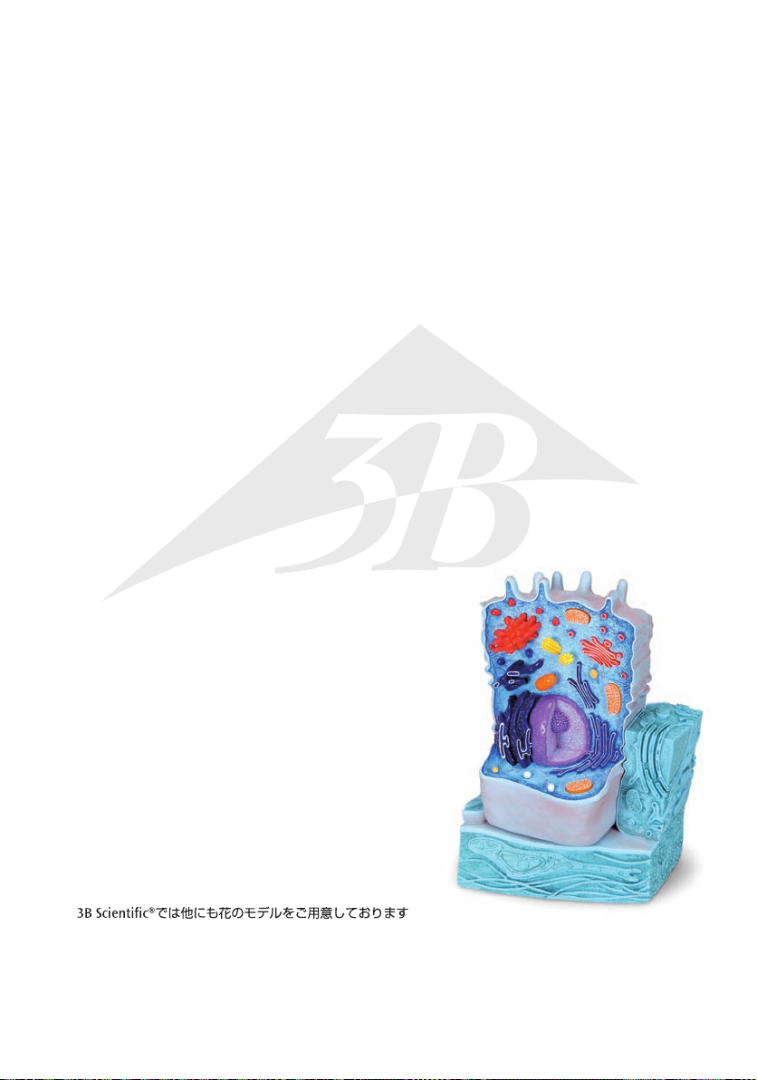
Chinese
8 线粒体
9 高尔基体
10 液泡
11 微粒体
12 细胞壁(分层结构)
Also available from 3B Scientific®:
R04 The Animal Cell
植物细胞模型
( 放大倍率约 500,000-1,000,000)
®
Ebenfalls bei 3B Scientific® erhältlich:
R04 Die tierische Zelle
Tambien disponible en 3B Scientific®:
R04 La Célula Animal
Egalement disponible auprès de 3B Scientific® :
R04 La cellule animale
Também disponível na 3B Scientific®:
R04 A célula animal
Disponibile anche presso 3B Scientific®:
R04 La cellula animale
也可以在 3B Scientific®获得:
R04 动物细胞
R04
Page 36

R05-08/06-1
®
3B SCIENTIFIC
3B Scientific GmbH
Rudorffweg 8 • 21031 Hamburg • Germany
Tel.: + 49-40-73966-0 • Fax: + 49-40-73966-100
www.3bscientific.com • 3b@3bscientific.com
© Copyright 2006 for instruction manual and design of product:
3B Scientific GmbH, Germany
PRODUC TS
 Loading...