

Page 1
… g o i n g o n e s t e p f u r t h e r
A79
(1000154)
Page 2

2
Page 3

®
Latin
1 Substantia trabecularis
2 Substantia compacta
3 Periosteum
4 Fibrae perforantes
5 Lamella circumferentialis externa
6 Osteon
7 Lamellae osteoni
8 Osteocytus
9 Vasa sanguineum (osteon)
10 Lamella interstitialis
11 Canalis perforans
12 Lamella circumferentialis interna
13 Endosteum
14 Medulla ossium (substantia spongiosa)
3
Page 4

®
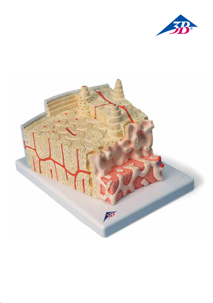
Bone Structure Model
English
This model shows a section of a lamellar bone, as
found in the human skeleton as a basic structure of
a tubular bone (approx. 80 times enlarged).
Compared to other bone types, tubular bones
contain few bone trabeculae (spongy substance or
substantia spongiosa) (1) and a thick compact layer
(compact substance or substantia compacta) (2).
The bone is covered by a membrane, the periosteum (3). The inner layer of the periosteum (osteogenic layer) consists of many cells and contains resting
precursor cells of the bone cells (osteoblasts) that
ensure regeneration in case of bone fractures. The
outer layer is made of firm, collagenous connective
tissue (fibrous layer). Bundles of collagenous fibers
pass directly from the periosteum into the connective tissue of the bone (perforating fibers, Sharpey
fibers) (4).
The next layer is the cortical layer (substantia corticalis) with its lamellar stratification of the intercellular substance (thus lamellar bone). Located at the
very outside are the circumferential lamellae (5)
running parallel to the periosteum. The basic structure of the lamellar bone is formed by the osteons,
also referred to as Haversian systems (6). The lamellae are layers of anorganic substance (approx. 65 %
of the complete substance), mainly hydroxyapatite,
and organic substance (over 90 % collagen). Since
the collagen fibers of the individual lamellae of
an oesteon run at various, respectively opposite
angles of inclination (7), the compound structure of
organic and anorganic components is additionally
stabilised against pull and push forces. The bone
cells (osteocytes) (8) are located between the lamellae, held in lacunae. The cells are nourished by the
Haversian vessels (9), because all osteocytes are
linked to one another and with the Haversian canal
through canaliculi containing their cell extensions.
The intermediate lamellae are located between the
Haversian systems (osteons) without any connection
to blood vessels (10).
disrupted by the transverse or oblique perforation
through the Volkmann canals. The cortical layer is
followed in a fluid transition by the trabecular layer
(spongy substance or substantia spongiosa), a spongy trabecular structure consisting of thin plates and
rods (1). In the direction of the medullary cavity,
the bony substance is limited by the inner circumferential lamella (12) and the inner membrane
called endosteum (13). The endosteum consists of
a connection of flat cells that can form new bones.
Red bone marrow (14) can be found between the
spongy trabeculae of the bone ends, and yellow
bone marrow or fatty marrow in the tubular part
of the bone. We thank Prof. Richard H. W. Funk,
MD, Director of the Institute of Anatomy of the Carl
Gustav Carus Medical Faculty, Dresden, Germany,
for his support in the development of this model.
Bone structure
1 Trabeculae of bone (spongy substance)
2 Compact substance or substantia compacta
3 Periosteum
4 Perforating fibers, Sharpey fibers
5 Outer circumferential lamella
6 Osteons
7 Lamellae of the osteons
8 Osteocytes
9 Haversian vessels
10 Intermediate lamellae
11 Volkmann canals
12 Inner circumferential lamella
13 Endosteum
14 Bone marrow (spongy substance)
Their thickness and stratification corresponds to
that of former Haversian systems, however, the
Haversian systems have built up over them in
the course of the continuous restructuring of the
bone that also continues after the growth period.
Running from the periosteum, the Volkmann
canals contain vessels connecting the vessels of the
Haversian canals (11). The Volkmann canals are
bony canals that are not surrounded by circular
lamellae, as opposed to the Haversian canals. The
systematic order of the Haversian canals is not
4
Page 5

®
Deutsch
Knochenstrukturmodell
Das Modell zeigt einen Ausschnitt aus einem
Lamellenknochen wie er im menschlichen Skelett
als Grundstruktur eines Röhrenknochens vorkommt
(ca. 80-fache Vergrößerung).
Im Röhrenknochen findet man im Vergleich zu
anderen Knochenformen wenig Knochenbälkchen
(Substantia spongiosa oder Spongiosa) (1) und eine
dicke kompakte Schicht (Substantia compacta oder
Kompakta) (2). Der Knochen wird von Knochenhaut
(Periost) überzogen (3). Dabei ist die innere Schicht
der Knochenhaut (Stratum osteogenicum) zellreich
und enthält ruhende Vorstufen der Knochenzellen
(Osteoblasten), die bei Knochenbrüchen die
Regeneration besorgen. Straffes, kollagenes
Bindegewebe
bildet die äußere Schicht (Stratum fibrosum). Vom
Periost gehen Bündel von Kollagenfibrillen direkt
in
das Bindegewebe des Knochens über (Fibrae perforantes, Sharpeysche Fasern) (4).
Es folgt die Rindenschicht (Substantia corticalis
oder „Kortikalis“) mit ihrer lamellären Schichtung
der Interzellularsubstanz (daher Lamellenknochen).
Ganz außen liegen die parallel zur Knochenhaut
liegenden Generallamellen (5). Die Grundstruktur
des lamellären Knochens sind die Knochensäulchen
(Osteone),
auch Haverssche Lamellensysteme oder
Speziallamellen genannt (6). Die Lamellen sind
schichtweise aus anorganischer Substanz (ungefähr
65 % der Gesamtmasse), hauptsächlich
Hydroxylapatit und organische Substanz (davon 90
% Kollagen) aufgebaut. Da die Kollagenfasern der
einzelnen Lamellen eines Osteons unterschiedliche,
jeweils gegensinnig laufende Steigungswinkel aufweisen (7), wird die Verbundstruktur von organischen und anorganischen Komponenten zusätzlich gegenüber Zug- und Druckkräften stabilisiert.
Jeweils zwischen den Lamellen liegen die
Knochenzellen (Osteozyten) (8) in Lakunen eingemauert.
Die Zellen werden von den Haversschen Gefäßen (9)
ernährt, denn untereinander und zum Haversschen
Kanal stehen alle Osteozyten durch Kanälchen
(Canaliculi), in denen ihre Zellausläufer liegen, in
Verbindung. Zwischen den Haversschen
Lamellensysteme (Osteonen) befinden sich
Schaltlamellen ohne Beziehung zu Blutgefäßen
(10). Sie entsprechen in Stärke und Schichtung ehemaligen Haversschen Systemen, dabei sind sie
durch den ständigen Umbau des Knochens, der
auch nach der Wachstumsphase fortbesteht, von
den Haversschen Systemen überbaut worden. Vom
Periost kommend enthalten die Volkmannschen
Kanäle Gefäße, die jene Gefäße der Haversschen
Kanäle verbinden (11).
Die Volkmannschen Kanäle sind Knochenkanäle,
die im Gegensatz zu den Haversschen Kanälen nicht
von zirkulären Lamellen umgeben sind. Die systematische Ordnung der Haversschen Kanälen wird
von der queren oder schrägen Perforation durch
die Volkmannschen Kanäle nicht gestört. Auf die
Rindenschicht folgt im fließenden Übergang die
Bälkchenschicht (Substantia spongiosa oder
„Spongiosa“), ein schwammartiges Trabekelwerk
aus dünnen Platten und Stäben (1). Zum
Knochenmarksraum grenzt sich die Knochensubstanz über die innere Generallamelle (12) und
die innere Knochenhaut (Endost) (13) ab. Das
Endost besteht aus einem Verband abgeflachter
Zellen, die neuen Knochen bilden können.
Zwischen den Spongiosabälkchen der
Knochenenden findet man rotes Knochenmark (14)
im röhrenförmigen Teil des Knochens gelbes
Knochenmark oder Fettmark. Wir danken Herrn
Prof. Dr. med. Richard H. W. Funk, Leiter des
Instituts für Anatomie der Medizinischen Fakultät
Carl Gustav Carus, Dresden/Deutschland, für die
Unterstützung bei der Entwicklung dieses Modells.
Knochenstruktur
1 Knochenbälkchen (Trabekel) der Spongiosa
2 Kompakta
3 Periost
4 Fibrae perforantes, Sharpeysche Fasern
5 Äußere Generallamelle
6 Osteone
7 Lamellen der Osteone
8 Osteozyten
9 Haverssche Gefäße
10 Schaltlamellen
11 Volkmannsche Kanäle
12 Innere Generallamelle
13 Endost
14 Spongiosa mit Knochenmark
5
Page 6

®
EspañolModelo de la estructura del hueso
El modelo muestra el fragmento de un hueso compacto tal como aparece en el esqueleto humano
como estructura básica de un hueso largo (aumentado 80 veces aprox.).
Los huesos largos contienen menos trabéculas
óseas (Substancia spongiosa o esponjosa) (1) que
otras formaciones óseas y una capa gruesa compacta (Substantia compacta o compacta) (2). Los
huesos están revestidos por el periostio (3). La parte
interna del periostio (Stratum osteogenicum) es
rica en células y contiene células precursoras de las
células óseas (osteoblastos), que se encargan de la
regeneración de las fracturas óseas. La parte externa está formada por un tejido conjuntivo colágeno
tenso (Stratum fibrosum). Del periostio salen haces
de fibrillas de colágeno que se dirigen al tejido
conjuntivo del hueso (Fibrae perforans o fibras de
Sharpey) (4). A continuación se encuentra la corteza
(Substantia corticalis o „cortical“) con la estratificación lamelar de su sustancia intercelular (por
ello denominados huesos compactos). Más externamente se hallan las laminillas circunferenciales
dispuestas paralelamente al periostio (5). La unidad
básica de estructura del hueso compacto son los
osteones, también denominados sistema de laminillas de Havers o laminillas basales (6). Las laminillas están formadas por sustancia inorgánica (casi
el 65% de la masa total), principalmente hidroxilapatita, y sustancia orgánica (90% colágeno). Ya que
las fibras colágenas de cada una de las laminillas se
diferencian en un solo osteón, y como en cada caso
se disponen en un ángulo de inclinación adecuado, la estructura de conexión de los componentes
orgánicos e inorgánicos permite una estabilización
suplementaria frente a las fuerzas de tracción y
de presión. Entre las laminillas se encuentran las
células óseas (osteocitos) (8) encajadas en lagunas.
Las células se nutren de los vasos sanguíneos (9) en
los conductos de Havers, además, todos los osteocitos están agrupados y se dirigen hacia el conducto
de Havers a través de canalículos en los que se
encuentran las prolongaciones celulares. Entre el
sistema de laminillas de Havers (osteones) se hallan las laminillas intersticiales sin relación con los
vasos sanguíneos (10). Corresponden en fuerza y
estratificación a los antiguos sistemas de Havers, y
constituyen una supraestructura de este sistema a
partir de la trasformación continua del hueso y que
se continúa después de la fase de crecimiento.
Cada uno de los vasos de los conductos de
Volkmann que provienen del periostio, se comunican con los vasos de los conductos de Havers
(11). Los conductos de Volkmann son conductos
nutricios, y a diferencia de los conductos de Havers
no están rodeados de laminillas circulares. El orden
sistemático de los conductos de Havers no se altera
por la perforación oblicua o trasversal de los conductos de Volkmann.
En la corteza continúa en fluída transición la capa
trabecular (substancia espongiosa), un entramado
esponjoso trabecular constituído por finas placas
y bastones (1). La médula ósea queda delimitada
en la cavidad de la médula ósea por encima de la
laminilla circunferencial interna (12) y el endostio
interno (13). El endostio está constituído por un
conjunto de células planas, capaces de construir
hueso nuevo. Entre las capas espongiosas de las
epífisis óseas se encuentra la médula ósea roja (14),
Agradecemos la colaboracíón del Prof. Dr. med.
Richard H.W. Funk, Director del Instituto Anatómico
de la Facultad de Medicina Carl Gustav Carus de
Dresden, Alemania en el diseño y desarrollo de este
modelo.
Estructuras óseas
1 Trabéculas del tejido esponjoso
2 Substantia compacta o compacta
3 Periostio
4 Fibrae perforantes, fibras de Sharpey
5 Laminilla circunferencial externa
6 Osteones
7 Laminillas de los osteones
8 Osteocitos
9 Vasos de los conductos de Havers
10 Laminilla intersticial
11 Conductos de Volkmann
12 Laminillas circunferenciales
13 Endostio
14 Tejido esponjoso con médula ósea
6
Page 7

®
Français Modèle de la structure osseuse
Le modèle montre une section d’un os lamellaire
tel qu’il se présente dans le squelette humain en
tant que
structure de base d’un os long (agrandissement env.
80 fois).
Par comparaison avec d’autres types d’os, on ne
trouve que peu de travées osseuses, la substance
spongieuse (Substantia spongiosa) (1) dans l’os
long et une grosse couche compacte (Substantia
compacta) (2). L’os est revêtu du périoste (3). La
couche interne du périoste (Stratum osteogenicum)
est riche en cellules et contient des précurseurs au
repos des cellules osseuses (ostéoblastes) qui assurent la régénération en cas de fractures osseuses.
Le tissu conjonctif collagène ferme forme la couche
externe (Stratum fibrosum). A partir du périoste,
des faisceaux de fibrilles collagènes atteignent
directement le tissu conjonctif de l’os (Fibrae perforantes, fibres de Sharpey) (4).
Vient alors la couche corticale (Substantia corticalis)
avec sa stratification lamellaire de la substance
intercellulaire (d’où le nom os lamellaire). Les
lamelles générales (5) situées parallèlement au
périoste se trouvent complètement à l’extérieur. Les
ostéons, également appelés systèmes lamellaires de
Havers ou lamelles spéciales, constituent la structure de base de l’os lamellaire (6). Les lamelles sont
disposées en couches de substance inorganique
(environ 65 % de la masse totale), principalement
d’hydroxylapatite et de substance organique (dont
90 % de collagène). Etant donné que les fibres de
collagène des différentes lamelles d’un ostéon
présentent des angles de montée situés en sens
inverse (7), la structure de liaison des composants
organiques et inorganiques est en plus stabilisée
contre les forces de traction et de pression. Les cellules osseuses (ostéocytes) (8) sont emmurées dans
des lacunes situées entre les lamelles. Les cellules
sont nourries par les canaux de Havers (9) car tous
les ostéocytes sont reliés entre eux et avec le canal
de Havers par des petits canaux (Canaliculi) où se
trouvent leurs stolons cellulaires.
Entre les systèmes lamellaires de Havers (ostéons)
se trouvent les lamelles intermédiaires sans relation
avec les vaisseaux sanguins (10). Au point de vue de
l’épaisseur et de la stratification, elles correspondent à d’anciens systèmes de Havers ; elles sont
cependant enveloppées par des systèmes de Havers
par la restructuration permanente de l’os, persistant également après la phase de croissance.
Les canaux de Volkmann, émanant du périoste,
contiennent des vaisseaux reliés aux vaisseaux des
canaux de Havers (11). Les canaux de Volkmann
sont des canaux osseux qui contrairement aux
canaux de Havers ne sont pas entourés de lamelles
circulaires. L’ordre systématique des canaux de
Havers n’est pas perturbé par la perforation transversale ou oblique des canaux de Volkmann.
A la couche corticale succède directement la couche
spongieuse (Substantia spongiosa), un réseau de
travées osseuses constitué de fines plaques et de
bâtonnets (1). La substance osseuse se délimite
de l’espace de la moelle osseuse par la lamelle
générale interne (12) et la peau osseuse interne
(endoste) (13). L’endoste est composé d’une association de cellules affaiblies pouvant former de nouveaux os. Entre les travées spongieuses des extrémités osseuses, on trouve la moelle osseuse rouge
(14), dans la partie longue de l’os la moelle osseuse
jaune. Nous remercions le Professeur Richard H. W.
Funk, docteur en médecine et directeur de l’Institut
d’anatomie de la faculté de médecine Carl Gustav
Carus, Dresde/Allemagne, pour l’aide apportée lors
du.
Structure osseuse
1 Ossification (trabécule) de la substance spongieuse
2 Substance compacte
3 Périoste
4 Fibres perforantes, fibres de Sharpey
5 Lamelle générale externe
6 Ostéons
7 Lamelles des ostéons
8 Ostéocytes
9 Vaisseaux de Havers
10 Lamelles intermédiaires
11 Canaux de Volkmann
12 Lamelle générale interne
13 Endoste
14 Substance spongieuse avec moelle épinière
7
Page 8

11
4
3
5
8
10
2
6
12
1
14
8
7
4
13
13
12
3
11
14
9
8
Page 9

9 7 8
3
12
13
14
4
11
9
7
13
14
9
Page 10

®
PortuguêsModelo de la estructura del hueso
El modelo muestra el fragmento de un hueso compacto tal como aparece en el esqueleto humano
como estructura básica de un hueso largo (aumentado 80 veces aprox.). Los huesos largos contienen
menos trabéculas óseas (Substancia spongiosa o
esponjosa) (1) que otras formaciones óseas y una
capa gruesa compacta (Substantia compacta o
compacta) (2). Los huesos están revestidos por el
periostio (3). La parte interna del periostio (Stratum
osteogenicum) es rica en células y contiene células
precursoras de las células óseas (osteoblastos), que
se encargan de la regeneración de las fracturas
óseas. La parte externa está formada por un tejido
conjuntivo colágeno tenso (Stratum fibrosum). Del
periostio salen haces de fibrillas de colágeno que se
dirigen al tejido conjuntivo del hueso (Fibrae perforans o fibras de Sharpey) (4).
A continuación se encuentra la corteza (Substantia
corticalis o „cortical“) con la estratificación lamelar
de su sustancia intercelular (por ello denominados
huesos compactos). Más externamente se hallan
las laminillas circunferenciales dispuestas paralelamente al periostio (5). La unidad básica de estructura del hueso compacto son los osteones, también
denominados sistema de laminillas de Havers o
laminillas basales (6). Las laminillas están formadas
por sustancia inorgánica (casi el 65% de la masa
total), principalmente hidroxilapatita, y sustancia
orgánica (90% colágeno). Ya que las fibras colágenas
de cada una de las laminillas se diferencian en un
solo osteón, y como en cada caso se disponen en
un ángulo de inclinación adecuado, la estructura
de conexión de los componentes orgánicos e inorgánicos permite una estabilización suplementaria
frente a las fuerzas de tracción y de presión. Entre
las laminillas se encuentran las células óseas
(osteocitos) (8) encajadas en lagunas. Las células se
nutren de los vasos sanguíneos (9) en los conductos
de Havers, además, todos los osteocitos están agrupados y se dirigen hacia el conducto de Havers a
través de canalículos en los que se encuentran las
prolongaciones celulares.
Entre el sistema de laminillas de Havers (osteones)
se hallan las laminillas intersticiales sin relación
con los vasos sanguíneos (10). Corresponden en
fuerza y estratificación a los antiguos sistemas de
Havers, y constituyen una supraestructura de este
sistema a partir de la trasformación continua del
hueso y que se continúa después de la fase de
crecimiento.
Cada uno de los vasos de los conductos de
Volkmann que provienen del periostio, se comunican con los vasos de los conductos de Havers
(11). Los conductos de Volkmann son conductos
nutricios, y a diferencia de los conductos de Havers
no están rodeados de laminillas circulares. El orden
sistemático de los conductos de Havers no se altera
por la perforación oblicua o trasversal de los conductos de Volkmann.
En la corteza continúa en fluída transición la capa
trabecular (substancia espongiosa), un entramado
esponjoso trabecular constituído por finas placas
y bastones (1). La médula ósea queda delimitada
en la cavidad de la médula ósea por encima de la
laminilla circunferencial interna (12) y el endostio
interno (13). El endostio está constituído por un
conjunto de células planas, capaces de construir
hueso nuevo. Entre las capas espongiosas de las
epífisis óseas se encuentra la médula ósea roja (14),
y en la parte tubular de los huesos la médula ósea
amarilla o grasa.
Agradecemos la colaboracíón del Prof. Dr. med.
Richard H.W. Funk, Director del Instituto Anatómico
de la Facultad de Medicina Carl Gustav Carus de
Dresden, Alemania en el diseño y desarrollo de este
modelo.
Estructuras óseas
1 Trabécula óssea
2 Substantia compacta o compacta
3 Periostio
4 Fibrae perforantes, fibras de Sharpey
5 Laminilla circunferencial externa
6 Osteones
7 Laminillas de los osteones
8 Osteocitos
9 Vasos de los conductos de Havers
10 Laminilla intersticial
11 Conductos de Volkmann
12 Laminillas circunferenciales
13 Endostio
14 Osso esponjoso com medula óssea
10
Page 11

®
Italiano Modello di struttura ossea
Questo modello mostra una sezione dell’osso
lamellare come si trova nello scheletro umano,
in funzione di struttura base di un osso tubolare
(ingrandito circa 80 volte).
Rispetto agli altri tipi di osso, le ossa tubolari
contengono poche trabecole (sostanza spugnosa)
(1) e uno strato compatto e spesso (sostanza compatta) (2). L’osso è ricoperto da una membrana, il
periostio (3). Lo strato interno del periostio (strato
osteogenico) è composto da numerose cellule e
contiene cellule precursori dormienti (osteoblasti)
che assicurano la rigenerazione in caso di frattura
dell’osso. Lo strato esterno è costituito da tessuto
connettivo solido e collagenoso (strato fibroso).
Fasci di fibre collagenose passano direttamente
dal periostio nel tessuto connettivo dell’osso (fibre
perforanti, fibre di Sharpey) (4). Lo strato successivo
è quello corticale (sostanza corticale) con la sua
stratificazione lamellare della sostanza intercellulare (da qui il nome osso lamellare). La sezione più
esterna è costituita dalle lamelle circonferenziali
(5), che corrono parallelamente al periostio. La
struttura di base dell’osso lamellare è formata dagli
osteoni, denominati anche sistemi Haversiani (6).
Le lamelle sono strati di sostanza anorganica (circa
il 65% della sostanza completa), principalmente
idrossiapatite, e di sostanza organica (più del 90%
collagene). Poiché le fibre di collagene delle singole
lamelle di un osteone corrono ad angoli di inclinazione diversi e rispettivamente opposti (7), la struttura dei componenti organici e anorganici è ulteriormente stabilizzata contro le forze di tensione e
pressione. Le cellule ossee (osteociti) (8) si trovano
tra le lamelle, contenute nelle lacune. Le cellule
vengono alimentate dai canali Haversiani (9), poiché tutti gli osteociti sono collegati gli uni agli altri
e con il canale Haversiano attraverso canalicoli che
contengono le loro estensioni cellulari.
ultimi non viene modificato dalla perforazione
trasversale o
obliqua dei canali di Volkmann.
Lo strato corticale è seguito in una transizione
fluida dallo strato trabecolare (sostanza spugnosa),
una struttura spugnosa trabecolare costituita da
sottili placche e aste (1). In direzione della cavità
midollare,
la sostanza ossea è delimitata dalla lamella circonferenziale interna (12) e da una membrana
interna chiamata endotelio (13). Quest’ultimo è
costituito da una rete di cellule piatte in grado di
formare nuove ossa. Tra le trabecole spugnose delle
estremità dell’osso si trova midollo osseo rosso
(14), mentre nella parte tubolare dell’osso si trova
midollo osseo giallo, tale a causa della presenza di
tessuto adiposo.
Si ringrazia il Prof. Richard H. W. Funk, MD, direttore dell’istituto di anatomia della facoltà di medicina Carl Gustav Carus di Dresda, per il suo contributo nello sviluppo di questo modello.
Struttura ossea
1 Trabecole ossee (Tessuto spugnoso)
2 Sostanza compatta
3 Periostio
4 Fibre perforanti, fibre di Sharpey
5 Lamella circonferenziale esterna
6 Osteoni
7 Lamelle degli osteoni
8 Osteociti
9 Canali Haversiani
10 Lamelle intermedie
11 Canali di Volkmann
12 Lamella circonferenziale interna
13 Endotelio
14 Midollo osseo
Le lamelle intermedie sono collocate tra i sistemi
Haversiani (osteoni) senza collegamento ai vasi
sanguigni (10). Il loro spessore e la loro stratificazione corrispondono a quelli di precedenti sistemi
Haversiani, tuttavia, questi ultimi sono cresciuti
sopra di esse nel corso della continua ristrutturazione dell’osso che continua anche dopo il periodo
di crescita.
I canali di Volkmann, che partono dal periostio,
contengono vasi che collegano i vasi dei canali
Haversiani (11). I canali di Volkmann sono canali
ossei non avvolti da lamelle circolari, a differenza
dei canali Haversiani. L’ordine sistematico di questi
11
Page 12

骨の構造モデル
日本語
骨の構造モデル
このモデルはヒトの大腿骨にみられる長管骨の基本構造である層板骨の断面を約80倍大で示している。
他の骨にみられる構造と較べると,長管骨の構造は次の特徴をもつ。(1):海綿質,(2):緻密質(厚い緻密の
層),(3):骨膜(骨を囲む膜)である。骨膜は二層からなっていて,その内層(骨形成層)は多くの細胞を
含み,特に骨芽細胞と呼ばれる骨細胞の前駆細胞は骨折などの際,骨組織の再生を促す。外層は固い膠原性結
合組織からなる(線維層)。膠原性線維の束はシャーピー線維(4)と呼ばれ,直接骨膜から骨の結合組織に侵
入する。
次の深層は皮質層で骨内の細胞質が層状に取り囲まれているのが特徴である(よって層板骨と呼ぶ)。その最
外層は外基礎層板(5)で,骨膜と平行に走る周状の層である。
層板骨の基本構造はハバース系(6)とも呼ばれるオステオン(骨単位)によりつくられる。層板骨は主にハイ
ドロキシアパタイトの無機物質(全体のおよそ65%を占める)と,有機物質(90%以上コラーゲン=膠原)
からなる。オステオンにある個々の層の膠原線維走行はさまざまで,それぞれ違った方向に走っているため(7)
,この複合構造は骨の耐久性の向上に役立っている。骨細胞(8)は骨層板間の小腔(lacunae)に見られる。
骨細胞はハバース管(9)により栄養を供給され,全ての骨細胞はハバース管とその小管を介して互いに連絡し
ている。
介在層板(10)はハバース系の間にあリ血管との連絡はない。介在層板の厚さと層板はハバース系のそれに比例
するが,ハバース系は成長期の終了後も続く骨の再成とともに介在層板にかぶさっていく。
骨膜より続くフォルクマン管(11)の中を通る血管はハバース管を通る血管同士をつないでいる。フォルクマン
管はハバース管とは異なり層板に取り囲まれてはいない。また,ハバース管の規律的な配列は横または斜めに
走るフォルクマン管により乱されることはない。
骨髄腔へ向かって,骨質は内基礎層板(12)と内骨膜(13)と呼ばれる内膜で仕切られる。内骨膜は新生骨を作
る平らな細胞のつながりである。赤色骨髄(14)は長管骨骨端の海綿質にあり,黄色骨髄(脂肪骨髄)は長管骨
の骨体にある。
モデル製作協力
日本語
Prof. Richard H. W. Funk, MD, Director of the Institute of Anatomy of the Carl Gustav Carus Medical Faculty, Dresden, Germany
骨の基本構造
1 骨梁(海綿質)
2 緻密質
3 骨膜
4 シャーピー線維(貫通線維)
5 外基礎層板
6 オステオン(骨単位)
7 層板(オステオンの層)
8 骨細胞
9 ハバース管
10 介在層板
11 フォルクマン管
12 内基礎層板
13 内骨膜
14 骨髄(海綿質)
Page 13

®
Русский Модель строения кости
На модели показан разрез пластинчатой
кости, основной структуры трубчатой кости
человеческого скелета (приблизительно
80-кратное увеличение).
По сравнению с другими типами костей трубчатые
кости содержат немного костных трабекул
(губчатое вещество) (1) и толстый компактный
слой (компактное вещество) (2). Кость покрыта
мембраной, надкостницей (3). Внутренний
слой надкостницы (остеогенный слой) состоит
из множества клеток и содержит клеткипредшественницы костных клеток в состоянии
покоя (остеобласты), за счет которых происходит
регенерация в случае перелома кости.
Наружный слой состоит из твердой коллагеновой
соединительной ткани (фиброзный слой). Пучки
коллагеновых волокон проходят напрямую через
надкостницу в соединительную ткань кости
(прободающие, или шарпеевы, волокна) (4).
Следующий слой – кортикальный (кортикальное
вещество) с пластинчатой слоистостью
внутриклеточного вещества (отсюда название —
пластинчатая кость). На периферии параллельно
надкостнице расположены циркулярные
пластинки (5). Основная структура пластинчатой
кости образована остеонами, также известными
под названием гаверсовой системы (6).
Пластинки – это слои неорганического вещества
(приблизительно 65% всего вещества), в основном
гидроксиапатита, и органического вещества (более
90% коллагена). Поскольку коллагеновые волокна
отдельных пластинок остеона проходят под
различными, взаимно противоположными углами
наклона (7), сложная структура органических
и неорганических компонентов придает
дополнительную устойчивость к действию сил
растяжения и давления. Клетки кости (остеоциты)
(8) располагаются между пластинками, в лакунах.
Клетки получают питание через гаверсовы сосуды
(9), поскольку все остеоциты связаны друг с
другом и с гаверсовым каналом канальцами с
клеточными выростами. Промежуточные пластинки
располагаются между гаверсовыми системами
(остеонами) без какой-либо связи с кровеносными
сосудами (10). Их толщина и слоистость
соответствует таковым прежних гаверсовых систем,
однако гаверсовы системы нарастают на них в
ходе непрерывного изменения структуры кости,
которое продолжается и после периода роста.
Отходящие от надкостницы фолькмановские
каналы содержат сосуды, соединяющие сосуды
гаверсовых каналов (11). Фолькмановские каналы
представляют собой костные каналы, в отличие от
гаверсовых каналов не окруженные циркулярными
пластинками. Систематический порядок
гаверсовых каналов не нарушается поперечным
или косым прободением фолькмановских
каналов. Кортикальный слой плавно переходит
в трабекулярный слой (губчатое вещество),
губчатую трабекулярную структуру, состоящую из
тонких пластин и столбиков (1). В направлении
полости костного мозга вещество кости ограничено
внутренней циркулярной пластинкой (12) и
внутренней мембраной, называемой эндостом
(13). Эндост состоит из группы плоских клеток,
способных образовывать новые кости. Красный
костный мозг (14) находится между губчатыми
трабекулами концов кости, а желтый, или
жировой, костный мозг – в трубчатой части кости.
Мы благодарим проф. Ричарда Фанка (Prof.
Richard H. W. Funk, MD), директора Института
анатомии, медицинский факультет им. Карла
Густава Каруса, Дрезден, Германия, за помощь в
разработке этой модели.
Структура кости
1 Трабекулы кости (губчатое вещество)
2 Компактное вещество
3 Надкостница
4 Прободающие волокна, шарпеевы волокна
5 Наружная циркулярная пластинка
6 Остеоны
7 Пластинки остеонов
8 Остеоциты
9 Гаверсовы сосуды
10 Промежуточные пластинки
11 Фолькмановские каналы
12 Внутренняя циркулярная пластинка
13 Эндост
14 Костный мозг (губчатое вещество)
13
Page 14

®
中文骨结构模型
该模型显示了板层骨的部分结构,该结构是最
近发现的作为人体骨骼单位的管状骨的基本结
构,
(该模型大约是真实尺寸放大的80倍)。
与其他类型的骨骼相比,管状骨是由很少的骨
小梁结构(疏松状或海绵状结构)(1)与一层
较厚的致密层构成(致密质或骨密质)(2)。
而且骨头上通常附有一层薄膜及骨膜(3)。
骨膜内层也就是生骨层,通常由较多的细胞构
成,并且该层内还有较多静息状态的骨前体细
胞(格根包尔氏细胞),这些细胞可以保证骨
折后的骨再生。外层是由坚硬的胶原结缔组织
层构成(纤维组织层)。这些胶原纤维束纵穿
骨外膜,进入骨的结缔组织内(穿通纤维,夏
皮纤维)(4)。
之下为皮质层,该层内有细胞间质的分层现象
(因此又称板层骨)。皮质层的最外围又称环
状层(5),该层平行于骨外膜。板层骨是由最
基本的股单位,也称哈弗氏系统构成(6)。板
层骨中的层主要是由大部分羟基磷灰石等无机
质(大约占全部物质的65%),与少量部分有机
质构成(而有机质中90%以上的是胶原)。由于
骨质各层之间的胶原纤维有不同的相反的倾角
(7),因此骨质中的有机质与无机质之间的稳
定性得到进一步的加强,这些结构可以抵抗外
力的推拉。骨细胞(8)位于各板层之间,保持
陷窝状。由于所有的骨细胞之间相互连接在一
起,并且与哈弗氏管系统紧密接触,所以所有
的骨细胞均是由哈弗氏管供养(9)。
我们要衷心的感谢德国德雷斯顿的卡尔古斯塔
夫卡斯医学院,解剖教研室主任Richard H. W.
Funk教授,同时他也是一名医生,对该模型的
大力支持。
骨结构
1 骨小梁(海绵体)
2 致密质
3 骨外膜
4 穿通纤维、夏皮纤维
5 外环骨板
6 骨单位
7 骨单位薄层
8 骨细胞
9 哈弗氏系统
10 中间层骨板
11 福尔克曼管
12 内层的环状层
13 骨内膜
14 骨髓(海绵体)
中间层位于哈弗氏系统之间,并且不与任何血
管相连接(10)。尽管他们的厚度与分层情况
均与前哈氏弗系统明显相关,但是哈弗氏系
统是在骨持续重建过程中基于这些中间层形成
的,而且这些哈弗氏系统在生长期之后同样会
继续发育。
福尔克曼管内有脉管,其在穿过骨外膜之后与
哈弗氏系统内的脉管(11)相互交通。福尔克曼
管为骨性管道,周围并没有环状层,而且其与
哈弗氏管相反。哈弗氏管系统的顺序并不会由
于其横贯或斜穿福尔克曼管而被打乱。
皮质层遵循一种流动的过渡小梁层特点,主要
由薄的板及棒构成的小梁状构成(1)。在骨髓
腔方向内,骨质受内层的环状层(12)与内膜层
(又叫骨内膜)(13)限制。骨内膜是由一系
列扁平的细胞连接构成,这些细胞可以形成新
生骨。红骨髓(14)位于骨端的骨小梁内,黄
骨髓又叫脂肪骨髓位于骨的管状部。
14
Page 15

®
15
Page 16

A79 (1000154)-10/12-35001963
3B Scientific
A wo r l d w i d e g ro u p o f c o mp a n i e s
3B Sci entific GmbH
Rudorffweg 8 • 21031 Hamburg • Germany
Tel.: + 49-40-73966-0 • Fax: + 49-40-73966-100
www.3bscientific.com • 3b@3bscientific.com
© Copyright 2002 / 2011 / 2012 for instruction manual and design
of product: 3B Scientific GmbH, Germany
 Loading...