

Page 1
Product Information
Version 2.0
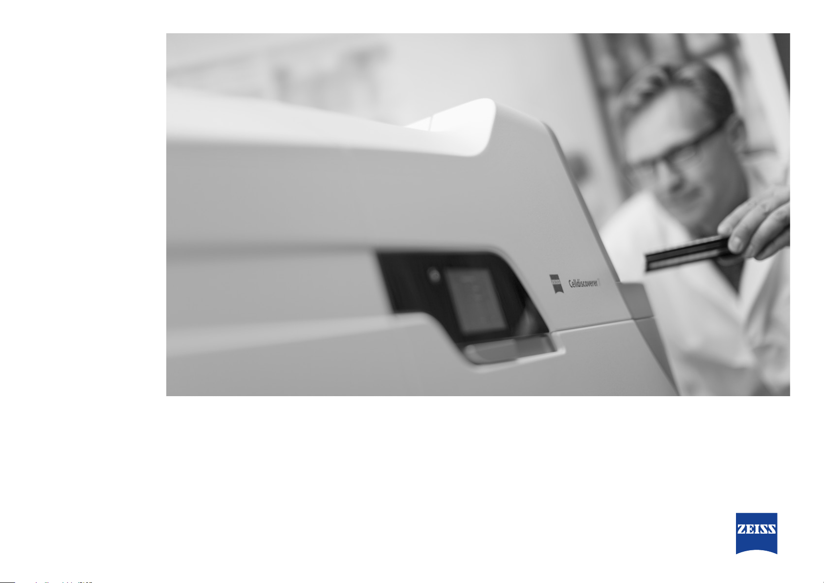
ZEISS Celldiscoverer7
Your Automated Platform for Live Cell Imaging
Page 2

Your Automated Platform for Live Cell Imaging
Animation
› In Brief
› The Advantages
› The Applications
› The System
› Technology and Details
› Service
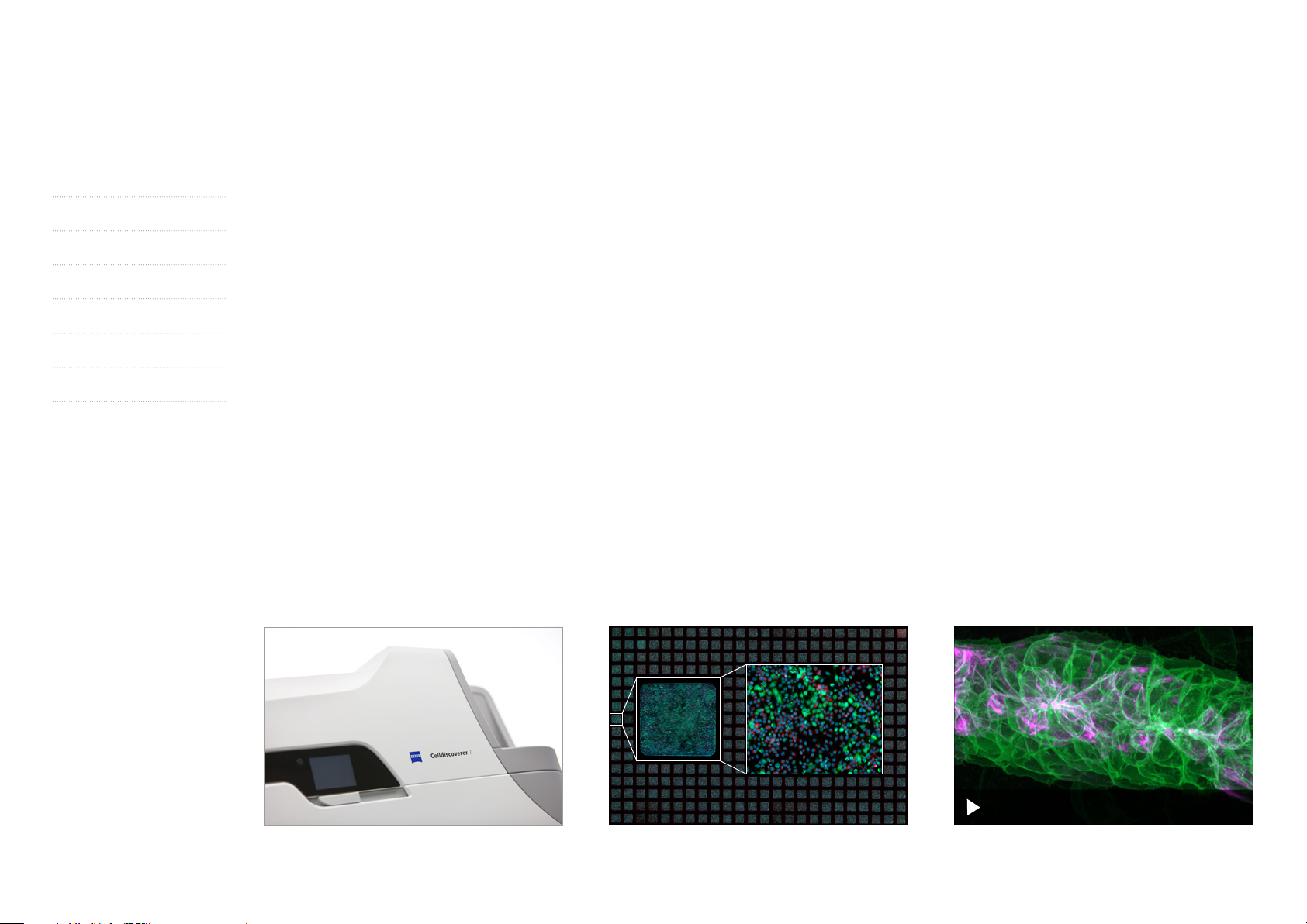
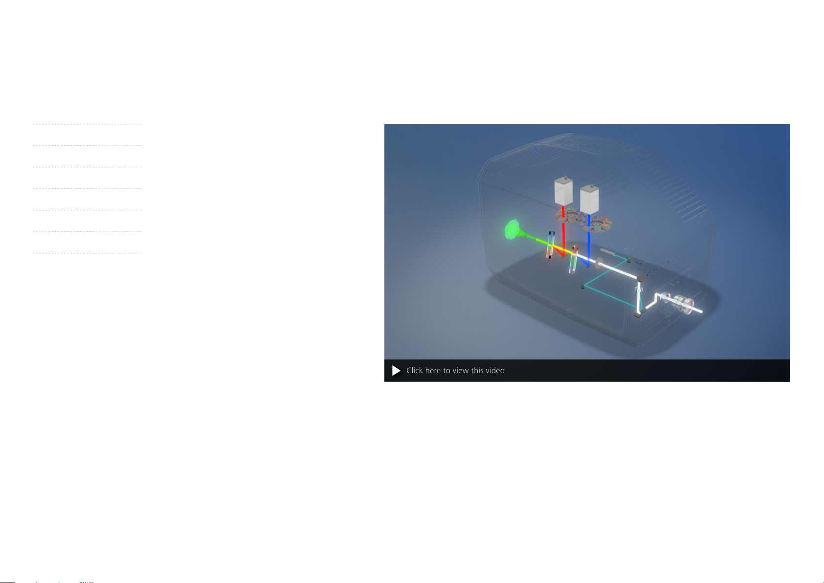
Often in life sciences research, the data you are after will only be revealed using
multiple runs of experiments and complex equipment. Automation can be the
only way to get there. With Celldiscoverer7, you can combine the easy-to-use
automation of a boxed microscope with the image quality and flexibility of a
classic inverted research microscope. Celldiscoverer7 calibrates itself, then
detects and focuses on your samples while the optics adjust themselves.
Leaving you free to get on with other projects. Whether working with 2D or 3D
cell cultures, tissue sections or small model organisms, you will acquire better
data in shorter times with this reliable automated research platform. What’s
more, you can enhance your Celldiscoverer7 with optical sectioning to get more
information from your three-dimensional samples. It’s your choice whether you
opt for confocal imaging with LSM900 and Airyscan2 or fast GPU deconvolution.
Click here to view this video
2
Page 3
Simpler. More Intelligent. More Integrated.
› In Brief
› The Advantages
› The Applications
› The System
› Technology and Details
› Service
A Flexible Platform
Celldiscoverer7 is a fully integrated high-end
imaging system. It comes with various incubation
and detection options so you can tailor the system
to your applications. Go for fast, sensitive sCMOS
or EMCCD cameras when performing your most
demanding live cell experiments and rapid
time-lapse recordings. To get better data from
your 3D samples, simply add the optional
LSM900 with Airyscan2 for confocal imaging,
or fast GPU-based deconvolution. Get all these
benefits and more with the in-built flexibility of
Celldiscoverer7.
Top Quality Data from Your Samples
For demanding long-term, time-lapse imaging,
Celldiscoverer7 gives you the advantage of Auto-
immersion and a hardware-based focus that finds
and keeps the focus automatically after detecting
the thickness and optical properties of the sample
carrier. Autocorr objectives then correct spherical
aberrations to deliver crisp contrast and high
resolution every time. Get image quality like
you‘ve never seen before – no need to adjust
manually. Keep your cells happy and they’ll deliver
unbiased data: Celldiscoverer7 provides a range
of integrated incubation options to create just the
right environment. The improved optical design
resolves more details in large fields of view.
Reproducible Results Made Easy
As soon as you start imaging, automatic calibration
routines take over to ensure reproducible results.
Check the current status and follow progress of
your experiments on the touchscreen.
With barcode recognition you can identify your
sample, sample carrier and even the type of
experiment. If you don‘t work with barcodes, an
automatic preview scan will identify the sample
carrier and calibrate it. ZEISS predictive service
offers lasting and optimal instrument performance
for increased system uptime and reliable results.
2,5×
20×
Click here to view this video
3
Page 4
Your Insight into the Technology Behind It
› In Brief
› The Advantages
› The Applications
› The System
› Technology and Details
› Service
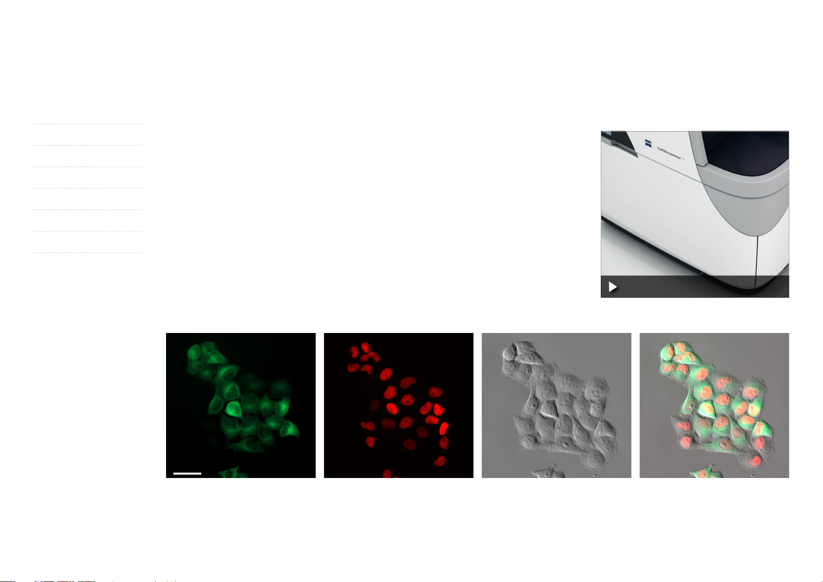
An Easy-to-Use Integrated Microscope
Observing live samples over a number of days or imaging lots of multiwell plates really puts your microscope
through its paces. To get reproducible, unbiased data, you must control environmental conditions such as
light, temperature, CO
etc. That’s why Celldiscoverer7 brings you a unique combination of a stable box,
2
darkroom and integrated inverted research microscope with optional incubation. It simplifies your laboratory
setup and makes work more comfortable.
All Celldiscoverer7 components are optimized for hassle-free automated imaging. New users and multi-user
facilities especially will enjoy the in-built automation and usability features when setting up complex
experiments. You’ll systematically avoid accidental hardware changes that might lead to biased data or even
damage your microscope. And Celldiscoverer7 can make you more productive, too: expect better data
in shorter times, with less training and maintenance. What’s more, as your needs grow you can expand
Celldiscoverer7 with confocal technology, external cameras, deconvolution, additional environmental
control – whatever you need for the challenge of live cell observation.
Click here to view this video
50 µm
72 h cell growth assay using a waterimmersion objective. HeLa Kyoto cells expressing H2B-mCherry Tubulin eGFP (Neumann et al., Nature 2010 Apr.1.; 464(7289):721-7) imaged every 15 minutes for 72 hours using
Autoimmersion; individual channels of the green (eGFP) and red (mCherry) fluorescence and the phase-gradient-contrast as well as an overlay. Sample courtesy of I. Charapitsa, Chemical Biology Core Facility,
EMBL, Heidelberg, Germany
4
Page 5

Your Insight into the Technology Behind It
› In Brief
› The Advantages
› The Applications
› The System
› Technology and Details
› Service
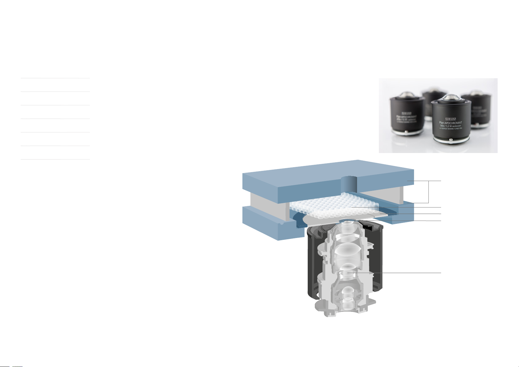
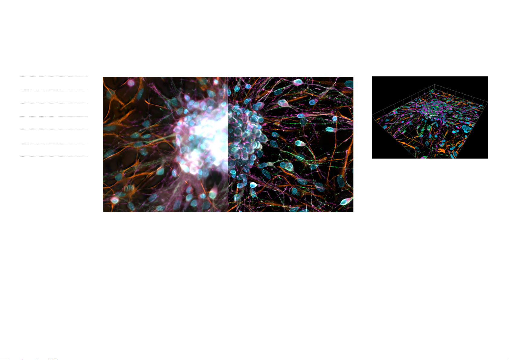
ZEISS Celldiscoverer7 Recognizes and Adapts Automatically to Your Samples
Live cell imaging requires objectives with high numerical apertures. Those objectives will only deliver high
contrast and sensitivity if their optics can adapt to variations in bottom thickness or to the material of
different sample carriers. With Celldiscoverer7 you’re now free to use Petri dishes, chamber slides, multiwell
plates, plastic or glass, thin or thick vessel bottoms, low skirt or high skirt plates. Automatic sample
recognition detects all relevant vessel features while loading your sample. Then Autocorr adjusts the
correction ring of the objective to compensate for spherical aberrations. Find Focus automatically places
your sample in focus and Definite Focus keeps it there. It’s never been easier to get crisp images with low
phototoxicity from deep inside your sample.
Left image shows spherical aberration due to unadjusted optics.
Right image shows the same structure using an Autocorr objective.
The correction results in increased contrast, resolution and
intensity, providing low phototoxicity. The images show tubulin
in FluoCell prepared slide #1. Sample courtesy of Invitrogen,
Thermo Fisher Scientific Inc.
5
Page 6
Your Insight into the Technology Behind It
› In Brief
› The Advantages
› The Applications
› The System
› Technology and Details
› Service
There Is No Life Without Water …
… and no live cell imaging without water immer-
sion. In life sciences, cell biology or screening
applications, your samples mostly consist of water
and / or will be mounted in aqueous solutions.
Celldiscoverer7 combines an outstanding water
immersion objective with rapid automated immer-
sion supply and removal.
A unique elastic silicon membrane fits perfectly
between the objective and sample chamber.
The silicon membrane simultaneously seals the
sample chamber to avoid unnecessary airflow
while protecting the system from potential liquid
spillage. Just select the water immersion objective
and water is supplied instantly to the front lens.
Within seconds the immersion is building up and
the lens is ready to use. When you switch back
to one of Celldiscoverer7’s dry objectives, the
immersion water is automatically removed.
Until now, automated imaging systems often
struggled as the immersion water quickly evapo-
rated. Celldiscoverer7 solves that problem by
automatically monitoring the immersion and
adding water in regular intervals, as needed.
With Celldiscoverer7 you can perform unbiased
live cell experiments at 37 °C over several days or
carry out extensive scanning processes on multi-
well plates.
By adapting the refractive index of your imaging
system to the samples, you’ll achieve more
efficient light collection and increased sensitivity.
And less phototoxicity significantly increases
viability of even your most challenging living
samples.
Heating element of
sample chamber
Sample
Silicone membrane
Membrane opening
with heated objective front
Water immersion objective
A silicone membrane allows automatic water immersion and seals the sample chamber.
6
Page 7
Your Insight into the Technology Behind It
› In Brief
› The Advantages
› The Applications
› The System
› Technology and Details
› Service
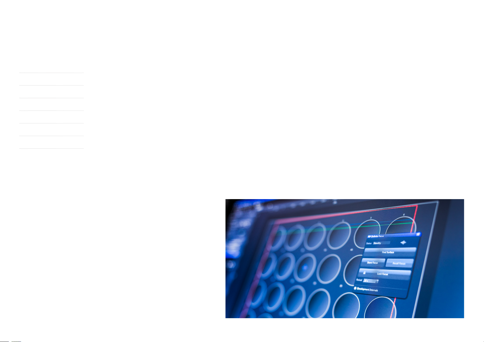
Get in Focus, Then Stay in Focus
Use the hardware-based Find Focus function to
automatically focus your sample and find your
region of interest quickly with just a single click.
This significantly reduces the time to your first
image and minimizes sample illumination.
Then select Definite Focus to maintain the focal
position throughout your experiments, whether
it takes a few seconds or several days.
Or combine both methods with the powerful
content-based autofocus of ZEN imaging soft-
ware. Celldiscoverer7 can automatically create
focus maps for multiple positions in long-term
time-lapse experiments. Simply choose the best
focus strategy for the experiment at hand.
Move to the Edge ...
… but not one step more, thanks to the Adaptive
Lens Guard. High optical performance often
compromises on the possible scanning area.
Celldiscoverer7 with its Adaptive Lens Guard
protects the objective from collisions with your
sample vessel or hardware components, automati-
cally maximizing the available scanning area.
Bottom thickness, skirt height and lateral dimen-
sions are important geometrical features of the
different sample carrier types – especially when
working with multiwell plates. Celldiscoverer7
automatically detects these features and adapts
accordingly. It also calculates the maximal possible
scanning area automatically, depending on the
individual sample carrier, objective and current
focus position in your experiment. The available
scanning area is always indicated on your monitor.
Change your experimental parameters and the
scanning area will adapt automatically, in real
time.
7
Page 8
Your Insight into the Technology Behind It
› In Brief
› The Advantages
› The Applications
› The System
› Technology and Details
› Service
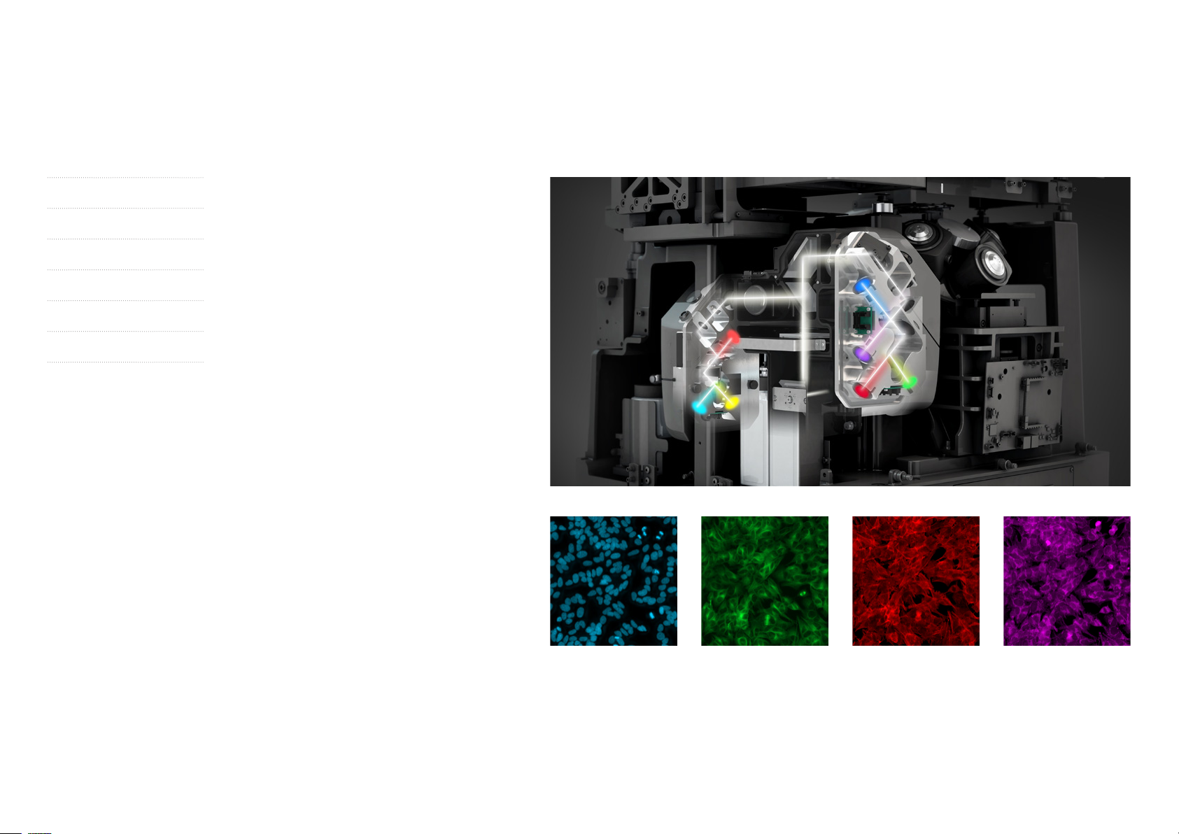
Capitalize on LED-Technology for
Live Cell Imaging
Celldiscoverer7 brings you all the advantages of
LED-technology for efficient illumination with low
phototoxicity, fast switching times and long-term
stability. That’s what delivers gentle imaging,
increased throughput and reproducible results.
The fluorescence excitation unit combines up to
seven LEDs for maximum flexibility in the choice
of dyes – from deep blue to far red. All LEDs are
hardware-triggered for precise, fast illumination.
During sample navigation LEDs are tightly synchro-
nized with camera frame rates. An automated
rectangular excitation field stop illuminates only
the active field of view, greatly reducing photo-
toxicity and fluorescence bleaching. Use high-
efficiency multi-bandpass filter sets for fast
acquisition of multiple fluorescent channels.
Celldiscoverer7 simply switches LEDs on / off –
without moving any mechanical parts – so you
get high-speed multi-channel imaging, even when
combined with transmitted light.
SH-SY5Y cells cultured on a 384 microwell plate. Multichannel image at a single position using the 20× / 0.95 objective. Extended depth
of focus from Z-stack. Hoechst – Chromatin (blue), anti-alpha-tubulin antibody FITC for alphas tubulin (green), Phalloidine for actin (red),
MitoTracker Deep Red for mitochondria (purple). Sample courtesy of P. Denner, Core Research Facilities, German Center of Neurodegenerative Diseases (DZNE), Bonn, Germany.
8
Page 9
› In Brief
› The Advantages
› The Applications
› The System
› Technology and Details
› Service
Your Insight into the Technology Behind It
100 µm
Click here to view this video
Use a Novel Transmitted Light Contrast
With Celldiscoverer7 you can use transmitted
light brightfield and phase gradient contrast.
This novel relief contrast adapts automatically to
the sample carrier geometry, providing excellent
contrast to the very edge of the vessel. It’s fully
compatible with all objectives, filter sets and
sample carriers. This contrasting method stays
robust, even against liquid meniscus or plastic lids.
Use the far-red transmitted light LED for gentle
imaging at very high speeds. You can perform
applications based on label-free assays or let the
system automatically combine transmitted light
with multiple fluorescence channels. All multi-
bandpass filter sets support the combination of
transmitted light and fluorescence, without
reducing sensitivity or speed. On top of that, this
unique motorized transmitted light unit allows
dispensing directly on the optical axis, without
SH-SY5Y cells cultured on a 384 microwell plate. Timelapse
has been acquired using 20× magnification and phase gradient
contrast. Sample and assay courtesy of P. Denner, Core Research
Facilities, German Center of Neurodegenerative Diseases
(DZNE), Bonn, Germany.
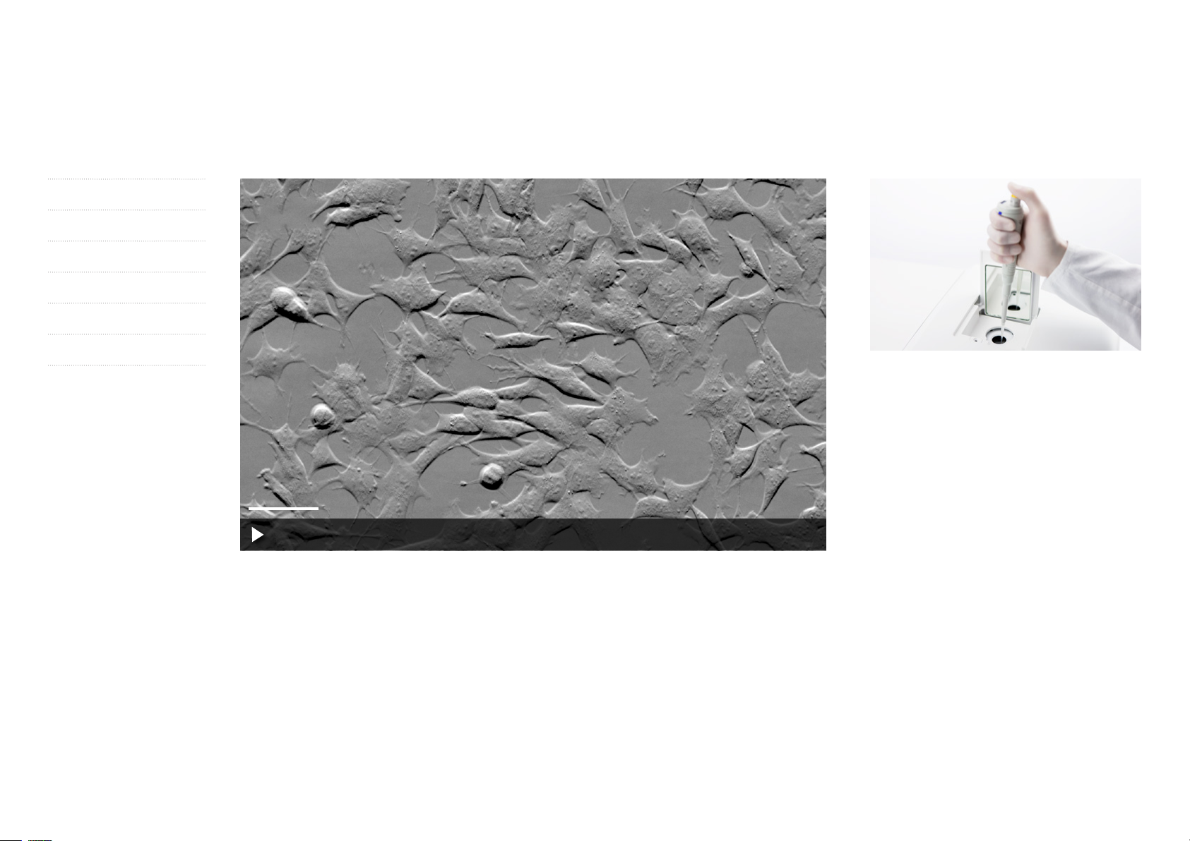
disturbing the environmental conditions. The dis-
pensing unit is always integrated. As soon as you
open the hatch on top of your Celldiscoverer7,
the transmitted light unit will automatically
change place with the dispensing unit. You now
have direct on-axis access to the specimen for
pipetting. You can add agents while maintaining
continuous physiological conditions.
9
Page 10
Expand Your Possibilities
› In Brief
› The Advantages
› The Applications
› The System
› Technology and Details
› Service
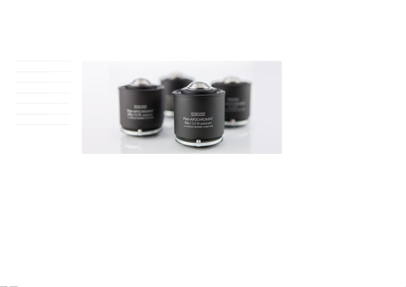
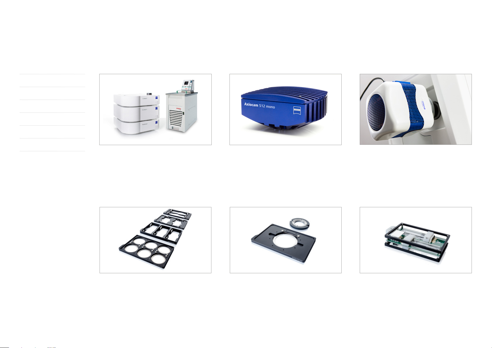
ZEISS Plan-APOCHROMAT 5× / 0.35
Objective
This objective is your choice for efficient sample
navigation. It creates impressive overview images
by delivering an unparalleled information density
in a single shot, especially in combination with
the microscope camera Axiocam 512 mono.
Many screening applications will strongly benefit
from the high resolution on large fields. The
objective easily handles thin and thick vessel
bottom made of glass or plastic.
In combination with the built-in magnification
changer it combines the benefits of three different
objectives into one: 2.5× / 0.12, 5× / 0.25 and
10× / 0.35 – at a fixed working distance.
ZEISS Plan-APOCHROMAT 20× / 0.7
Autocorr Objective
From thin to thick, from plastic to glass – this
objective adapts automatically to every sample
you load on your Celldiscoverer7. It delivers an
unparalleled numerical aperture of 0.7 through
1.2 mm plastic bottom without compromising
image resolution and contrast. This tremendous
flexibility will make the lens your multipurpose
objective, especially if you would like to image
cells, which can only grow on plastic bottom.
In combination with the built-in magnification
changer this objective combines the benefits of
three different objectives into one: 10× / 0.35, 20× /
0.7 and a 40× / 0.7 – at a fixed working distance.
ZEISS Plan-APOCHROMAT 20× / 0.95
Autocorr Objective
This objective delivers high numerical apertures
without applying immersion. It is optimized for thin
vessel bottoms. No matter if your cells prefer glass
or plastic – this objective will adapt to bottom
material and thickness variations. With the increased
sensitivity this objective is ideal to generate crisp im-
ages on large areas or multiple positions at high
speed. In combination with the built-in magnifica-
tion changer this objective combines the benefits
of three different objectives into one: 10× / 0.5,
20× / 0.8 and 40× / 0.95 – at a fixed working distance.
ZEISS Plan-APOCHROMAT 50× / 1.2 W
Autocorr and Autoimmersion Objective
This objective delivers high light collection effi-
ciency and resolution. In combination with the
Autoimmersion function it matches perfectly to
samples in aqueous solution. Since it reduces
phototoxicity to a minimum, it‘s your choice for
your most demanding life cell imaging applica-
tions, e.g. long-term imaging of subcellular struc-
tures. Optimized for thin bottoms it adapts auto-
ma tically to the bottom material and thickness.
No matter which field of view you prefer, this
objective will deliver a constant numerical aperture
of 1.2 and combines the benefits of three different
objectives into one: 25× / 1.2, 50× / 1.2 and
100× / 1.2 – at a fixed working distance.
10
Page 11
Expand Your Possibilities
Click here to view this video
› In Brief
› The Advantages
› The Applications
› The System
› Technology and Details
› Service
Perform Automated Gentle and
Fast Confocal 3D Imaging
Life happens in 3D – and your research often calls
for optical sectioning to image your samples
with best possible contrast and resolution. Now
you can add LSM900 with Airyscan2 to your
Celldiscoverer7. You get the best of both worlds:
ease of use and automation from a fully integrat-
ed microscope platform and the superb confocal
image quality and flexibility of the LSM9 family
with Airyscan 2. You perform superresolution 3D
imaging with up to 1.5× resolution improvement.
And you easily separate multiple labels with
spectral imaging. You can now analyze dynamic
processes with photomanipulation for FRAP,
FRET or related techniques. It’s never been easier
to precisely connect widefield and confocal
images. Fast mixed-mode acquisition simplifies
and speeds up your workflow and gives you
unique insights into your sample.
The elegant beam path design of your LSM900
with Airyscan2 gives you spectral flexibility.
Each single component is optimized for highest
sensitivity and contrast. Instead of losing light at a
closed pinhole, Airyscan2 collects more emission
light and extracts more spatial information than
classic confocals. You can use Airyscan2 with all
high NA dry or water immersion objectives.
Your live cell imaging benefits from superb image
quality with greatly reduced phototoxicity.
Use low magnification widefield to quickly pre-scan
your complete sample, then identify regions of
interest. It's so easy to then image those regions
with LSM900 and Airyscan2 for optical section-
ing with superresolution. The new Multiplex mode
for Airyscan2 employs smart detection schemes
for this unique area detector. This parallelization
allows to image two times faster, while keeping
best resolution and SNR. Use this mode to image
dynamic processes, or to achieve higher through-
put and productivity.
11
Page 12
› In Brief
› The Advantages
› The Applications
› The System
› Technology and Details
› Service
Expand Your Possibilities
Rat cortical primary culture. Antibody staining of bIII-tubulin (Cy2,
green), Nestin (Cy3, red) and DCX (Cy5, purple), nuclei stained with
DAPI (blue). 3D reconstruction of the deconvolved Z-stack (shadow
projection). Sample courtesy of H. Braun, LSM Bioanalytik
GmbH, Magdeburg, Germany.
Comparison between widefield (left) and deconvolved (right) Z-stack projection using GPU-based Deconvolution.
Get More Details with Deconvolution
When imaging three-dimensional samples, out-
of-focus light sometimes blurs your structure of
interest. For these images, you need deconvolution –
a combined optical and mathematical method – to
increase contrast and improve the signal-to-noise
ratio and resolution. With Celldiscoverer7 it is
easier than ever before to first acquire a Z-stack
of your samples and then deconvolve the image
to reassign all detected photons to their origin.
With ZEN imaging software you use advanced
deconvolution algorithms, including a novel
approach with depth variant point-spread-func-
tions for deep imaging. Combine this with
Celldiscoverer7’s unique Autocorr objectives
and you will get excellent results from thicker
samples, e.g. 3D-cell culture. And you will get
them up to 30 times faster than with the traditional
technology that works on your processing PC’s
RAM, thanks to Celldiscoverer7’s new GPU-
accelerated, parallel CUDA processing. Use the
increased speed to extract maximum information
from the large datasets you acquired in those
demanding long-term, time-lapse or multiwell
screening applications.
12
Page 13
› In Brief
› The Advantages
› The Applications
› The System
› Technology and Details
› Service
Expand Your Possibilities
Easily achieve stable environmental conditions
for your demanding live-cell experiments. You can
control the temperature with the optional heating
unit or a Julabo cooling circulator. In combination
with a humidifier, optional CO
you control athmospheric conditions.
and / or O2 module
2
Depending on your most common imaging needs,
you can now choose between Axiocam 506 mono
or Axiocam 512 mono.
No matter if you choose a ZEISS Axiocam micro-
scope or a third party camera – if you have to
increase acquisition speed and sensitivity for special
applications, Celldiscoverer’s additional camera
port provides the flexibility you need.
Your Celldiscoverer7 can load multiwell plates,
dishes, chamber-slides or standard slides.
All sample holders are optimized for large scan-
ning areas, fully compatible with water immersion
and autoclavable.
Celldiscoverer7 allows you to run perfusion
experiments efficiently, while maintaining homo-
genous and stable environmental conditions.
Celldiscoverer7 offers an effective way to keep
the sample chamber clean. The insert plate for
UV disinfection is automatically recognized
by the system and you start the disinfection
workflow via the touchscreen.
13
Page 14

Expand Your Possibilities
› In Brief
› The Advantages
› The Applications
› The System
› Technology and Details
› Service
ZEN Imaging Software Shortens the Path
to Your Goal
ZEN – ZEISS Efficient Navigation – is the single
user interface you will see on all imaging systems
from ZEISS. ZEN imaging software leads you
simply and quickly to the result.
At all times you see which options the system
is making available to you and which step is
appropriate to take next. ZEN makes it easy to
operate every imaging system from ZEISS correctly
and intuitively. As a result you save time, reduce
training and support costs, and get faster answers
to your questions.
With Celldiscoverer7 you profit from advanced
automation features:
• Simple and intuitive carrier-based navigation
via mouse and keyboard
• A dedicated automation wizard to create scan
profiles for routine or reoccurring tasks
• A range of hardware- and software-based
focus strategies to set up even complex multi-
position experiments
• Fast overview images. Create an overview of
your cells just once, then there’s no need to
expose them to unnecessary light doses during
experiment setup.
• Cell viability put first with samples illuminated
only as long as the camera acquires an image
• An optimized CZI file format for large datasets
and seamless integration
into existing image analysis workflows
• Open interfaces. Use your CZI dataset in all
major software packages that use the
BioFormats library, e.g., Fiji, Python, Matlab,
Icy, Knime, Imaris, Arivis.
14
Page 15
Expand Your Possibilities
› In Brief
› The Advantages
› The Applications
› The System
› Technology and Details
› Service
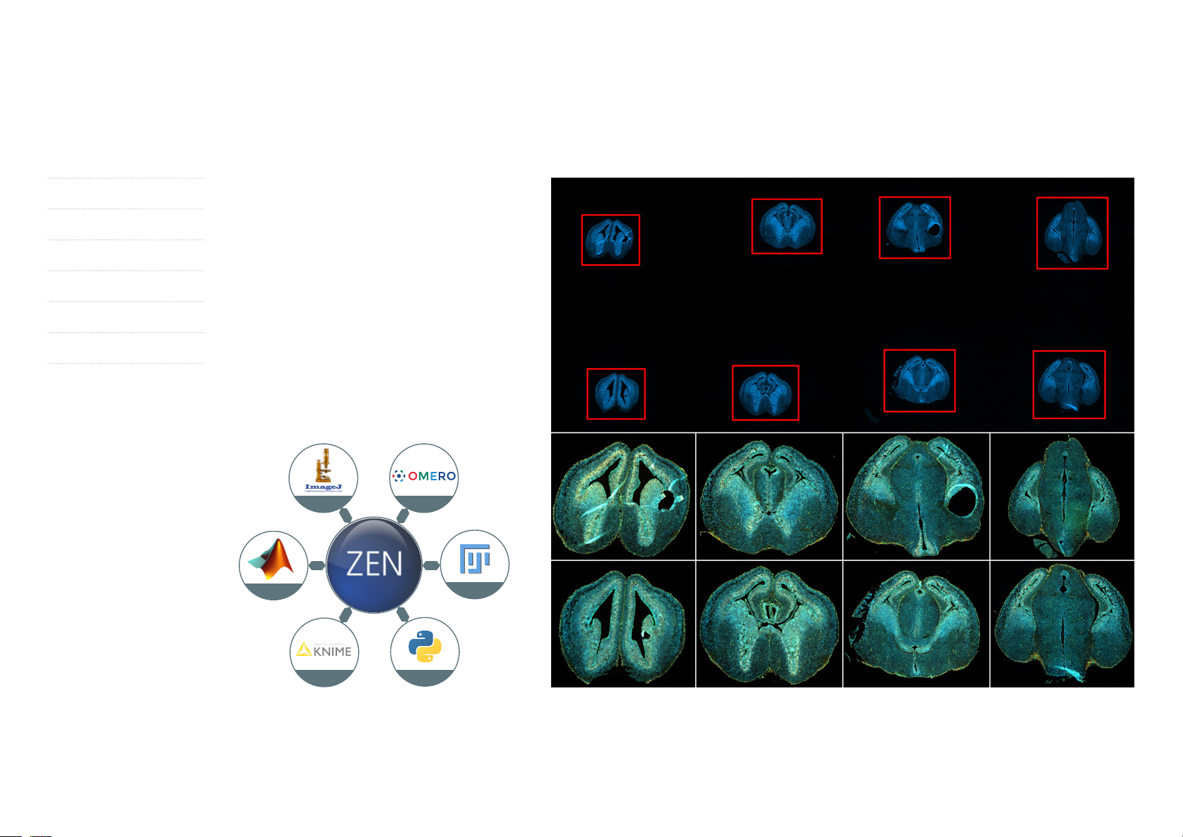
OAD is Your Interface
to ZEN Imaging Software
• Use Python scripts to customize and automate
your workflows.
• Integrate external image analysis applications
into your workflows.
• Exchange image data with external programs
like ImageJ, Fiji, MATLAB, KNIME or Python.
• Use feedback for smart experiments.
• Get more reliable data in less time.
It's your choice.
ImageJ
MATLAB
Omero
FIJI
KNIME
OAD enables the analysis of data acquired with ZEN imaging
software by other programs like ImageJ. Transfer your results
back to ZEN for further analysis and display.
Python
The result of overview scan using low magnification (top panel) was used to automatically detect the brain slices via image analysis.
The results (XYZ position and the height / width of detected objects) were used in a automated subsequent scan using a high NA objectives,
where the system carried out an individual tile scan for every detected object in a complete automated fashion without any additional
user interaction. Sample courtesy of P. Grigaravicius, FLI – Leibniz Institute on Aging, Jena, Germany.
15
Page 16

Expand Your Possibilities
Click here to view this video
› In Brief
› The Advantages
› The Applications
› The System
› Technology and Details
› Service
ZEISS ZEN Connect Lets You Overlay and Organize Images from Any Source.
Connect All Your Multimodal Data
to Expand Correlative Microscopy
Expanding classic correlative microscopy, ZEN
Connect is open to all your images: you can load
complex multidimensional images as easily as
simple overview images from your mobile phone.
It makes no difference whether your imaging
technology is from ZEISS or from third parties.
All image data can be aligned, overlayed and
shown in context. So long as your external images
adhere to the well-established Bio-Formats stan-
dard, ZEN Connect will even keep their metadata.
Acquire Overview Images for Easy
Navigation
Image your sample with a ZEISS stereo micro-
scope or any other low magnification system.
Then move to your high-resolution system of
choice. With ZEN Connect you only need to align
it once, then use the overview image to navigate
and find your ROIs. All subsequent high-resolution
images will be shown in context as you zoom in
and out across the borders of resolution domains
and imaging technologies. A single click on the
overview image brings your stage to the right
position to examine or reevaluate any of your
ROIs with the full image overlay.
Smart Data Management
All the images you acquire with ZEN Connect are
saved in well-structured database projects, com-
plete with an intuitive label attached automatically
to each image file. You'll always stay on top of
things – during your experiments as well as months
afterwards when analyzing your work. It's easy to
find all your overlay images and their connected
datasets. You can even search for microscope type
and imaging parameters with the new filter func-
tion of ZEN Connect.
16
Page 17

ZEISS
Enterprise Server
Expand Your Possibilities
› In Brief
› The Advantages
› The Applications
› The System
› Technology and Details
› Service
ZEISS Predictive Service
Maximizes System Uptime
Once connected to your network and activated,
this advanced technology will automatically track
the health status of your instrument and collect
system log files in the background to improve
remote diagnosis.
Relevant technical data such as operating hours,
cycle counts or voltages are periodically moni-
tored via a secure connection to our data center.
The ZEISS Predictive Service application evaluates
the performance of your microscope as system
data can be received and analyzed.
Our support engineers will diagnose any issues by
analyzing data on the Enterprise Server – remotely
and without interruption to your operation. • Maintain highest system availability
Your Network
Increase your uptime through close monitoring
of the system’s condition as remote support
can often provide immediate solutions
• Data security
Ensure highest data security standards using well
established technologies like PTC Thingworx and
Microsoft Azure Cloud. No personal or image
data is uploaded, only machine data
HTTPS
HTTPS
• Fast and competent support
Use secure remote desktop sharing to easily
get an expert connected
• Optimum instrument performance
As the status of your system is monitored,
necessary actions can be planned before they
become urgent
17
Page 18

Tailored Precisely to Your Applications
› In Brief
› The Advantages
› The Applications
› The System
› Technology and Details
› Service
Typical Applications Task ZEISS Celldiscoverer7 Offers
Multiwell plates for Live Cell or fixed
endpoint assays
Label free assays Perform label free growth curve assays over several days. Transmitted light source: high-speed IR-LED (725 nm) offering low phototoxicity
High-Content Screening Acquire high resolution images of multi-labelled
Transfected and non-modified
Live Cell Cultures
Evaluate and document cell culture from multiwell plates. Transmitted light – phase gradient contrast for high-resolution images through glass and plastic vessels
Up to 7 LED excitation wavelengths
Low magnification, large field of view – high numerical aperture lenses
Automatic sample carrier detection and calibration
Scan the maximum area of a multiwell plate at different
magnifications and resolutions.
cell culture from multiwell plates quickly.
Pharmacological or chemical or drug screening. Option to add a plate loader
Evaluate and document transfection rate and
transfection stability using fluorescent markers.
Work with different sample carriers. Automatic measurement of sample carrier bottom thickness and Autocorr Objectives for enhanced
Adaptive Lens Guard and automatic sample carrier calibration ensure maximized scan area depen ding
on the plate type 100 % plate scanning from 2.5× to 100× is possible whole well – single shot
Stable Incubation with temperature (heating / cooling), CO
Simple and reproducible Hardware Autofocus for focus drift compensation
Autoimmerson for water immersion lens
Up to 7 LED excitation wavelengths
Autocorr objectives for automated aberration correction
Adaptive Lens Guard and automatic sample carrier calibration ensure maximized Scan area
Barcode reader for easy sample identificiation
Preview Scan
Open Application Developement for Python scripting – open access to third party analysis tools
Fast Multibandpass Main Beam Splitter and Emmission Filter Wheels
Large working distance enables higher / better 3D content screening
Transmitted light – phase gradient contrast for high-resolution images through glass and plastic vessels
Stable Temperature and O
Autoimmerson for water immersion lens
contrast and resolution
Adaptive Lens Guard and automatic sample carrier calibration ensure maximized scan area
/ CO2 controlled enviroment
2
and O2 control
2
18
Page 19

Tailored Precisely to Your Applications
› In Brief
› The Advantages
› The Applications
› The System
› Technology and Details
› Service
Typical Applications Task ZEISS Celldiscoverer7 Offers
Label-free fixed and thin tissue slices or
small organisms
Fixed fluorescently labelled tissue,
cell culture samples or small organisms
Multi-labelled living tissue section, organs,
small organisms, organotypic-, spheriod or
cell culture preparations
Stimulus-induced responses of cells,
tissue or whole organisms
Document and evaluate cell and tissue morphology
and growth state.
Change quickly between large overview scans and
high resolution imaging.
Identification, quantification and qualification of
cell types, pathological and pharmacological
pathways using cell-, tissue and protein markers in
2D and 3D samples.
Short-term or long-term observation of physiological
and morphological parameters in 2D / 3D during growth,
differentiation, motility and interaction.
Analyse the embryogenesis of small model organisms. Large working distances of 5× and 20× / 0.7 objectives offer fast, high resolution and deep imaging
Observation of stimulus-induced responses of
cells, tissue or organisms without disturbing the
environmental control.
Transmitted light – phase gradient contrast for high-resolution images through glass and plastic vessels
Quick change of field of view using triple magnification changer
Large working distances of 5× and 20× / 0.7 objectives offer fast, high resolution and deep imaging
Up to 7 LED excitation wavelengths
GPU-accelerated 3D-Deconvolution
Large working distances of 5× and 20× / 0.7 objectives offer fast, high resolution and deep imaging
Autoimmerson for water immersion lense
Autocorr objectives for automated aberration correction
Stable incubation with temperature (heating / cooling), CO
LED illumination unit with up to 7 excitation wavelengths
Experiment Feedback for adaptive experiments
GPU-accelerated 3D-Deconvolution
Large working distances of 5× and 20× / 0.7 objectives offer fast, high resolution and deep imaging
GPU-accelerated 3D-Deconvolution
Semi-automatic dispensing work flow
Dispensing unit allows to add compounds into the field of view
Option to install a perfusion chamber
and O2 control
2
19
Page 20

Tailored Precisely to Your Applications
› In Brief
› The Advantages
› The Applications
› The System
› Technology and Details
› Service
Typical Applications, Typical Samples Task ZEISS Celldiscoverer7 with LSM900 Offers
Antibody stained tissue slices Document morphological relations of structures Airyscan2 with GaAsP detector for imaging
Live cell culture Study the motility of vesicles and organelles Up to 8 frames per second confocal time lapse imaging
Live cell culture with two labels Study the motility of subcellular structures Airyscan2 with GaAsP detector and Multiplex mode for time lapse imaging in
2D or 3D at up to 8 frames per second
Explore the interaction of two proteins exploiting the
Förster Resonance Energy Transfer effect
Live cells with multiple labels Image over a long time in an automated way Experiment Designer software tool combined with three parallel spectral channels
Live or fixed cells with multiple labels
and overlapping emission signals
Cellular structures with weak labels Image subcellular structures at physiological expression levels LSM900 with GaAsP detector or Airyscan2
Study molecular dynamics Photomanipulation FRAP analysis tool, available in ZEN (black edition), classical timed bleaching or flexible
Plant roots Follow the changes of subcellular structures over time
Model organisms, e.g. Zebrafish, Drosophila
or C. elegans, Arabidopsis
Cleared samples Image whole organs or entire organisms Specialized objective with long working distance and autocorrection for bottom material
Examine the interplay of multiple proteins Parallel acquisition of all signals with three spectral channels and linear unmixing
with high resolution
See fine details of the organization and dynamics
of endogeneously expressed FP proteins
FRET analysis tool, available in ZEN (black edition)
interactive bleaching strategies
Airyscan2 with GaAsP detector for high resolution imaging beyond 40 μm deep into
tissue with up to 6 full frames per second (512 × 512 pixel)
Airyscan with GaAsP detector for high sensitivity imaging and increased resolution
beyond 40 μm deep into tissue.
and thickness available (20× 0,7)
20
Page 21

› In Brief
› The Advantages
› The Applications
› The System
› Technology and Details
› Service
ZEISS Celldiscoverer7 at Work
500 µm
Whole well, single shot.
384 microwell plate imaged with 2.5× magni fication
in 3 channels. Each well fits into one single image.
You avoid time-consuming scanning of wells and
subsequent stitching and increase your through-
put. He overall image quality and resolution allows
e.g. segmentation of single cell nuclei and there-
fore counting of cells.
20 µm
50 µm
Sample courtesy of P. Denner, Core Research Facilities, German
Center of Neurodegenerative Diseases (DZNE), Bonn, Germany.
21
Page 22

› In Brief
› The Advantages
› The Applications
› The System
› Technology and Details
› Service
ZEISS Celldiscoverer7 at Work
50 µm
50 µm 50 µm 50 µm
SH-SY5Y cells cultured on a 384 microwell plate. Five channel image at a single position using Plan-APO CHROMAT 20× / 0.95; EDF from Z-stack; Hoechst-Chromatin (blue), anti-alpha-tubulin antibody FITC for alpha
tubulin (green), Phalloidine for actin (red), MitoTracker deepRed for mitochondria (purple), phase gradient contrast, overlay image. Sample courtesy of P. Denner, Core Research Facilities, German Center of
Neurodegenerative Diseases (DZNE), Bonn, Germany.
50 µm 50 µm
22
Page 23

ZEISS Celldiscoverer7 at Work
› In Brief
› The Advantages
› The Applications
› The System
› Technology and Details
› Service
15 (out of 24) living cricket embryos mounted in
low-melt agarose. Cells are expressing nuclear-
localized GFP. The overview image shows a multi-
position experiment. At each position two
embryos fit into the field of view. Acquired within
30 seconds incl. Z-stacks of 17 images each (thick-
ness 350 µm, 2.3 seconds). This enables imaging
of multiple crickets in a synchronized way.
The resulting spatio-temporal image resolution
allows characterization of movement and division
of single cells throughout the embryo during
development. Magnification: 2.5× using short
exposure times of 35 ms.
1000 µm
200 µm
Sample courtesy of S. Donoughe, Biological Labs, Harvard University, Cambridge, USA
23
Page 24

ZEISS Celldiscoverer7 at Work
› In Brief
› The Advantages
› The Applications
› The System
› Technology and Details
› Service
0 h
500 µm
36 h
72 h
Click here to view this video
12 h
48 h
84 h
24 h
60 h
96 h
Five days long-term imaging of cricket embryo-
genesis. The development of an eGFP-expressing
cricket embryo mounted in low-melt agarose was
imaged every 5 minutes for a total length of 5 days.
During the first day the retraction of the yolk and
dorsal closure can be seen followed by further
growing of the embryo. EDF-images created from
Z-stacks; acquired with 2.5× magnification using
short exposure times of 35 ms.
Z-stacks were 350 μm thick and were acquired
within 2.3 seconds.
Sample courtesy of S. Donoughe, BioLabs Building 2087, Harvard
University, Cambridge, USA
24
Page 25

› In Brief
› The Advantages
› The Applications
› The System
› Technology and Details
› Service
ZEISS Celldiscoverer7 at Work
Click here to view this video
Calcium imaging in beating cardiomyocytes stained in green using a Calcium kit; imaging with 8 fps using
Plan-APOCHROMAT 50× / 1.2 W with Auto immersion; the green fluorescence changes intensity upon con-
traction of the cells; frequency of individual contractions analyzed with ZEN MeanROI tool; diagram shows
delayed contraction in regular intervals caused by component given to the cells.
Sample courtesy of Sanofi-Aventis Deutschland GmbH, R&D IDD /
in vitro Biology, Frankfurt, Germany
25
Page 26

ZEISS Celldiscoverer7 at Work
› In Brief
› The Advantages
› The Applications
› The System
› Technology and Details
› Service
0 d
100 µm
3 d
1 d
4 d
2 d
5 d
Click here to view this video
GFP HEK (Human Embryonic Kidney) cells, transiently expressing eGFP. Imaged through a 1 mm plastic
bottom; images taken every 5 minutes for a total of 5 days; imaging started shortly after induction of the
expression via Tetracyclin treatment. Overlay of phase gradient contrast and green (eGFP) fluorescence:
• After one day: cells are subconfluent and start to express eGFP. Due to the transient transfection and the
Tetracyclin treatment some round and dead cells are visible.
• After two days: cells have recovered from the transfection and start to grow again.
• At the end of the time series: cells are confluent and bright green due to eGFP expression.
Sample courtesy of Sanofi-Aventis Deutschland GmbH;
R&D IDD / in vitro Biology, Frankfurt, Germany
26
Page 27

ZEISS Celldiscoverer7 at Work
› In Brief
› The Advantages
› The Applications
› The System
› Technology and Details
› Service
0 h
100 µm
0 h
22 h
22 h
45 h
45 h
Click here to view this video
48 h Cell Proliferation Assay Control vs.
AuroroB Kinase siRNA Knockdown
HeLa Kyoto cells (Neumann et. Al., Nature 2010
Apr.1.; 464(7289):721-7) expressing H2B-mCherry
were imaged every 30 minutes for 48 hours in a
96 well plate using Plan-APOCHROMAT 10× / 0.5.
Top row: A series of images showing untreated
control cells. The lack of dead cells and the
healthy shape of the nuclei (arrows indicate
methodic cells) clearly demonstrates the stability
and homogeneity of the incubation, the stable
focus, low phototoxicity as well as virtually no
photobleaching.
Bottom Row: A series of images showing cells
treated 24 h before acquisition with a siRNA
against AuroraB Kinase on the same plate as the
control (top row). The slower proliferation and the
misshaped nuclei (arrows and insets) demonstrate
the mitotic defects caused by the knockdown.
Sample courtesy of S. Reither, Advanced Light Microscopy
Facility, EMBL, Heidelberg, Germany
27
Page 28

ZEISS Celldiscoverer7 at Work
› In Brief
› The Advantages
› The Applications
› The System
› Technology and Details
› Service
Expansion Microscopy in Mouse Brain
The goal of Expansion Microscopy is to make
small structures visible that could otherwise not
be observed with conventional or superresolution
microscopy. Here, a protein-retention expansion
technique was applied to expand the tissue. The
sample is enlarged by a factor of 4.5 to 5 – up
to several mm in X / Y dimensions and several
hundred µm in the Z dimension. Especially the
5× / 0.35 and the 20× / 0.7 objectives of
Celldiscoverer7 are well suited to image such
samples as they have a large field of view, high
resolution and a large working distance.
Top: Whole brain
Bottom left: Axon bundles
Bottom right: Pyramidal cells
5000 µm
The images shown here are extended-depth-of-
focus images created from Z-stacks acquired with
a 2.5× magnification imaged through 1.2 mm of
polystyrene. Staining: YFP expressing neurons.
Sample courtesy of S. Asano, Boyden lab, MIT, Cambridge, USA
500 µm 500 µm
28
Page 29

› In Brief
› The Advantages
› The Applications
› The System
› Technology and Details
› Service
ZEISS Celldiscoverer7 at Work
Autofluorescence Imaging of Arachnids
Small Arachnids where collected from tropical leaves
in South America. Imaging with Celldiscoverer7
saves time, since the low magnification objectives
(5× / 0.35 and 20× / 0.7) deliver finest details in
large fields of view.
A combination of several wavelengths was used
to observe autofluorescence. The images shown
here are extended-depth-of-focus images created
from Z-stacks.
Left: Genital of the third leg of Huitaca sp. imaged
with a 20× magnification.
Center: Same as before but excited with a different
combination of wavelengths.
Right: Microgavia oviformis imaged with 2.5×
magnification.
200 µm200 µm 200 µm
Sample courtesy of L. Benavides, Giribet Lab, Harvard University,
Cambridge, USA
29
Page 30

ZEISS Celldiscoverer7 at Work
› In Brief
› The Advantages
› The Applications
› The System
› Technology and Details
› Service
Application for Label-Free Measurement
of Cell Proliferation
The growth of cultured cells has been imaged in
long-term time-lapse movies over 72 hours using
phase gradient contrast (image 1).
To quantify proliferation, cell region (image 2,
red overlay) was detected automatically using
supervised machine learning (random forests)
in each time frame.
The growth curve (image 3) shows the relative
cell coverage over time, averaged for all images
in one well. The assay allows image based cell
proliferation measurements.
By using label-free imaging in phase gradient
contrast, cell growth is not affected by phototoxicity
or any further sample processing.
This approach offers several advantages:
• Very low disturbance, non-invasive monitoring
of cells
• Kinetic live cell data, no single end point.
• Compatible to standard micro-well plates
(e.g. 96well or 384well).
• Applicable for screening cell-based applications.
Sample and assay courtesy of P. Denner, Core Research Facilities, German Center of Neurodegenerative Diseases (DZNE), Bonn, Germany.
30
Page 31

› In Brief
ZEISS Celldiscoverer7 at Work
› The Advantages
› The Applications
› The System
› Technology and Details
› Service
Rat cortical primary neuron culture. Antibody staining of bIIItubulin (Cy2, green), Nestin (Cy3, red) and DCX (Cy5, purple),
nuclei stained with DAPI (blue). Maximum intensity projection of
a Z-stack.
Top row: Conventional widefield images.
Bottom row: Deconvolved images using GPU-based
deconvolution. Deconvolution algorithm: constrained iterative
using a depth variant point-spread function.
Sample courtesy of H. Braun, LSM Bioanalytik GmbH,
Magdeburg, Germany.
31
Page 32

› In Brief
› The Advantages
› The Applications
› The System
› Technology and Details
ZEISS Celldiscoverer7 at Work
› Service
100 µm
100 µm
Fixed starlet sea anemone (Nematostella vectensis)
stained with Hoechst (nuclei) and Phalloidin (actin).
Side view imaged with a combination of camera
based phase gradient contrast mode (top) and
high sensitivity mode with Airyscan2 (bottom).
Maximum intensity projection of 19 z-planes.
50 µm
Click here to view this video
Sample courtesy of A. Stokkermans, Ikmi Group, EMBL, Heidelberg, Germany
Fine image details and high signal to noise ratio
can clearly be seen on the insert in the top
right image, showing an enlarged view of a
tentacle area.
Video: Top view of a young animal, showing
mouth and four tentacle buds. Maximum intensity
projection of 69 z planes imaged with Airyscan2
Multiplex. Images were acquired using the water
immersion objective with a total magnication of
25× and a numerical aperture of 1.2.
32
Page 33

ZEISS Celldiscoverer7 at Work
› In Brief
› The Advantages
› The Applications
› The System
› Technology and Details
› Service
Click here to view this video
Lateral line primordium migration and deposition
of immature neuromasts in a Zebrafish embryo
A
B
500 µm
A
200 µm
B
(Danio rerio). Animals were anesthetized and
embedded using low concentrated agarose in a
glass bottom petridish.
Initial camera based imaging allowed for a quick
and easy sample navigation (top) combining
Phase Gradient Contrast with fluorescence acqui-
sition.
Subsequent high resolution imaging with Airyscan2
in Multiplex mode was done on individual positions
identified in the widefield image (white boxes).
A) Maximum intensity projections of an immature
neuromast (127 z planes).
B) Maximum intensity projections of the lateral
line primordium tip migrating through the
animal (155 z-planes).
Green: LYN-eGFP (mebranes);
Red: tagRFP-T-UTRCH (actin).
The gentle and fast image acquisition that is
inherent to the Airyscan2 Multiplex mode is very
benificial for this kind of application. The animal is
unperturbed by the imaging while images with a
very high signal to noise ratio as well as level of
detail can be acquired at the same time.
Click here to view this video
20 µm
Sample courtesy of J. Hartmann and D. Gilmour, EMBL,
Heidelberg, Germany
33
Page 34

ZEISS Celldiscoverer7 at Work
› In Brief
› The Advantages
› The Applications
› The System
› Technology and Details
› Service
Primary lung fibroblasts stained with mitotracker
red (mitochondria) and a DNA marker (nuclei).
The acquisition seemlessly combines two imaging
modes – the fluorescent channels where captured
in confocal mode using highly sensitive GaAsP
detectors while the Phase Gradient Contrast is
camera based.
A timelapse of 2.5h was acquired using a 40×
magnification with a numerical aperture of 0.95.
50 µm
Click here to view this video
Sample courtesy of A. Hocke, Charité, Berlin, Germany
34
Page 35

ZEISS Celldiscoverer7 at Work
› In Brief
› The Advantages
› The Applications
› The System
› Technology and Details
› Service
Organoid from a human breast cancer cell line.
The cells express GFP-labeled H2B (nucelei) and
mCherry (cytoplasmic staining depicted here in
grey for better visualization).
Several organoids where grown in a multiwell
plate with Matrigel. Initial sample navigation was
performed using the transmitted light at a low
magnification of 2.5× to identify interesting
organoids.
Subsequently, high resolution images were
acquired using the water immersion objective
with a total magnification of 50×. 61 z-planes
were acquired using ZEISS Celldiscoverer 7 with
LSM900 and Airyscan2 in Multiplex mode.
One can clearly appreciate the robustness of the
imaging given that Matrigel is not an ideal optical
medium and the organoid was imaged at a dis-
tance of several micrometers from the coverglass.
Click here to view this video
Sample courtesy of S. Gawrzak and M. Jechlinger, EMBL, Heidelberg, Germany
35
Page 36

ZEISS Celldiscoverer7 at Work
› In Brief
› The Advantages
› The Applications
› The System
› Technology and Details
› Service
Trachea system in a living fruitfly embryo (Droso-
phila melanogaster) imaged using ZEISS Celldis-
coverer 7 with LSM900 and Airyscan2 in Multi-
plex mode. A water immersion objective with a
magnification of 25× and numerical aperture of
1.2. in combination with multi-tile acquisition
(8 tiles, 143 z-planes) was used.
CD4-mIFP under a tracheal promoter color coded
for depth.
Click here to view this video
Sample courtesy of D. Rios-Barrera, Leptin Group, EMBL, Heidelberg, Germany
36
Page 37

ZEISS Celldiscoverer7 at Work
› In Brief
› The Advantages
› The Applications
› The System
› Technology and Details
› Service
Caenorhabditis elegans germline. Decapitated
nematodes where localized in widefield mode
using a low magnification of 2.5× (transmitted light
and fluorescence, DAPI; left). This allowed for an
easy and convenient workflow to identify areas of
interest for subsequent fast high resolution imaging
in Multiplex mode for ZEISS Celldiscoverer 7 with
LSM900 and Airyscan2 (right). A 25× magnifac-
tion with water immersion and NA 1.2 was used to
generate a z-stack of 62 planes.
Individual chromosomes in different meiotic cells
are clearly distinguishable – see magnified box.
Blue: DAPI (DNA);
Green: Alexa 488 (cross-over sites);
Orange: Alexa 546 (synaptonemal complex);
Red: Alexa 647 (chromosome axis).
5 µm
Click here to view this video
Sample courtesy of S. Köhler, EMBL, Heidelberg, Germany
20 µm
Click here to view this video
37
Page 38

› In Brief
› The Advantages
Your Flexible Choice of Components
› The Applications
› The System
› Technology and Details
› Service
21 34
1 Microscope
• ZEISS Celldiscoverer7
• Automatic sample container recognition
• Barcode reader
• Focus stabilization
• Magnification changer 0.5× / 1× / 2×
• Apochromatic FL beampath with adaptive field stop
• ZEISS Axiocam 506 mono or Axiocam 512 mono
• Additional camera port
• On-axis access for dispensing
• UV-disinfection
2 Objectives
• Plan-APOCHROMAT 5× / 0.35
• Plan-APOCHROMAT 20× / 0.7 autocorr
• Plan-APOCHROMAT 20× / 0.95 autocorr
• Plan-APOCHROMAT 50× / 1.2 W autocorr
autoimmersion
5
3 Illumination
• Transmitted light unit:
IR-LED (725 nm) brightfield, oblique contrast,
phase gradient contrast
• Fluorescence:
LEDs 385, 420, 470, 520, 567, 590 and 625 nm
High-efficiency multibandpass filter sets
Additional emission filter wheel
4 Imaging Systems
• LSM900 with Airyscan2
5 Accessories
• Temperature and atmospheric control
(heating / cooling; CO
, O2)
2
• Insert plates and perfusion chambers for dishes,
multi-chamber slides and standard slides
• Additional recommended cameras
• ZEISS Axiocam 512 mono
• ZEISS Axiocam 702 mono
• Photometrics EMCCD evolve 512 delta
• Hamamatsu Orca Flash 4.0
• Photometrics Prime 95B
6 Software
• ZEN celldiscoverer includes modules for multi-
dimensional image acqusition, Tiles & Positions,
Experiment Designer, advanced image process-
ing and analysis tools
• Recommended additional modules:
• GPU-based deconvolution (GPU-DCV)
®
• 3Dxl Viewer – powered by arivis
• Open application development (OAD)
38
Page 39

› In Brief
System Overview
› The Advantages
› The Applications
› The System
› Technology and Details
› Service
Axiocam 512 mono
Axiocam 702 mono
Trigger cable
Axiocam 503/506/512/702, LEMO
(required in addition, without fig.)
EMCCD Camera
evolve 512 delta
Trigger cable
evolve 512 delta, LEMO
(required in addition, without fig.)
Hamamatsu
Orca Flash 4.0 V3
supported by
Celldiscoverer 7
Photometrics
Prime 95B
Trigger cable
Prime 95B, LEMO
(required in addition,
without fig.)
Camera Adapter 60N-C 1" 1,0x
Filter set 90 HE
Filter set 91 HE
Filter set 92 HE
Filter set 93 HE
Filter set 94 HE
LED Illumination unit FL
(already included in the
delivery of Celldiscoverer 7)
LED Set 01
for excitation wavelengths
385, 470, 567, 625 nm
(each system 1x required)
Axiocam 506 mono
Axiocam 512 mono
Extension housing for
Celldiscoverer 7 system unit
1
LED Illumination unit FL
extension for Celldiscoverer 7
LED Set 02
for excitation wavelengths
420, 520, 590 nm
O2 Module S1 for Celldiscoverer 7
CO2 Module S1 for Celldiscoverer 7
TempModul S1 for Celldiscoverer 7
Circulator S1 (100 V)
(without fig.)
Circulator S1 (230 V)
(without fig.)
2
3
Celldiscoverer 7 system unit
Celldiscoverer 7 LCI system unit
Celldiscoverer 7 confocal system unit
Celldiscoverer 7 LCI confocal system unit
Power supply
unit
1
LSM 900
Scanning module
LSM 900
with:
LSM 900 MA-PMT detection module,
LSM 900 GaAsP-PMT detection module,
2-Channel Deflecting optics,
Additional GaAsP-detector,
Additional MA-PMT detector
Objective Plan-Apochromat 5x/0.35
(already included in the delivery of
Celldiscoverer 7 system unit)
LSM 900 adapter kit
Celldiscoverer 7
Objective Plan-Apochromat 20x/0.7
autocorr
Objective Plan-Apochromat 20x/0.95
autocorr
Component rack
LSM 900
Objective Plan-Apochromat 50x/1.2 W
autocorr with autoimmersion
LSM 900
Laser module
LM URGB
Airyscan 2 module
Additional Detector
LSM 900
2
Insert plate for UV disinfection
(already included in the delivery of
Celldiscoverer 7)
for perfusion with POC-R2
3
Insert plate
for 6 Petri dishes 35
Insert plate
for 1 Petri dish 35/60
Insert plate
for 2 slides 76x26 mm
Insert plate
for 2 slides/Lab-Tek™
chambers 57x26 mm
Insert plate for 3 slides 76x26 mm Insert plate
(already included in the delivery of
Celldiscoverer 7)
Storage and Analysis PC 36 TB
Memory Upgrade (64 GB)
for Data Analysis PC
Microscopy Workstation Premium Intel Xeon multilingual
for Celldiscoverer 7
with:
System configuration 5 image and OS for HP Z6
Processor Intel Xeon Gold 5122 (hp Z6)
Memory 32 GB (1x32) DDR4-2666 MHz ECC registered RAM
Graphics Card NVIDIA Quadro P4000 8GB DP or
Graphics Card NVIDIA Quadro P6000 24GB DP
PC Interface Microscope and Trigger
Hard Drive Extension 8TB RAID10 (hp Z6)
Network Adapter 2x10 GbE RJ45 (hp Z6)
Language Pack English US
Monitor TFT 32" HP Z32
System table, small,
air damped,
level regulated,
with breadboard, 900 mm x 750 mm
(for Celldiscoverer 7 system unit
without Extension housing or LSM)
System table, mid-size,
air damped,
level regulated,
with breadboard,
1200 mm x 900 mm
(for Celldiscoverer 7 system unit
with Extension housing or LSM 900)
System table, large,
air damped, level regulated,
with breadboard, 2000 mm x 750 mm
(for Celldiscoverer 7 system unit
combined with Extension housing
rd
and 3
party accessories)
39
Page 40

Technical Specifications
› In Brief
› The Advantages
› The Applications
› The System
› Technology and Details
› Service
Dimensions Width (approx.) Depth (approx.) Height (approx.) Weight (approx.)
Celldiscoverer7 710 mm 640 mm 700 mm 136 kg
Footprint Celldiscoverer7 585 mm 560 mm
Incl. Extension housing 1270 mm 640 mm 700 mm 187 kg
Footprint incl. Extension housing 1170 mm 560 mm
Celldiscoverer7 incl. LSM900 1310 mm 690 mm 705 mm
Component rack 400 mm 550 mm 600 mm 35 kg
Airyscan2 400 mm 250 mm 145 mm 5 kg
Power Supply 400 mm 250 mm 145 mm 6 kg
Laser module 400 mm 250 mm 145 mm 10 kg
Technical data
Celldiscoverer7 and Extension housing Noise emission According to EN 55011 class A
Noise immunity According to DIN EN 61326-1
Protection class 1
Ingress protection rating IP 20
Radio interference suppression To EN 55011 Class A
Type of operating site Closed room facility
Electrical safety To DIN EN 61010-1 (IEC 61010-1) conforming to CSA and UL regulations
Degree of pollution 2
Overvoltage category II
Celldiscoverer7 Line input voltage; max. current 100 V to 240 V ± 10 %; 6A ~
Line frequency 50 Hz – 60 Hz
Celldiscoverer7 incl. LSM900 /
Extension housing
Input for connection of Celldiscoverer7 100 V to 240 V ± 10 %, 50 Hz – 60 Hz, max. 4.0 A ~
Output to internal 6 sockets 100 V to 240 V ± 10 %, 50 Hz – 60 Hz
Permissible total current on 6 internal sockets Max. 4.0 A ~
The internal sockets can be connected via the software
The extension housing is powered by Celldiscoverer7
40
Page 41

Technical Specifications
› In Brief
› The Advantages
› The Applications
› The System
› Technology and Details
› Service
Environmental requirements
Storage (in packaging) Permissible ambient temperature +5 °C to +40 °C
Permissible relative air humidity (no condensation) max. 75 % at +35 °C
Transport (in packaging) Permissible ambient temperature –20 °C to +55 °C
Permissible relative air humidity (no condensation) max. 75 % at +35 °C
Operation Permissible ambient temperature +15 °C to +35 °C
Recommended ambient temperature (e.g. for incubation) +18 °C to +25 °C, optimally +22 °C
Warm-up time 1 h for standard imaging; ≥ 4 h for high-precision and / or long-term measurements
Permissible relative air humidity max. 65 % at 30 °C
Atmospheric pressure 800 hPa to 1060 hPa
XYZ motorization
Motorized xy-scanning stage
Motorized z-drive
Optical specifications
Nosepiece
Magnification changer, afocal
Travelling range 300 mm × 140 mm
Reproducibility ± 1 µm
Absolute precision ± 5 µm
Resolution 0.1 µm
Reproducibility ± 0.025 µm
Absolute precision 0.14 µm
Resolution ± 0.01 µm
• 4× motorized nosepiece
• in combination with the 3× magnification changer this offers the
functionality of 12 objectives
• 0.5×, 1×, 2× magnification, providing three different magnifications
for each objective
• depending on the objective configuration it offers a magnification
range from 2.5× – 100×
• switching between magnifications ~1 sec
• enables constant working distances for each magnification
Component always included Component optionally available
41
Page 42

Technical Specifications
› In Brief
› The Advantages
› The Applications
› The System
› Technology and Details
› Service
Optical specifications
Plan-Apochromat 5× / 0.35
Plan-Apochromat 20× / 0.7 autocorr
Plan-Apochromat 20× / 0.95 autocorr
Plan-Apochromat 50× / 1.2 W autocorr, autoimm.
Adaptive Lens Guard
Temperature control
Adaptive Autocorr
Autoimmersion, water
Magnification changer
0.5× 1× 2×
M = 2.5×
NA = 0.12
M = 10×
NA = 0.35
M = 10×
NA = 0.5
M = 25×
NA = 1.2
• automatically maximizes scanning area, while protecting the objectives from collisions with other hardware or sample vessels
• scanning range is indicated and updated automatically via control software
• all objectives are equipped with heating elements for temperature control
• in combination with the optional heating unit, objective temperature is adjusted automatically,
depending on the user-defined sample temperature
• enables stable and homogeneous temperature within the sample chamber
• automatic correction of aberrations (for high magnification objectives)
• adapts objectives automatically to vessel bottom material and thickness
• enables correction of aberration due to high penetration depths and refrective index mismatch of the sample
(5× objective is not sensitive to variations of bottom thickness and material and does not require a correction)
• comes along with the Plan-Apochromat 50× / 1.2 W objective
• enables automatic supply and removal of water immersion
• water level is automatically indicated in the control software and on the display
• upgradable in the field
M = 5×
NA = 0.25
M = 20×
NA = 0.7
M = 20×
NA = 0.8
M = 50×
NA = 1.2
M = 10×
NA = 0.35
M = 40 ×
NA = 0.7
M = 40 ×
NA = 0.95
M = 100×
NA = 1.2
Auto-
correction
– –
Auto-
immersion
–
–
Temperature
control
Thick vessel
bottom up to
1.2 mm PS
–
–
2
Thin vessel bottom
0.13 – 0.21 mm
glass / COC
0.15 – 0.21 mm PS
1
Working
2
distance
5.10 mm
2.20 mm
0.76 mm
0.84 mm
Optional LSM 900 with Airyscan2 for up to 1.5× resolution improvement
Objective Plan-Apochromat 5× / 0.35 Plan-Apochromat 20× / 0.7 Plan-Apochromat 20× / 0.95 Plan-Apochromat 50× / 1.2 W
Magnification changer 0.5× 1× 2× 0.5× 1× 2× 0.5× 1× 2× 0.5× 1× 2×
Usage with Airyscan MPLX + + + + + - + ++ ++ ++ ++ -
Usage with Airyscan HS + + + + + + + ++ ++ ++ ++ +
Component always included Component optionally available 1 Cycloolefincopolymer 2 Polystyrene
42
Page 43

Technical Specifications
› In Brief
› The Advantages
› The Applications
› The System
› Technology and Details
› Service
Focus
Hardware-based focus finder
Hardware-based focus stabillization
Software-based autofocus
Transmitted light and contrasting techniques
Transmitted light unit
Lightsource
Contrast techniques
• automatically focusses on the sample (lower side of sample)
• a user-defined offset can be used to change the default position
• enables automatic generation of focus maps for microwell plates
• compatible with every objective and filter set
• can be combined with focus stabilization and ZEN blue software autofocus
• focus stabilization system maintains focus position over long-term
• compatible with every objective and filter set
• hardware and software support for multi-position and multi-offset stabilization
• can be combined with focus finder and ZEN blue software autofocus
• focusses automatically on user-defined structures and regions of interest based on the image content
• can be combined with focus finder and focus stabilization
• fully compatible with fluorescent applications, environmental control, dispensing and perfusion option
• enables label-free imaging or provides additional information in combination with fluorescent applications
• high-speed IR-LED (725 nm) offering low phototoxicity
• brightfield
• oblique contrast
• adaptive phase gradient:
adapts automatically to vessel geometry providing excellent contrast to the edges of the vessels
• all contrast techniques are compatible with all objectives, filter sets and sample vessels, i.e. plastic and glass incl. lids
Component always included Component optionally available
43
Page 44

Technical Specifications
› In Brief
› The Advantages
› The Applications
› The System
› Technology and Details
› Service
Fluorescence illumination
Fluorescence illumination unit
LEDs are synchronized with image acquisition
LEDs are synchronized with the live-window
Automated excitation field stop
Switching time between FL channels • switching between fluorescence channels using high-efficient multi-bandpass filter sets <1 ms
5-position beamsplitter wheel
Emission filter wheel
Filter sets
• apochromatic excitation beampath incl. adaptive field stop
• up to seven LEDs (385 / 420 / 470 / 520 / 567 / 590 / 625 nm)
• life time of LEDs >10,000 h
• switching between LEDs <1 ms
Sample is only exposed during image acqusition (acquisition trigger mode) thus reducing phototoxicity.
Sample is only exposed during live-window update (live-window trigger mode), significantly reducing phototoxicity during sample navigation.
A motorized field stop adapts automatically to the current field of view thus reducing phototoxicity effectively.
• switching 5-position beamsplitter wheel <80 ms
• 5× position beamsplitter wheel
• switching time <80ms
• 7× motorized emission filter wheel
• user accessible
• fits 25 mm emission filters
• switching emission filter wheel <80 ms
Filter set 90 HE
• quad-band filter set for 385 nm, 470 nm, 567 nm, 625 nm LED and IR-TL LED
• beamsplitter RQFT 405+493+575+653; emission filter QBP 425 / 30+514 / 30+592 / 25+709 / 100
• additional band for transmitted light
Filter set 91 HE
• triple-band filter set for 420 nm, 520 nm, 590 nm LED and IR-TL LED
• beamsplitter RTFT 450+538+610; emission filter TBP 467 / 24+555 / 25+687 / 145
• additional band for transmitted light
Filter set 92 HE
• triple filter set for 385 nm, 470 nm, 590 nm LED and IR-TL LED
• beamsplitter RTFT 405+493+610; emission filter TBP 425 / 30+524 / 50+688 / 145
• additional band for transmitted light
Filter set 93 HE
• double bandpass for 470 nm, 567 nm and IR-TL LED
• beamsplitter RDFT 493+575; emission filter TBP 514 / 32+605 / 50+730 / 60
• additional band for transmitted light
Filter set 94 HE
• double filter set for 385 nm, 520 nm and IR-TL LED
• beamsplitter RDFT 405+538; emission filter TBP 444 / 69+581 / 77+730 / 60
• additional band for transmitted light
Component always included Component optionally available
44
Page 45

Technical Specifications
› In Brief
› The Advantages
› The Applications
› The System
› Technology and Details
› Service
Fluorescence illumination
LED Set 1
LED Set 2
TL IR
Channel
Filter sets
LEDs [nm]
LED 385
BP 385 / 30
LED 470
BP 469 / 38
LED 567
BP 555 / 30
LED 625
BP 631 / 33
LED 420
BP 423 / 44
LED 520
BP 511 / 44
LED 590
BP 591 / 27
IR LED
725 / 50
90 HE
quad
91 HE
triple
92 HE
triple
93 HE
dual
× × ×
× × ×
× ×
×
×
× ×
× ×
× × × × ×
94 HE
dual
DAPI, Hoechst 33342 & 33258, Alexa 350 & 405, ATTO 390, True Blue, EBFP, T-Sapphire,
CellTracker Blue, LysoTracker Blue, wtGFP (uv), Aminocoumarin, Cascade Yellow
Alexa 488, Fluorescein, eGFP, Calcein, Fluo-4, Fluo-8, JC-1, mKaede, NBD,
TagGFP, LysoTracker Green, ATTO 465, ATTO 490, Oregon Green Bapta, BOBO-1, Cytox
Green, MitoTracker Green, YoYo-1, YoPro-1
Cy3, Bodipy TMR, mBanana, mOrange, TurboRFP, tdTomato, TagRFP, DsRed2 ("RFP"),
TRITC, PAmCherry, PATagRFP, Alexa Fluor 555 & 546, DsRed monomer, SNARF, POPRO-3, Magnesium Orange, SYTO 82
Cy5, Alexa Fluor 610, 633, 635 & 647, ATTO 610 to 647N, ATTO Oxa12, ATTO Rho14,
Bodipy 630 / 650-X, Bodipy 650/665-X, CF™ 620R, CF™ 633, CF™ 640R, DyLight 633,
DyLight 649, PSmOrange (red), iRFP670
Alexa Fluor 430, ECFP, ATTO 425, ATTO 430LS, SpectrumAqua, Cerulean,
mCFP, CyPet, Y66W, mKeima-Red, LysoSensor™ Green DND-153, SYTOX Blue, Chromomycin A3, POPO-1, PO-PRO-1, SYTO 40, SYTO 41, SYTO 42, SYTO 43
Alexa 514 & 532, eYFP, Calcein, Fluo-4, Fluo-8, Bodipy 515, YoPro-1, YoYo-1, Calcium
Green, Syto 23, Thiazole Orange, LysoTracker
mEOS2.0, mCitrine, mVenus, Topaz
Alexa Fluor 594, Cy3.5, mPlum, mRaspberry, mNeptune, mCherry, pa-mRFP1, KFP1,
mEos2 (red), mEos3.2 (red), LipidTOX™ Red, Calcein red-orange,
CellTracker Red, ER-Tracker Red, CellTrace BODIPY
All filter sets offer an IR transmitted light bandpass. This bandpass enables
IR-brightfield contrast without switching any filter components and without
affecting FL-efficiency.
®
Green DND-26, mEos3.2 (green),
®
TR
Lasers
Laser module URGB
(pigtailed; 405, 488, 561, 640 nm)
Component always included × Component compatible Component optionally available
Single-mode polarization preserving fiber
Typical total dynamic range of 10.000:1; direct modulation 500:1
Diode laser (405 nm, 5 mW)
Diode laser (488 nm, 10 mW)
Diode (SHG) laser (561 nm, 10 mW)
Diode laser (640 nm, 5 mW)
45
Page 46

Technical Specifications
› In Brief
› The Advantages
› The Applications
› The System
› Technology and Details
› Service
Sample mounting
Insert plate for 1 Petri dish 35 / 60
Insert plate for 6 Petri dishes 35
Insert plate for 2 slides 76 × 26 mm
Insert plate for 3 slides 76 × 26 mm
Insert plate for 2 slides / Lab-Tek™ chambers 57 × 26 mm
Insert plate for perfusion with POC-R2
Detection options
Internal camera *
External camera port **
Additional / optional cameras
LSM900
• for mounting of Petri dishes
• fits one Petri dish d = 35 mm or d = 60 mm, autoclavable
• for mounting of Petri dishes
• fits six Petri dishes d = 35 mm, autoclavable
• for mounting of slides
• fits two slides 76 × 26 mm, autoclavable
• for mounting of slides
• fits three slides 76 × 26 mm, autoclavable
• fits two Lab-Tek™ chambers 57 × 26 mm, autoclavable
• fits for perfusion with POC-R2
Axiocam 506 mono, Axiocam 512 mono
• external, user accessible camera port to mount additional cameras
• motorized switching between internal and external camera <200 ms
Axiocam 512 mono
Axiocam 702 mono
Photometrics EMCCD evolve 512 delta
Hamamatsu Orca Flash 4.0
Photometrics Prime 95B
• two spectral detection channels, GaAsP (typical QE 45 %) or multialkali (MA) PMT (typical QE 25 %)
• one additional GaAsP PMT, MA PMT, or Airyscan detector
• Airyscan2 with spatial detection (32 channels GaAsP) with up to 1.5× improved resolution and 4 – 8× improved SNR.
Airyscan Multiplex [HS-2Y]: up to 8 images/sec with 512 × 512 pixels
Component always included Component optionally available * Select one internal camera ** Not available on systems with LSM900
46
Page 47

Technical Specifications
› In Brief
› The Advantages
› The Applications
› The System
› Technology and Details
› Service
Resolution and speed (examples)
Pixel resolution • depending on the magnification and camera:
• 1.82 µm @ 2.5× using Axiocam 506
• 0.23 µm @ 20× with Axiocam 506
• 1.24 µm @ 2.5× using Axiocam 512
• 0.03 µm @ 100× using Axiocam 512
Typical scan speeds • 96 well plate, four channels, exposure 50 ms per channel, full resolution, one position per well: <4 min
• 96 well plate, three confocal channels simultaneously (multicolor track), image size 512 × 512 px, bidirectional scan at max. speed,
one position per well: <2.5 min. (with optional LSM900)
• 384 well plate, single channel, exposure 100 ms, full resolution, 1 position per well (e.g. whole well single shot): < 6 min
• 384 well plate, whole well using a high resolution 20× objective, four channels, exposure 50 ms per channel, full resolution: <2.5 min
Automatic sample recognition
Pre-scan unit (incl. barcode reader)
• automatically detects vessel types before final mounting:
• slides
• Petri dishes (35 / 60 mm)
• LabTek-chamber slides (incl. number of wells)
• microwell plates incl. plate type, i.e. number of wells
• The following 1D barcodes are detected on slides and wellplates:
• Code 39 (3of9 und W / MOD43)
• Code128 Auto, Code128 A, Code128 B, Code128 C
• Interleaved 2of5
• UPC A und UPC E
• EAN 8 und EAN 13
• Codebar
• UCC / EAN 128
• on slides the following 2D barcodes are detected:
• DataMatrix
• QR-Code
Automatic vessel bottom recognition
Automatic plate calibration
Component always included Component optionally available 1 Cycloolefincopolymer 2 Polystyrene
• automatic detection of vessel bottom material (glass / COC
• automatically adjusts autocorr objectives to the material
• automatic detection of vessel bottom thickness
• automatically adjusts autocorr objectives to the thickness
• automatically measures vessel skirt height, e.g. the distance between the support area and the actual sample bottom
• delivers the skirt height to the Adaptive Lens Guard to update the scanning area
• automatically calibrates individual plates, i.e. well diameter and distance, plate length, height and rotation
1
and PS 2)
47
Page 48

Technical Specifications
› In Brief
› The Advantages
› The Applications
› The System
› Technology and Details
› Service
Environmental control
TempModule S1
Module S1
CO
2
Module S1
O
2
Humidifier unit
Circulator S1
Dispensing unit
Insert plate for UV disinfection
Component always included Component optionally available
l
• controls temperature of bottom and top plate of sample chamber
• temperature range within sample chamber: 30 – 45 °C
• temperature homogenity across a whole microwell plate: ± 0.6 @ 37 °C
• operated by ZEN blue control software
• generates a stable, user defined CO2 concentration within the sample chamber
• ensures an optimal and stable pH value in cell culture media over long term
• a built-in CO
• operated by ZEN blue control software
• O2-control device to achieve a stable, controlled decrease of the O2 concentration by displacement with N2 within the sample chamber
• a built-in O
• operated by ZEN blue control software
• prevents evaporation of culture medium during long-term experiments
• liquid level is indicated automatically
• cooling unit controls temperature of top plate of sample chamber
• temperature range = 14 – 28 °C
• temperature homogenity (microwell plate) = ± 2 °C
available for air objectives only
• offers on-axis access to specimen
• enables pipetting without disturbing environmental conditions
l
• allows sequential, semi-automatic pipetting of multi-positions
• incl. two UV bulbs, 1.0 W each
• emitting 254 nm
l
• fully automated disinfection process takes 23 min
• can be used on-demand or for preventive maintenance
sensor permanently monitors the CO2 concentration
2
sensor permanently monitors the O2 concentration.
2
48
Page 49

Count on Service in the True Sense of the Word
› In Brief
› The Advantages
› The Applications
› The System
› Technology and Details
› Service
Because the ZEISS microscope system is one of your most important tools, we make sure it is always ready
to perform. What’s more, we’ll see to it that you are employing all the options that get the best from
your microscope. You can choose from a range of service products, each delivered by highly qualified
ZEISS specialists who will support you long beyond the purchase of your system. Our aim is to enable you
to experience those special moments that inspire your work.
Repair. Maintain. Optimize.
Attain maximum uptime with your microscope. A ZEISS Protect Service Agreement lets you budget for
operating costs, all the while reducing costly downtime and achieving the best results through the improved
performance of your system. Choose from service agreements designed to give you a range of options and
control levels. We’ll work with you to select the service program that addresses your system needs and
usage requirements, in line with your organization’s standard practices.
Our service on-demand also brings you distinct advantages. ZEISS service staff will analyze issues at hand
and resolve them – whether using remote maintenance software or working on site.
Enhance Your Microscope System.
Your ZEISS microscope system is designed for a variety of updates: open interfaces allow you to maintain
a high technological level at all times. As a result you’ll work more efficiently now, while extending the
productive lifetime of your microscope as new update possibilities come on stream.
Profit from the optimized performance of your microscope
system with services from ZEISS – now and for years to come.
>> www.zeiss.com/microservice
49
Page 50

Carl Zeiss Microscopy GmbH
07745 Jena, Germany
microscopy@zeiss.com
www.zeiss.com/celldiscoverer
Not for therapeutic, treatment or medical diagnostic evidence. Not all products are available in every country. Contact your local ZEISS representative for more information.
EN_41_011_100 | CZ 03-2019 | Design, scope of delivery, and technical progress subject to change without notice. | © Carl Zeiss Microscopy GmbH
 Loading...