

Page 1
Leica M720 OH5
Premium Surgical Microscope
A New Dimension in Comfort
Living up to Life
Page 2

Comf
2
Page 3
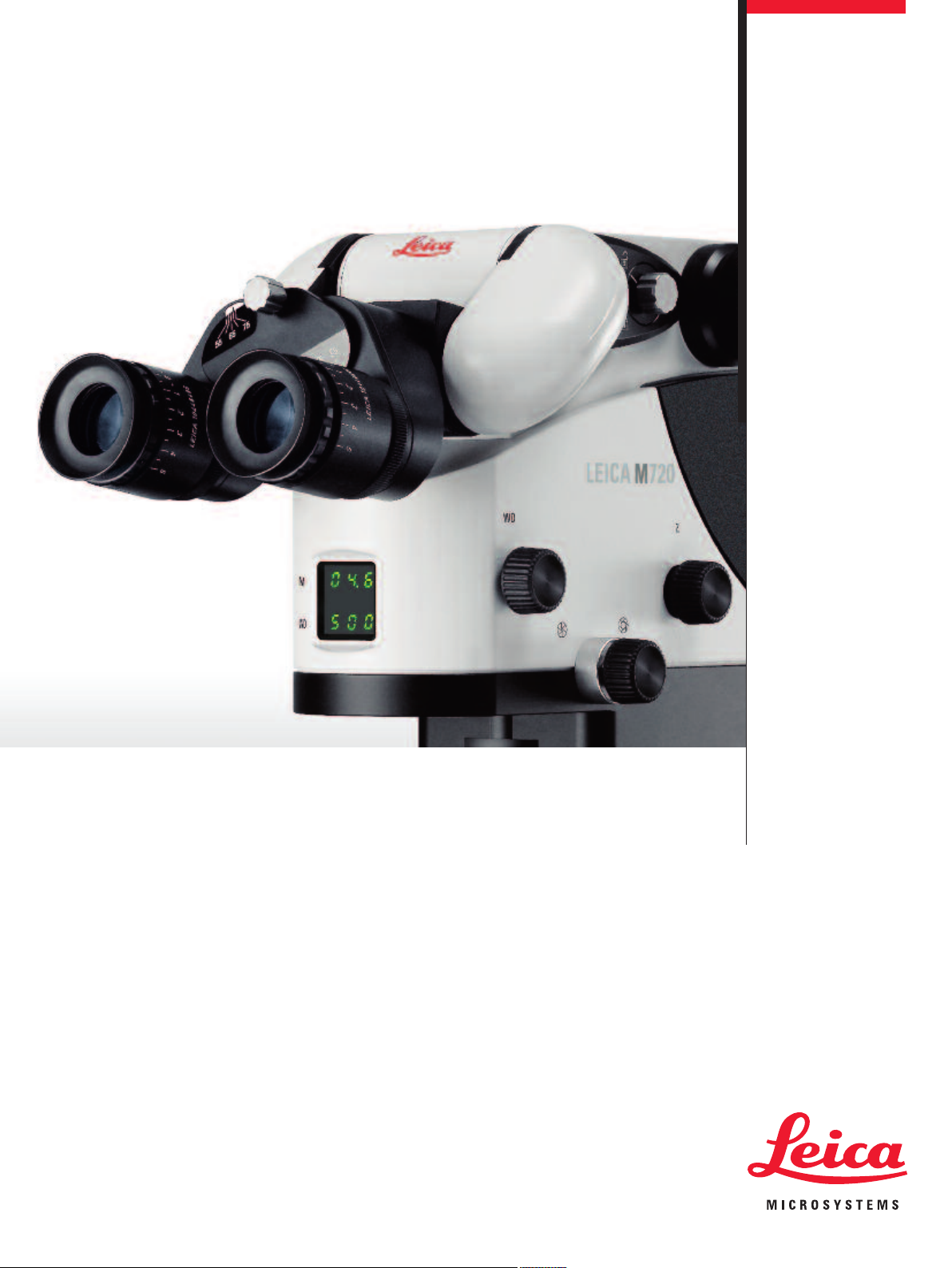
Excellence in optics and ease of use are the hallmarks of the Leica Microsystems brand.
The finest resolution, image contrast and color fidelity assure brilliant detail visibility, and
unprecedented ergonomics simplify your workflow in the operating room.
To this, Leica Microsystems now adds a new dimension in comfort. Our ingenious Horizontal Optics Technology and stand provide you with the most compact microscope with
the highest maneuvering precision and positioning flexibility on the market.
Your benefit: more room to work and improved viewing conditions make operating in all
positions more comfortable – for safe and successful surgical outcomes.
The Leica M720 OH5 – A new dimension in comfort.
3
Page 4
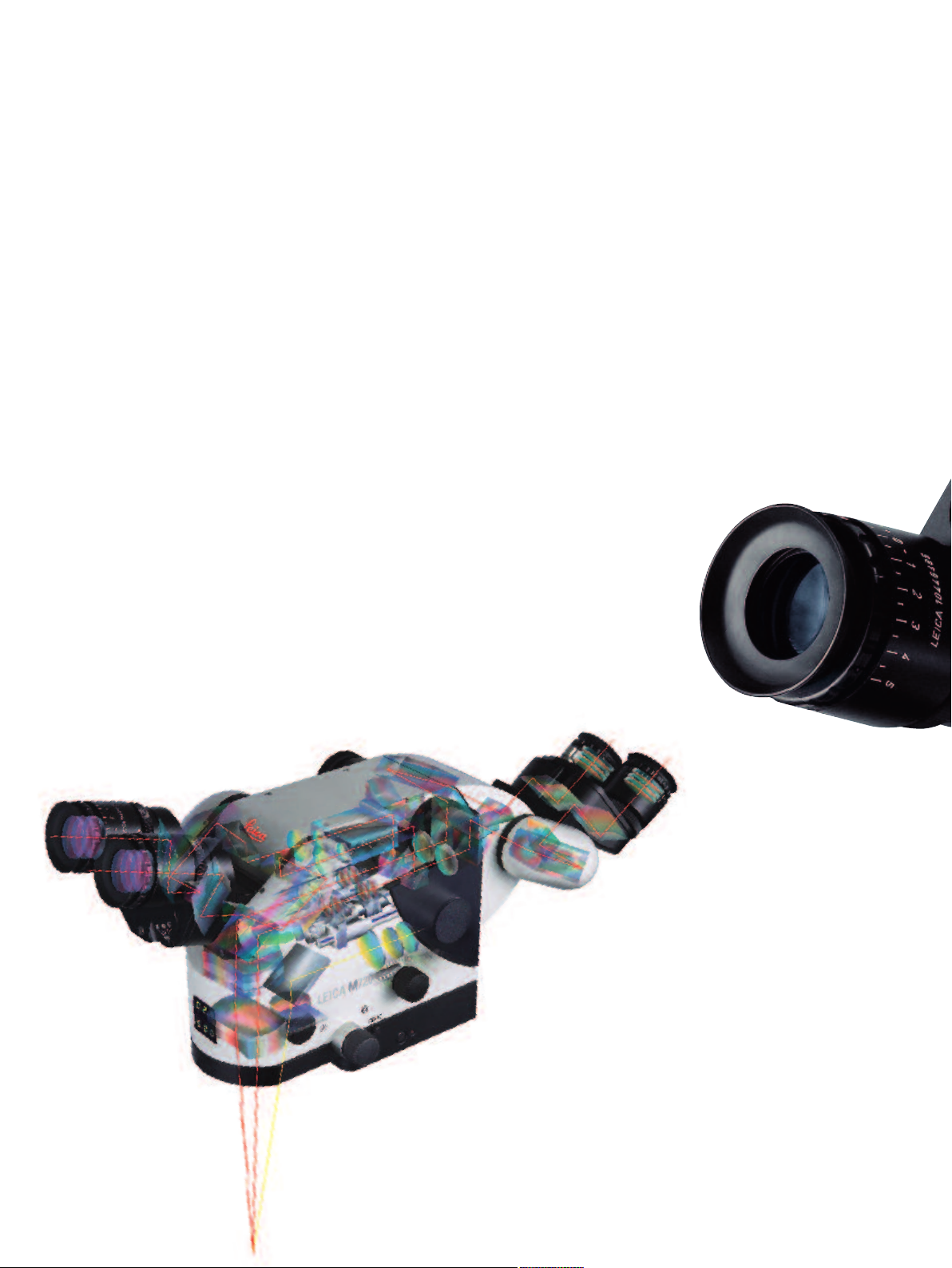
Breakthrough in Surgical Microscope Design
For years, surgeons have needed a surgical microscope with smaller, more compact
optics. Traditional microscope design has evolved over the years using large,
vertical optical zoom lens systems which have limited the surgeon’s working room
and ability to work comfortably. With the Leica M720 OH5, Leica writes a revolutionary new chapter in microscope design. The heart of the innovation: Horizontal
Optics Technology.
The new Leica M720 optical head of the OH5 is far more compact than today’s
surgical microscopes. Designed along a horizontal, as opposed to a vertical plane,
Horizontal Optics Technology provides a significant improvement in surgeon comfort, particularly for awkward procedures such as posterior fossa cranial surgery
where patients are positioned in an upright posture. Horizontal Optics Technology
therefore provides a substantial gain in free working distance for both cranial and
spine cases, allowing you unobstructed access to the surgical area.
Real innovation makes life easier – and the Leica M720 OH5 does exactly this.
The innovative Horizontal Optics
Technology reduces the size of the
optical head and gives surgeons
more room to work – dramatically
increasing their comfort level.
4
Page 5
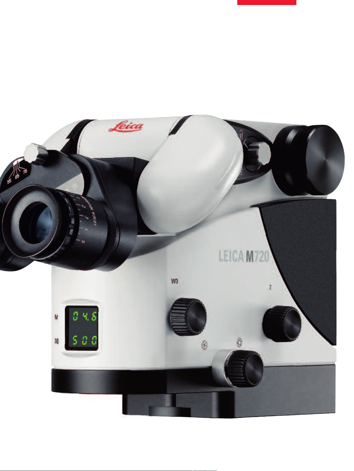
A New Dimension in Comfort
Actual size 1:1
5
Page 6
Page 7

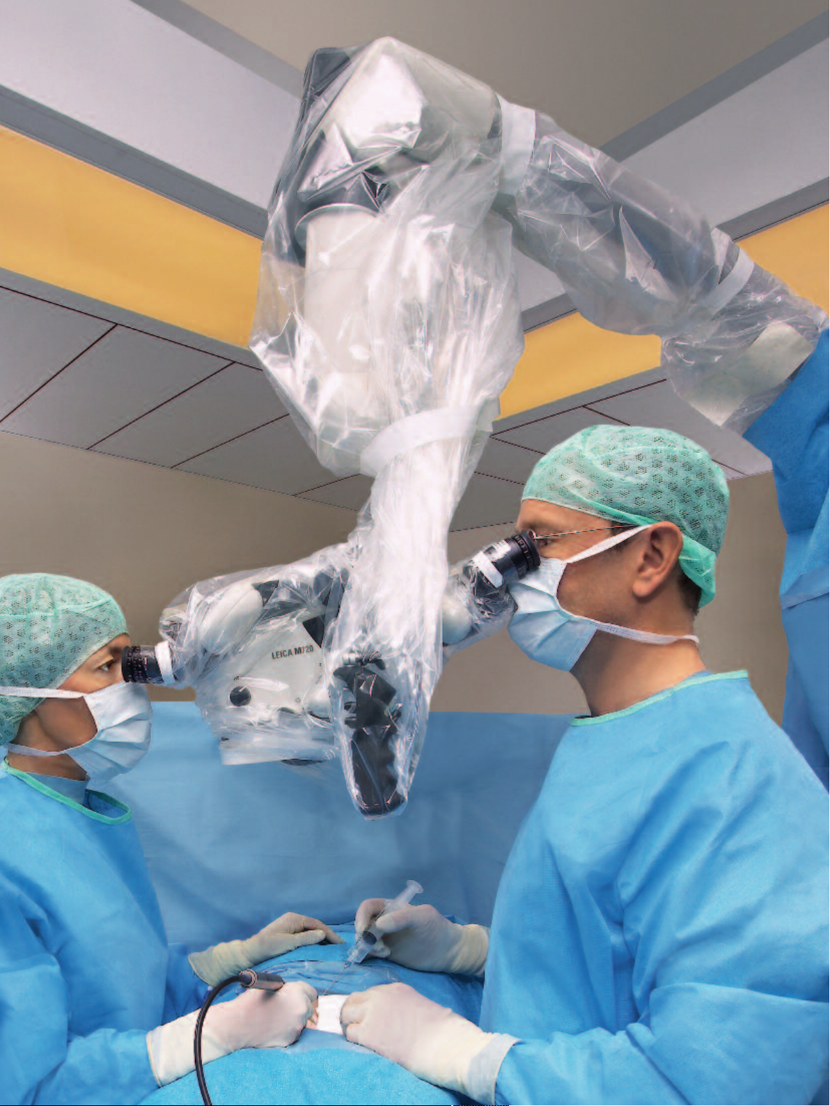
An Individual Fit
Leica Butterfly-Binoculars
Every person is unique in stature and physical requirements. Surgical microscope designers can
only accommodate different body sizes and changing operating conditions through the use of
individually adjustable components that create the right working conditions for different people of
different statures.
In addition to the optical system’s compact size, the new binocular tubes are designed with an
inclination range of 115°, which allows each individual to always work in comfort. They also feature
Leica’s innovative Butterfly-Binoculars, which enable the eyepieces to swing to a second viewing
plane, quickly and easily.
This extra flexibility creates ideal working conditions for both the surgeon and the assistant. Leica
Butterfly-Binoculars accommodate all body heights and the most challenging surgical positions.
During surgery, Leica ButterflyBinoculars ensure comfortable
positioning for all individuals.
7
Page 8

C O M P A C T D E S I G N
H I G H E S T O V E R H E A D C L E A R A N C E
8
Page 9

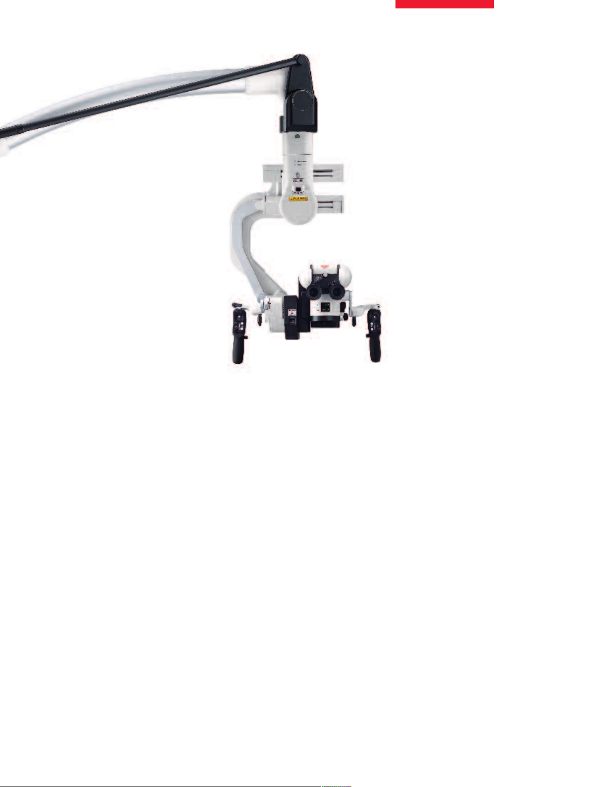
Freedom of Positioning
Superior Reach and Overhead Clearance
The Leica M720 OH5 allows perfect positioning for surgery and takes up very little space in the
operating room. The system provides the highest overhead clearance and the longest reach of any
surgical microscope on the market today. This superior reach gives the surgeon ultimate flexibility
to place the microscope wherever it best fits his or her surgical needs – behind the surgeon in the
unique overhead position, or positioned anywhere around or across the operating table.
S U P E R I O R R E A C H
9
Page 10

Page 11

Unrivalled Maneuverability
The ergonomic 150° range of
the incline angle combined
with the most compact optical
system provides the surgeon
with unmatched comfort, even
in the most difficult positions.
With a 100° range of lateral
movement, the surgeon can
easily achieve the most
challenging side views.
Precise Movement
The Leica M720 OH5 offers a greatly expanded range of movement in all dimensions for improved microscope maneuverability.
The microscope features robotic functions on two axes (X/Y)
to allow a very high degree of precise movement. The robotic
functions can be activated by hand and/or foot controls.
Just as the Leica OH4, it takes only half the force to move
the Leica OH5 compared to other high-end microscopes. The
system is vibration-free at all magnification levels. The stand’s
patented, advanced movement system achieves perfect rebalance in six axes and at all locations and angles of the microscope.
ErgoLock™
The main surgeon’s binocular tube is freely adjustable within a
range of 115°. Leica’s ErgoLock™ option enables the tube to be
easily locked in five defined positions. This feature ensures
stability of the binocular position especially when using the
mouth switch control.
Convenient Mouth Switch
Leica’s ergonomically-designed mouth switch control allows the
surgeon to easily position the microscope while leaving both
hands free during surgery.
The binocular tube is easily locked in five
defined positions with Leica’s ErgoLock™
option, ensuring stability.
Leica’s mouth
switch provides
the ultimate
hands-free
control.
The ergonomic design and
sturdy, all-metal construction
of the pistol grip ensure
comfort and stability when
moving the microscope.
11
Page 12

The Leica M720 OH5 offers innovative illumination solutions for
the benefit of the surgeon and the safety of the patient.
BrightCareTM– Working-distance-controlled illumination
As a microscope’s working distance decreases, the intensity of
the microscope light (without adjustments) increases. This can
pose a risk of tissue burns to patients. The BrightCareTMworkingdistance-controlled light intensity feature addresses this issue to
provide more safety for the patient by adjusting light intensity
based on the working distance.
Working Distance
Long
working distance
Illumination Settings
Decreased
working distance
creates burn potential
BrightCareTMAdjustedMax Max
M
BrightCare
auto adjustment for safer illumi-
nation levels (up to 60% decrease
based on the working distance)
T
AutoIrisTM– Magnification-controlled illumination
As magnification increases, the field of view becomes smaller,
but the illumination field remains the same. This can potentially
cause tissue burns. To provide additional safety for the patient,
the AutoIrisTMmagnification-controlled illumination diameter
automatically works with the zoom, providing a field of illumination that is only as wide as the surgeon’s field of view.
12
Microscope at LOW
Magnification
At low magnification, the
field of illumination (yellow)
fills the field of view (green).
Illumination Settings
Microscope at HIGH
Magnification
Previously, as magnification
increased, the field of view
became smaller, but the
illumination field remained the
same. The illumination outside
the field of view could potentially cause tissue burns (red).
Leica Microsystems
Microscope with AutoIris
Now, Leica’s AutoIris
automatically works with the
zoom, decreasing the field
of illumination as the field of
view decreases. There is
no peripheral illumination to
cause tissue burns outside
the field of view.
TM
TM
Page 13

The Leica M720 OH5 features
two independent 300W xenon
illumination systems to provide
continous illumination.
Safety without Compromise
Illumination in an Instant
The Leica M720 OH5 features two completely independent
300W xenon arc-lamp illumination systems. After notice, the second system automatically activates in the event of lamp failure in
the primary system, which gives the surgeon peace of mind that
surgery will not be jeopardized.
Fast System Reboot
If the power cable becomes disconnected for any reason, the
system reboots in less than 30 seconds, which is by far the fastest
reset time available today.
Anti-microbial Coating for Added Safety
AgProtect™, Leica’s antimicrobial nano silver coating, provides
outstanding protection to microscope users by reducing exposure to surface pathogens. The coating covers the microscope’s
outside surfaces and protects the operator and other individuals
in the work area by penetrating the membranes of microbes to
prevent replication. Leica Microsystems contributes added safety
for its customers, medical teams and their patients through
AgProtect™.
Protective Lens
For clear viewing in a sterile environment, there are two versions
of the protective lens for the Leica M720’s objective; both are
made of high-quality optical glass. One protective lens is sterilizable, reusable, and designed for use with sterile drapes available
on the market today. The other version is disposable and comes
with a protective lens welded into a sterile drape.
13
Page 14

Modular Design Supports all Applications
The Leica M720 OH5 offers the correct module for every surgical
application. Whether configured with the main binoculars only,
with assistant binoculars or with the Leica DI C700 dual imaging
module, the Leica M720 OH5 is always compact, elegantly
designed and easy to maneuver. The four simultaneous viewing
ports not only provide ultimate flexibility in surgery, but also provide an excellent platform for teaching applications.
Binocular tube replacement
Assistant attachment for spine
OpenArchitecture™ for IGS Integration
The Leica DI C700 dual imaging module allows the surgeon to
input data from external sources, such as high-resolution RGB
video signals, correlated data from IGS systems, and standard
CT or MRI data. With an IGS computer, the CT or MRI can be fully
correlated to the image in either eyepiece. The fully correlated
image can overlay the actual image or a shutter can be used,
allowing the surgeon to view the actual image through one
eyepiece and the fully correlated image through the other
eyepiece.
Tool Tracking Perfected
In combination with the tool tracking capabilities of an IGS
system, the Leica M720 OH5 microscope can track a surgical
instrument as it moves in the X, Y, and focus axis. Move the
instrument and the microscope follows with no need for the
surgeon to touch the handle grips and move his or her hands
out of the surgical field.
Neuro-endoscopy Images
Non-correlated images such as endoscopy images can be
projected with the highest resolution and contrast available on
the market today. With the Leica DI C700, the surgeon can view
the endoscopy image in whichever microscope eyepiece he or
she chooses.
Leica DI C700 dual imaging module
with assistant attachment for spine
14
In combination with
the tool tracking capabilities
of an IGS system, the optical
axis and focal plane of
the microscope follow the tip
of a navigated tool
in any lateral direction.
Page 15

Leica MDRS4 Digital Video System
Flexibility Built In
Ready for Future Imaging Technologies
The selection of video options changes continuously as imaging
technology evolves. The Leica M720 OH5 is an open architecture
system that will adapt to the newest video innovations as they
become available. The Leica MDRS4 digital video recording system, today’s most advanced video technology, is built into the
Leica M720 OH5 floor stand for convenience and easy accessibility.
Video Screen Integrated with the Floor Stand
The Leica M720 OH5 features a built-in, movable video screen
arm, with three rotation axes and an inclination axis to best
position the large video flat screen into the perfect position for all
viewers. In addition, all functions of the integrated Leica MDRS4
digital video recording system are conveniently and directly controlled via the large video screen (using a keyboard, touch pad or
touch screen option).
The video camera adapter allows
focus and magnification independently of the surgeon’s view.
The First Integrated High-definition Video
HDMD™ is a compact, high-definition (HD) recording system
matched and optimized for Leica Microsystems to integrate with
Leica surgical microscopes. Recording in a 4:3 format at 720p
(progressive) and 30fps (frames per second), it provides full image
coverage on a 24” HD flat screen (optional). The state-of-the-art
MPEG4 video compression allows reduced file sizes.
Full image coverage on a 24”
high-definition flat screen
15
Page 16

Fast Focus Guaranteed
TM
SpeedSpot
accurate focusing
of all viewing ports.
allows
SpeedSpot
TM
Fast, accurate microscope focusing is easy with the Leica
SpeedSpotTMand its two laser beams. And, as a focusing
reference, SpeedSpotTMhelps ensure that the image is always
sharply defined for all three viewing ports (surgeon, assistant,
and documentation).
True Auto-Balance
Leica’s single button auto-balance saves valuable time. With only
two pushes of one button, Leica’s patented auto-balance system
fully balances all six axes for precise positioning.
Intraoperative Re-Balance
A microscope may need re-balancing during surgery due to
changing needs for the surgeon’s and assistant’s positioning. It is
easy to re-balance the microscope intraoperatively, even through
a sterile drape. Pushing the AC/BC button conveniently located
above the optical head quickly and accurately re-balances the
microscope in seconds thus ensuring uninterrupted surgery.
Leica’s new graphical user
interface and hard keys
for illumination control and
auto-balancing.
16
Intraoperative re-balancing
for uninterrupted surgery.
Page 17

Invisible Becomes Visible
The Leica FL800*: A valuable option for the Leica M720 OH5
Leica FL800 provides state-of-the-art neurovascular fluorescence
for fluorescence-based angiography. With this option, surgeons
can determine the patency of vessels during surgery directly
through the surgical microscope eyepieces or on a video monitor.
* The Leica FL800 neurovascular device
has received FDA 510(k) clearance.
Please check the status of
Leica FL800 regulatory approval with
your local Leica representative.
ICG injection after 2 seconds:
Arterial view.
ICG injection after 5 seconds:
Capillary view.
ICG injection after 9 seconds:
Venous view.
17
Page 18

lectrical data
E
Power connection for 1600 VA 50/60 Hz
Leica M720 OH5 100 V (+10% / –15%), 120 V (+10% / –15%), 220 V (+10% / –15%), 240 V (+10% / –15%)
Protection class Class 1
Leica M720 OH5 Microscope
Magnification APO OptiChrome™-6:1 zoom, motorized, revolutionary new optical concept with horizontal zoom for maximum
compactness of the microscope
Focus Motorized or manual focusing via a multifocal lens
Eyepieces Wide field eyepieces for eyeglass wearers, 10× for main surgeon and opposite assistant,
12.5× for lateral assistant, dioptric setting +/– 5 with adjustable eye cup
Objective APO OptiChrome™ Multifocal lens, 200 mm to 500 mm variable working distance through motorized function,
with manual override
Illumination Continuously adjustable illumination field diameter with Gauss-shaped light distribution;
continuously adjustable brightness at a constant color temperature
AutoIris™ Built-in, automatic, zoom-synchronized illumination field diameter, with manual override and reset feature
Main light source High-performance 300 Watt xenon arc-lamp through fiber optic
Emergency lamp 300 Watt xenon arc-lamp on a separate electrical system
BrightCare™ Safety technology for safer illumination levels: the working distance is synchronized to the illumination control
SpeedSpot™ Dual laser focusing device for fast, precise microscope positioning
Binocular tubes Binocular tubes feature flexible butterfly ergonomic height adjustment for optimal body position at the
microscope; 115° variable angle: 0° to 115° range, for main surgeon, –55° to +60° for opposite assistant
ErgoLock™ Built in locking device to hold main surgeon’s binocular tube fixed in five predefined angles:
10°, 35°, 65°, 90°, and 115°
Compact dimensions Only 72 mm minimal height from the main surgeon’s binocular to the objective,
with the microscope in a horizontal position
Only 232 mm minimal length from the main surgeon’s binocular to the objective,
with microscope in posterior fossa seated patient position
Surface coating Covered with anti-microbial coating
Optical data
Magnification range 1.5× to 17.0× with 10× eyepiece
Field of view 12.5 mm to 143 mm with 10× eyepiece
Microscope carrier
Rotation of optics 540°
Lateral tilt 50° to left / 50° to right
Inclination tilt –30° to +120°
XY speed Zoom-correlated XY speed
Balancing A, B, C, and D axes are fully automatic, each can be manually balanced
Intraoperative re-balancing AC/BC button for automatic intraoperative re-balancing of the A and C axes, and for re-balancing the B and
C axes
Brakes One brake for A/B axis, one brake for C axis
Indicator LED for fluorescence mode status, LED for video record status
Accessories
Second observer Stereo attachment to beam splitter for second observer
Binocular tube Variable angle of 30° to 150° for the second observer
Video adapter Leica Video Zoom Adapter, 3:1 zoom, 35 mm to 100 mm focal length, c-mount, with fine focus,
Leica NIR Dual Video Adapter
18
Page 19

Technical Data
Leica M720 OH5
maging Leica DI C700 high-resolution, true color dual imaging module for correlated and non-correlated data display,
I
resolution 1024×768 pixels, color depth 24 bit, gray scale 256, contrast >= 1: 300,
color temperature 2500° – 9000°K
Asepsis Sterilizable protective glass cover for the objective, sterilizable components for all drive knobs,
drapes are available (specifically designed for the Leica M720)
Laser Laser micromanipulator from 3rdparty is in development
IGS
Interface / Compatibility Open architecture for IGS systems
Fluorescence
Vascular Fluorescence Optional Leica FL800 integrated vascular fluorescence is available in the USA, EU, and most countries
Leica M720 OH5 floor stand
Type Floor stand with six electromagnetic brakes
Base 720 mm × 720 mm with four 360° rotatable casters of 130 mm diameter each, one central single-step foot brake
Balancing New “no brake release” Auto-balance; One button / two pushes for complete automatic balancing
of stand and optics
Intraoperative re-balancing AC/BC button for automatic intraoperative AC axis re-balancing and for BC axis re-balancing
Swing arm Patented advanced movement system for perfect balance in six axes, new vibration-dissipating technology
Floor stand control unit New generation touch panel technology. The latest electronics control for the continuous operation of all
motorized functions and illumination intensity. Built-in BrightCare™ technology for working distance synchro-
nized illumination control. Data displayed via LCD. ISUS™ Intelligent Set-up System; menu selection based
on unique software for user-specific configuration, with built-in electronic auto-diagnosis and user support.
Software-independent hard keys for illumination and auto-balancing; indicator for main / backup illumination
and fluorescence mode, Open architecture for future software developments.
Light source 300 Watt dual xenon arc-lamp illumination system and built-in automatic lamp quick changer
Controls 10-function pistol grips for zoom, focus, all-free release of six brakes. Side button to control three user-defined
brakes, motorized lateral tilt and inclination (XY), and Leica DI C700 functions. All functions are freely
programmable with the exception of the all-free button; mouth switch for three brakes (XYZ), 12-function foot
control and hand switch.
Documentation integration Prepared for integration with video and digital recording systems, open architecture
Connectors Numerous built-in connectors for video, IGS, and control data transfer
Internal power 12 Volt DC, 19 Volt DC, and AC connections
Carrier for monitor 700 mm long, flexible arm with 4 axes for rotation and inclination to carry optional video monitor
Materials All-solid metal construction
Surface coating Covered with anti-microbial coating
Range cantilever Max. 1925 mm
Load Min. 8.0 kg and max. 11.7 kg of accessories attach to the microscope
Weight Approx. 310 kg as a fully configured system
Storage Dimensions 1945 mm (height) × 1395 mm (width) × 830 mm (depth)
Conformity
• Medical devices directive 93/42/EEC
Classification: Class I, in compliance with appendix IX, rule 1, with reference to rule 12 of the directive.
• Medical electrical equipment, Part 1: General requirements for safety IEC 60601-1; EN 60601-1; UL60601-1;
CAN/CSA-C22.2 NO. 601.1-M90
• Electromagnetic compatibility IEC 60601-1-2; EN 60601-1-2
The Surgical Division, within Leica Microsystems (Schweiz) AG, has the management system certificate
for the international standards ISO 9001:2000 / ISO 13485:2003 and ISO 14001:2004 relating to quality
management, quality assurance and environmental management.
19
Page 20

www.leica-microsystems.com
Leica Microsystems operates globally in four divi sions,
where we rank with the market leaders.
Life Science Division
The Leica Microsystems Life Science Division supports the
imaging needs of the scientific community with advanced
innovation and technical expertise for the visualization,
measurement, and analysis of microstructures. Our strong
focus on understanding scientific applications puts Leica
Microsystems’ customers at the leading edge of science.
Industry Division
The Leica Microsystems Industry Division’s focus is to
support customers’ pursuit of the highest quality end result.
Leica Microsystems provide the best and most innovative
imaging systems to see, measure, and analyze the microstructures in routine and research industrial applications,
materials science, quality control, forensic science investigation, and educational applications.
Biosystems Division
The Leica Microsystems Biosystems Division brings histopathology labs and researchers the highest-quality,
most comprehensive product range. From patient to pathologist, the range includes the ideal product for each
histology step and high-productivity workflow solutions
for the entire lab. With complete histology systems featuring innovative automation and Novocastra™ reagents,
Leica Microsystems creates better patient care through
rapid turnaround, diagnostic confidence, and close customer collaboration.
Surgical Division
The Leica Microsystems Surgical Division’s focus is to
partner with and support surgeons and their care of patients with the highest-quality, most innovative surgi cal
microscope technology today and into the future.
“With the user, for the user”
Leica Microsystems
The statement by Ernst Leitz in 1907, “with the user, for the user,” describes the fruitful collaboration
w
ith end users and driving force of innovation at Leica Microsystems. We have developed five
brand values to live up to this tradition: Pioneering, High-end Quality, Team Spirit, Dedication to
Science, and Continuous Improvement. For us, living up to these values means: Living up to Life.
Active worldwide
Australia: North Ryde Tel. +61 2 8870 3500 Fax +61 2 9878 1055
Austria: Vienna Tel. +43 1 486 80 50 0 Fax +43 1 486 80 50 30
Belgium: Groot Bijgaarden Tel. +32 2 790 98 50 Fax +32 2 790 98 68
Canada: Richmond Hill/Ontario Tel. +1 905 762 2000 Fax +1 905 762 8937
Denmark: Herlev Tel. +45 4454 0101 Fax +45 4454 0111
France: Nanterre Cedex Tel. +33 811 000 664 Fax +33 1 56 05 23 23
Germany: Wetzlar Tel. +49 64 41 29 40 00 Fax +49 64 41 29 41 55
Italy: Milan Tel. +39 02 574 861 Fax +39 02 574 03392
Japan: Tokyo Tel. +81 3 5421 2800 Fax +81 3 5421 2896
Korea: Seoul Tel. +82 2 514 65 43 Fax +82 2 514 65 48
Netherlands: Rijswijk Tel. +31 70 4132 100 Fax +31 70 4132 109
People’s Rep. of China: Hong Kong Tel. +852 2564 6699 Fax +852 2564 4163
Portugal: Lisbon Tel. +351 21 388 9112 Fax +351 21 385 4668
Singapore Tel. +65 6779 7823 Fax +65 6773 0628
Spain: Barcelona Tel. +34 93 494 95 30 Fax +34 93 494 95 32
Sweden: Kista Tel. +46 8 625 45 45 Fax +46 8 625 45 10
Switzerland: Heerbrugg Tel. +41 71 726 34 34 Fax +41 71 726 34 44
United Kingdom: Milton Keynes Tel. +44 1908 246 246 Fax +44 1908 609 992
USA: Bannockburn/lllinois Tel. +1 847 405 0123 Fax +1 847 405 0164
and representatives in more than 100 countries
4HE3URGICAL$IVISIONWITHIN,EICA-ICROSYSTEMS3CHWEIZ!'HOLDSTHEMANAGEMENTSYSTEM
CERTIFICATESFORTHEINTERNATIONALSTANDARDS)3/)3/AND)3/
RELATINGTOQUALITYMANAGEMENTQUALITYASSURANCEANDENVIRONMENTALMANAGEMENT
10 M1 151 Oen/B• © Leica Microsystems (Schweiz) AG • CH-9435 Heerbrugg, 2009 • Printed in Switzerland – IX.2009 – RDV – Illustrations, descriptions and technical data are not binding and may be changed without notice.
 Loading...