

Page 1
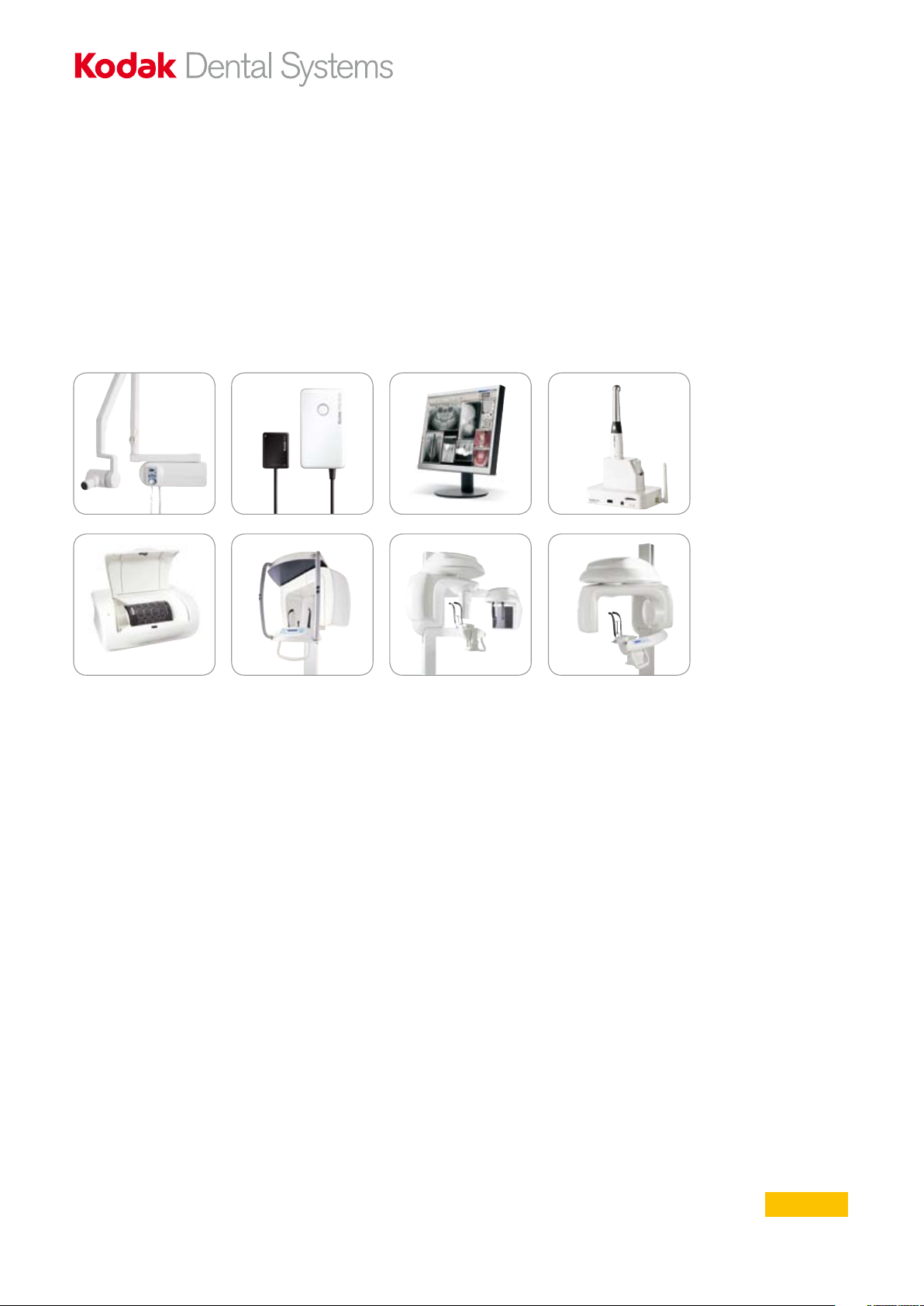
The complete guide to KODAK Dental Systems products
Product guide
Page 2
Why have we created
2
this product guide?
This brochure aims to help you choose the right
KODAK Dental Systems products for the dental
practice.
With this in mind, we have summarised the most
important advantages and selling points of our
digital radiography and imaging systems.
For further information please contact your local authorised dealer:
Register your product online and get an
additional year of warranty!
http://register.kodakdental.com/eamer
Visit our website: www.kodakdental.com
E-mail: europedental@cshdental.com
2
Page 3
...optimised
3
for digital!
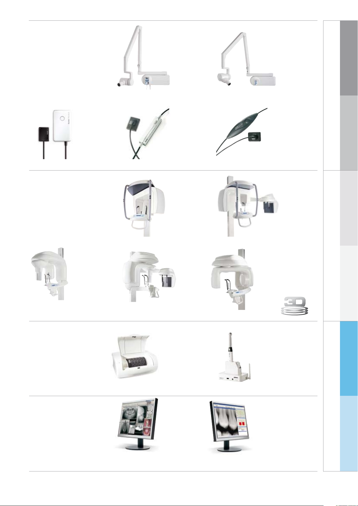
X-ray units
...the best
sensor - Wireless!
KODAK RVG 6500 System
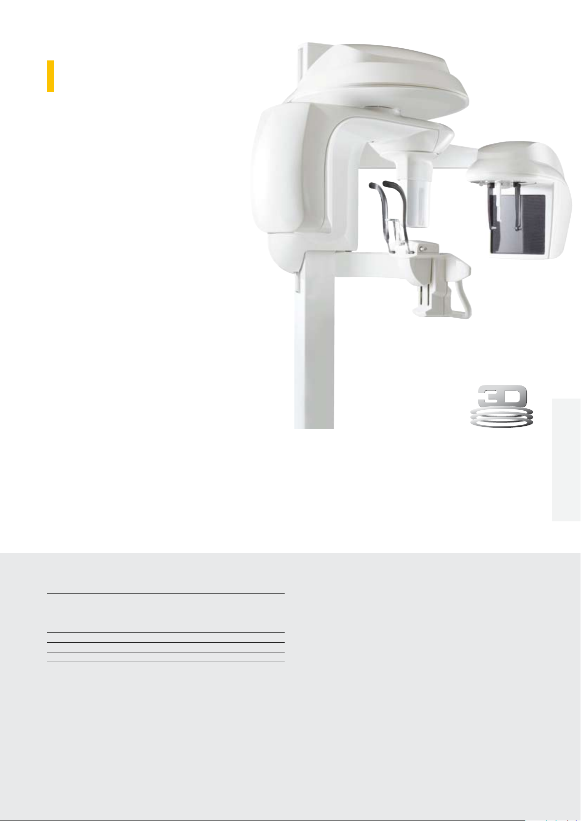
The 3-in-1
solution:
panoramic,
cephalometric
and 3D
KODAK 2200 System
...the best
sensor!
KODAK 8000 System
KODAK 2100 System
KODAK RVG 5100 SystemKODAK RVG 6100 System
Panoramic and
Ceph systems with
outstanding high
quality images
KODAK 8000C System
...large volume,
especially for oral
and maxillofacial
surgery, orthodontics
and implantology
KODAK RVG
radiography
Digital intraoral
6500/6100/5100
KODAK 8000/8000C
Digital extraoral radiography
KODAK 9000 System
KODAK 9000 3D System
KODAK 9000C System
A unique
intraoral and
panoramic
digital solution
KODAK CR 7400 System
...one
software
for all
products!
KODAK Dental Imaging
Software
KODAK 9500 SystemKODAK 9000C 3D System
Superb images at the
touch of a button - The
ideal conversation starter!
KODAK 1500 Camera
LOGICON Caries Detector
Software
KODAK 9000/9500
KODAK 1500
KODAK CR 7400
Intraoral camera
Computed radiography
Software
KDIS/LOGICON KODAK 2200/2100
Page 4
Intraoral radiography
4
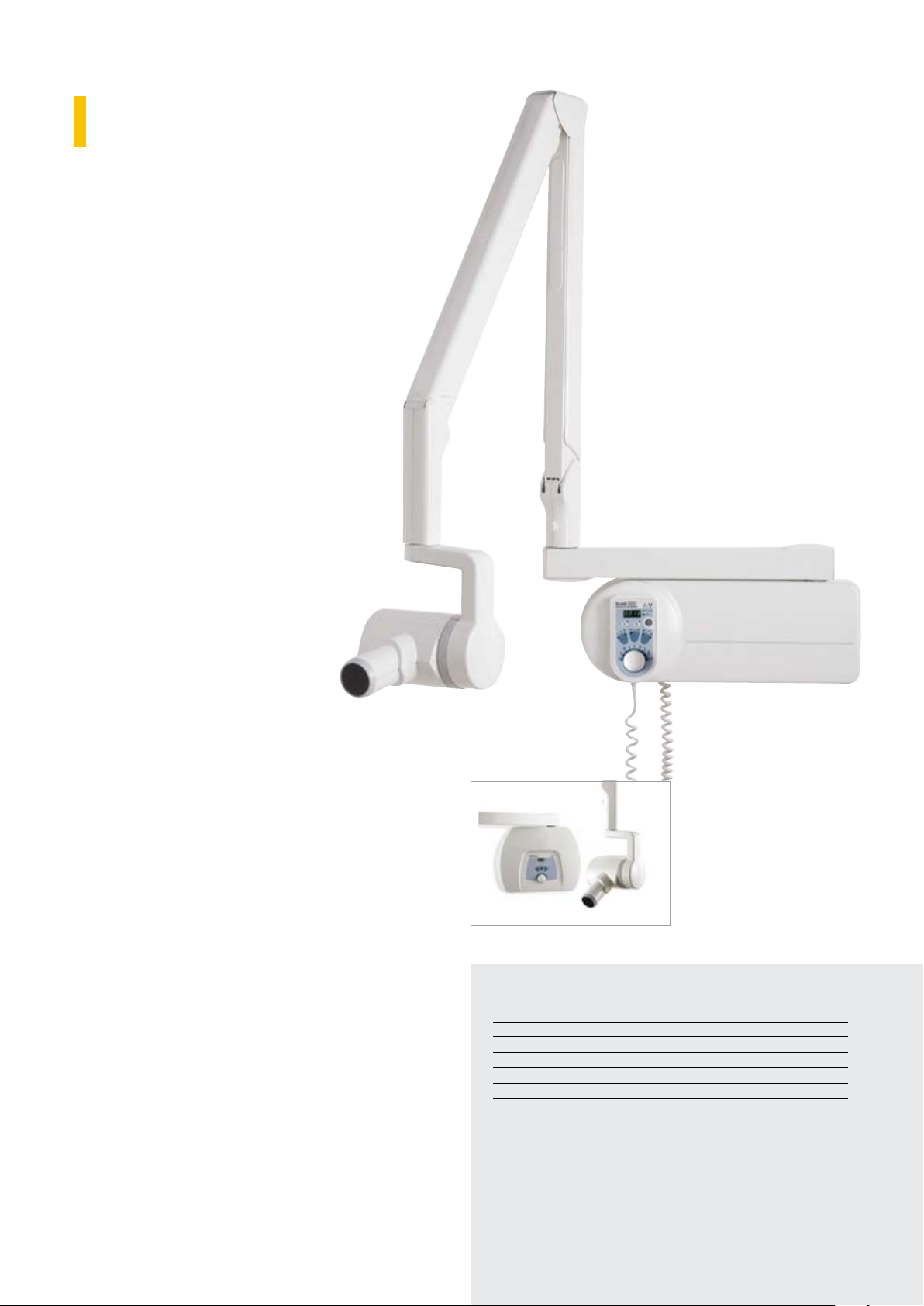
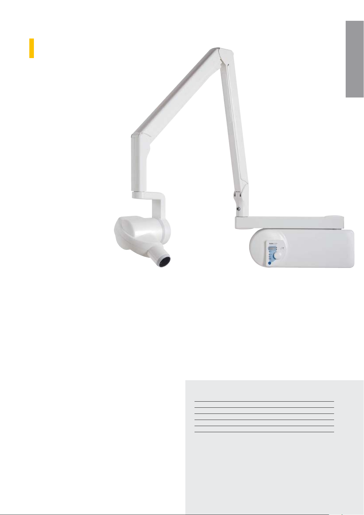
KODAK 2200
Intraoral X-Ray System
Ensure superior digital image quality in any environment with
the Kodak 2200 system. Designed for safety and
efficiency, the intuitive unit helps you make more accurate
diagnoses.
Features and benefits
Total control on tube voltage (60 or 70 kV) ●
for high contrasted or high latitude images
depending on examination requirements
Intuitive and easy-to-use thanks to its user’s ●
interface including a dental arch timer
Maximum image quality with minimum ●
exposure for better patient treatment
thanks to its pre-programmed control unit
Optimized for Kodak RVG sensors as well ●
as for Kodak film
High frequency for better patient safety - ●
reduces the radiation dose up to 25% in
comparison with a standard generator
Premium generator at an ●
exceptional price
After each exposure, the dose level ●
is displayed for simple dose
monitoring
Multiple configurations available, ●
including ceiling mount and a new
wall mounting option fully compatible with the
Irix installed base
Recommended resale price
Kodak 2200 system ¤ 2,999
Ordering information
Complete unit with wall
mount for replacing
existing IRIX systems as
well as a variety of other
popular manufacturers’
systems. Existing holes can
be used for wall mounting
Irix mount series
Kodak 2200 system for Irix replacement
with short arm (170 cm) Cat. No. 5159652
Kodak 2200 system for Irix replacement
with standard arm (188 cm) Cat. No. 5159645
Kodak 2200 system for Irix replacement
with long arm (205 cm) Cat. No. 5159660
Wall mount series
Kodak 2200 system
with short arm (170 cm) Cat. No. 5153580
Kodak 2200 system
with standard arm (188 cm) Cat. No. 5153572
Kodak 2200 system
with long arm (205 cm) Cat. No. 5153564
Technical specifications
Power supply 230 – 240 V
X-ray generator Very high frequency – DC (300 kHz)
Tube voltage: 60 kV, 70 kV
Tube current: 7 mA
Focal spot: 0.7 mm IEC
Focal spot/skin distance 200 mm (8 in)
X-ray units are also available with 100-110-130 V.
Page 5
Intraoral radiography
5
KODAK 2100
Intraoral X-Ray System
Obtain sharp, high contrast images quickly and easily
with this affordable high-frequency generator
- ideal for your basic intraoral needs.
Features and benefits
High-frequency DC technology at ●
the price of a conventional
generator
Easy-to-use and fast-setting ●
generator thanks to its
improved timer design
Sharp and high-contrast ●
images for easy diagnosis
Dose display after each ●
exposure
Ideal for digital sensors ●
or analog films
KODAK 2200/2100KODAK 2200/2100
Recommended resale price
Kodak 2100 system ¤ 2,599
Ordering information
Arm lengths available
Kodak 2100 system
with short arm (170 cm) Cat. No. 5153671
Kodak 2100 system
with standard arm (188 cm) Cat. No. 5153663
Kodak 2100 system
with long arm (205 cm) Cat. No. 5153655
Technical specifications
Power supply 230 – 240 V
X-ray generator Very high frequency – DC (300 kHz)
Tube voltage 60 kV
Tube current 7 mA
Focal spot 0.7 mm IEC
Focal spot/skin distance 200 mm (8 in)
X-ray units are also available with 100-110-130 V.
Page 6
Digital intraoral radiography
6
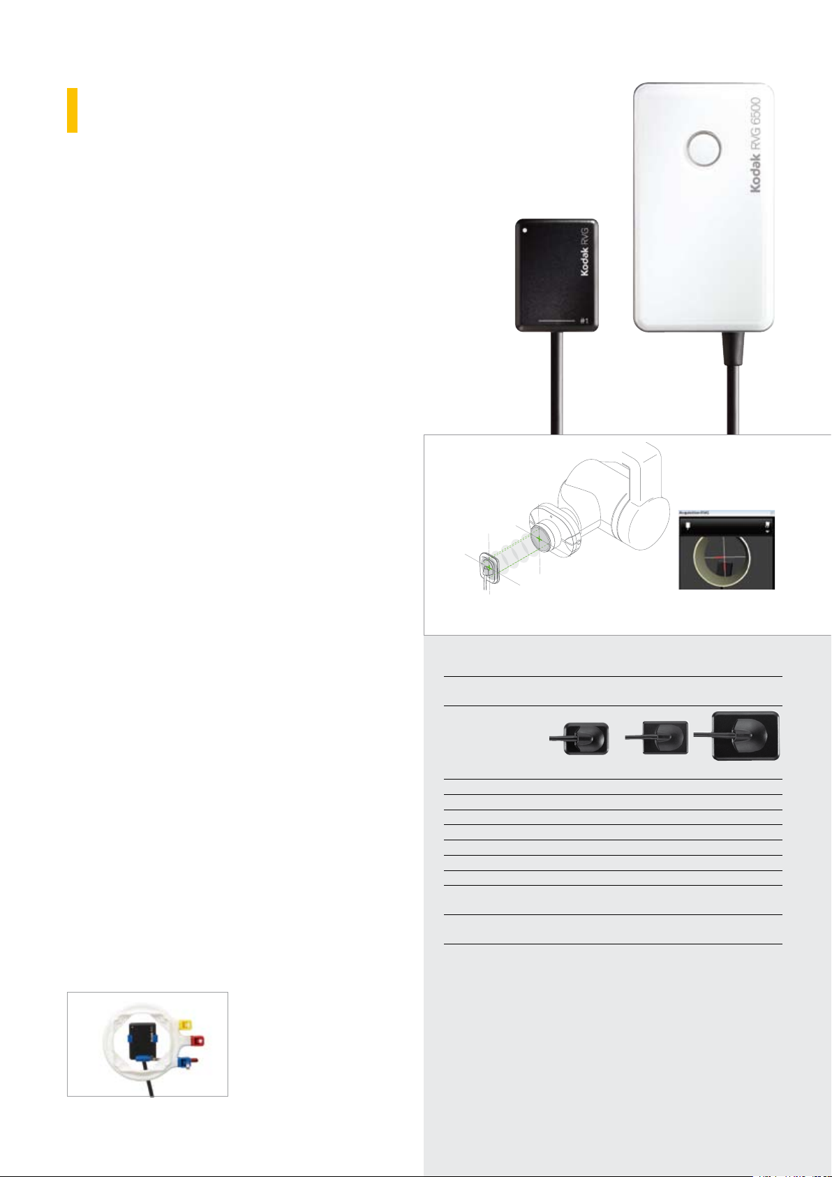
KODAK RVG 6500
System
As the first RVG sensor to offer Wi-Fi compatibility
and an Intelligent Positioning System (IPS), the
Kodak RVG 6500 system delivers the same exquisite
image quality as our wired models.
Features and benefits
Wi-Fi connectivity for faster and more secure ●
image transfer
Superior image quality for enhanced diagnostic ●
capability thanks to the highest true resolution in the
industry (20 line pairs/mm)
Accurate positioning thanks to an exclusive ●
Intelligent Positioning System (IPS) to help ensure the
x-ray beam and sensor are accurately aligned and
angulated
Improved positioning for less retakes and increased ●
productivity in the practice
Compatible with the iPod touch and iPhone for ●
enhanced portability in and outside the practice
Great patient comfort with sensor rounded corners ●
Sensors available in three sizes* ●
Robustness & Reliability ●
The IPS localizes the
position of the sensor
during positioning.
*IPS functionality not available with size 0 sensor.
Recommended resale price
RVG 6500 size 1 sensor ¤ 6.999
RVG 6500 size 1 sensor with IPS ¤ 7.999
24 months warranty extension - size 1 ¤ 499
RVG 6500 size 2 sensor ¤ 7.999
RVG 6500 size 2 sensor with IPS ¤ 8.999
24 months warranty extension - size 2 ¤ 599
Order information
RVG 6500 size 1 sensor Cat. No. 5159702
RVG 6500 size 1 sensor with IPS Cat. No. 5159728
Warranty extension Cat. No. 7423452
Hygienic sheaths size 1 sensor Cat. No. 5155239
RVG 6500 size 2 sensor Cat. No. 5159710
RVG 6500 size 2 sensor with IPS Cat. No. 5159736
Warranty extension Cat. No. 7423494
Hygienic sheaths size 2 sensor Cat. No. 5155221
Logicon software (see page 23)
The RVG 6500 system
comes with various accessories, including a sample
pack of disposable hygienic
sheaths, positioners, and
mounting holder.
The software displays real-time notifications regarding
the position of the x-ray beam in relation to the sensor.
Technical specifications:
Sensor technology CMOS, scintillator, optical fiber with shock-resistant
protective layer
Sensor size 0 Sensor size 1 Sensor size 2
True image resolution 15 lp/mm >20 lp/mm >20 lp/mm
Pixel size 18.5 µm 18.5 µm 18.5 µm
Outside dimensions 22.2 x 30.8 mm 27.5 x 37.7 mm 32.2 x 44.1 mm
Dimensions of active area 17 x 22 mm 22 x 30 mm 27 x 36 mm
Matrix dimensions (pixels) 900 x 1200 1200 x 1600 1440 x 1920
Wireless technology Wi-Fi 802.11 g
Battery Lithium
Control box dimensions
(non-IPS wireless)
Control box dimensions
(with IPS)
IMPORTANT NOTE:
Only Kodak dental imaging software version 6.12 or higher can operate
RVG 6500 system.
Operating system
• XP SP2/SP3 with DirectX 9.0c or higher, Windows Vista (Home
Premium, Business, Ultimate Editions) or Windows 7 32 bits or 64 bits
(Home Premium, Professional, Ultimate)
Important System Requirements: Minimum requirements: 2 GHz Intel
Duo Core, 2 GB RAM, Hard disk drive:1.2 GB for software installation,
80 GB free space for software
83 (H) x 47 (W) x 16 (D) mm
83 (H) x 47 (W) x 21 (D) mm
Page 7
Digital intraoral radiography
7
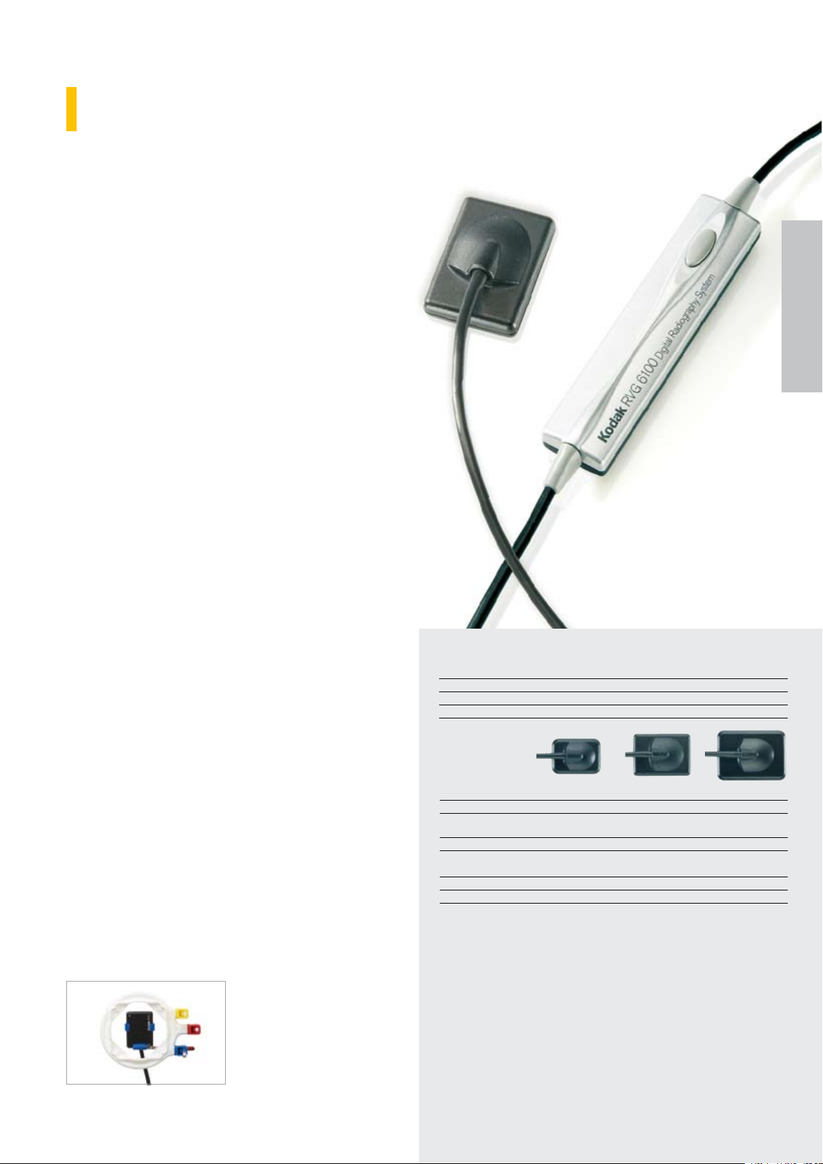
KODAK RVG 6100
Digital Radiography System
Can be used for all dental x-ray procedures but
is especially suited for endodontics where a high
degree of image detail is required.
Features and benefits
The best sensor technology in the market for ●
25 years
Highest measurable resolution (20 line pairs/mm) ●
for quick and confident diagnoses
The unrivalled detail reproduction you need for ●
complex examinations
Ergonomically designed for easy and accurate ●
positioning
Rounded corners for your patients’ ease ●
Pediatric size 0 with up to 40% less radiation than ●
larger sensors
USB cable – easy handling and flexible - for use in ●
multi chair practices
KODAK RVG
6500/6100/5100
Recommended resale price
RVG 6100 sensors sizes 1 and 0 ¤ 5,999
24-months warranty extension size 0 ¤ 899
24-months warranty extension sizes 1 ¤ 499
RVG 6100 sensor size 2 ¤ 6,999
24-months warranty extension size 2 ¤ 599
Ordering information
RVG 6100 size 1 sensor Cat. No. 5154828
Warranty extension Cat. No. 7423452
Hygienic sheaths size 1 sensor Cat. No. 5155239
RVG 6100 size 2 sensor Cat. No. 5154810
Warranty extension Cat. No. 7423494
Hygienic sheaths size 2 sensor Cat. No. 5155221
RVG 6100 size 0 sensor Cat. No. 5155114
Warranty extension Cat. No. 7423825
Hygienic sheaths size 0 sensor Cat. No. 5155205
Logicon software (see page 23)
The RVG 6100 system
comes with various accessories, including a sample
pack of disposable hygienic
sheaths, positioners, and
mounting holder.
Technical specifications
Theoretical resolution 27.03 lp/mm
Pixel size 18.5 micrometers
Technology Sensor with optical fiber technology
Connection USB 2
Actual image resolution
Purpose
External dimensions
Dimensions
Active area
Number of pixels
Delivery scope USB hub + 5 m connection cable
IMPORTANT NOTE:
Only Kodak dental imaging software version 6.12 or higher can operate RVG 6100
system.
Operating system
• XP SP2/SP3 with DirectX 9.0c or higher, Windows Vista (Home Premium,
Business, Ultimate Editions) or Windows 7 32 bits or 64 bits (Home Premium,
Professional, Ultimate)
• Mac OS on request
Important System Requirements: Minimum requirements: 2 GHz Intel Duo Core, 2
GB RAM, Hard disk drive:1.2 GB for software installation, 80 GB free space for
software
Sensor size 0
15 lp/mm
Pediatric
examinations
22.2 x 30.8 mm
17 x 22 mm
2
)
(374 mm
1.08 million
Sensor size 1
> 20 lp/mm
All-purpose sensor
27.5 x 37.7 mm
22 x 30 mm
2
)
(660 mm
1.92 million
Sensor size 2
> 20 lp/mm
Bitewing
examinations
32.2 x 44.1 mm
27 x 36 mm
2
)
(972 mm
2.76 million
Page 8
Digital intraoral radiography
8
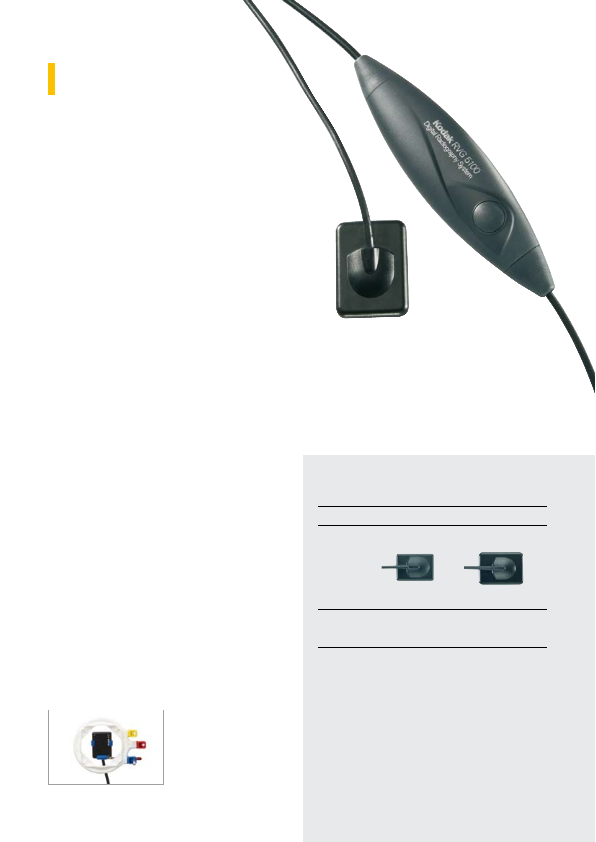
KODAK RVG 5100
Digital Radiography System
Affordable high quality digital imaging solution for
all applications.
Features and benefits
True resolution of 14 line pairs/mm - equivalent ●
to or better than most of the other manufacturers’
sensors
Second only to the RVG 6500 and RVG 6100 ●
systems in image quality
Cable positioned on the back of the sensor for ●
precise and effortless positioning
Rounded corners for increased patients’ comfort ●
Easy handling and flexibility thanks to USB cable ●
Excellent for use in multiple chair practices ●
Recommended resale price
RVG 5100 size 1 sensor ¤ 4,499
24-months warranty extension - size 1 ¤ 499
RVG 5100 size 2 sensor ¤ 5,499
24-months warranty extension - size 2 ¤ 599
Ordering information
RVG 5100 size 1 sensor Cat. No. 5154802
Warranty extension Cat. No. 7423361
Hygienic sheaths size 1 sensor Cat. No. 5155239
RVG 5100 size 2 sensor Cat. No. 5154794
Warranty extension Cat. No. 7423387
Hygienic sheaths size 2 sensor Cat. No. 5155221
Logicon software (see page 23)
The RVG 5100 system comes
with a RINN XCP-Ora kit.
Technical specifications
For both sensors
True image resolution 14 lp/mm
Theoretical resolution 27.03 lp/mm
Pixel size 18.5 micrometers
Technology Sensor with optical fiber technology
Connection USB 2
Purpose
External dimensions
Dimensions of active
area
Number of pixels
Delivery scope USB hub + 5 m connection cable
IMPORTANT NOTE:
Only Kodak dental imaging software version 6.12 or higher can operate
RVG 5100 system.
Operating system
• XP SP2/SP3 with DirectX 9.0c or higher, Windows Vista (Home
Premium, Business, Ultimate Editions) or Windows 7 32 bits or 64 bits
(Home Premium, Professional, Ultimate)
• Mac OS on request
Important System Requirements: Minimum requirements: 2 GHz Intel
Duo Core, 2 GB RAM, Hard disk drive:1.2 GB for software installation,
80 GB free space for software
• Mac OS on request
Sensor size 1
All-purpose sensor
27.5 x 37.7 mm
22 x 30 mm (660 mm
1.92 million
Sensor size 2
Bitewing examinations
32.2 x 44.1 mm
2
27 x 36 mm (972 mm
)
2.76 million
2
)
Page 9

× ×
9
×
×
N/A
KODAK RVG
6500/6100/5100
× × ×
ü
RVG 6500 System with IPS RVG 6500 System RVG 6100 System RVG 5100 System
ü ü
ü ü ü ü
ü ü ü ü
ü ü ü ü
ü ü
ü ü ü ü
ü ü ü
ü ü ü
N/A
Special Size 0 kit**
RINN type baskets
RINN XCP-ORA kit
Toothbrush type holders
lll lll ll lll ll
lll lll ll lll ll
lll l l l
lll ll lll ll
lll ll lll ll
lll ll l
Best
Comparative Matrix for RVG Range
Features
Intelligent Positioning System (IPS)
Connectivity Wi-Fi Wi-Fi USB2 USB2
True resolution > 20 lp/mm > 20 lp/mm > 20 lp/mm* 14 lp/mm
Sensor size 1 - 2 0 - 1 - 2 0 - 1 - 2 1 - 2
iPhone / iPod touch acquisition feature
iPhone / iPod touch review feature
Sensor waterproofness
Twain compatibility
Positioning accessories delivered with the system:
Applications Size 0 Size 1 Size 2
*Size 0 resolution: 15 lp/mm
**Only with size 0 sensor
Systems RVG 6500* / 6100 Systems RVG 6500 / 6100 Systems RVG 5100 System RVG 6500 / 6100 Systems RVG 5100 System
General diagnostics
Caries detection
Endodontics
Implantology
Pedodontics
Periodontics
Good ll Better lll
l
* Size 0 sensor not available with RVG 6500 system with IPS
Page 10

Digital extraoral radiography
10
KODAK 8000
Digital Panoramic System
The perfect solution for your everyday panoramic needs.
Features and benefits
Convenient and unique face-to-face patient ●
positioning provides easy access for all
patients, including those with disabilities
High image quality with less radiation ●
generation than conventional generators for
improved patient treatment
Programs available for all examination needs ●
–TMJ, panoramic, segmented panoramic and
maxillary sinus images, adult and paedriatric
applicatlons
Intuitive computer user interface offers simple ●
and precise image processing
Real-time diagnosis with instant acquisition ●
and visualization for time saving and higher
productivity
Reliable, affordable and easy to integrate ●
Upgradable to the Kodak 8000C system for ●
ceph
Ordering information
Recommended resale price
Kodak 8000 system ¤ 23,999
24-months warranty extension ¤ 999
Technical specifications
Unit dimensions 888 x 1,180 x 2,315 mm (L x D x H)
Positioning on the patient Frontal with 3 laser beams
and four-point head cover
Image acquisition Direct and in real time
Gray scale 16384: 14 bits
Radiography mode Panoramic images
Segmented panoramic images
Maxillary sinus images
TMJ, 2 lateral views
TMJ, 4 lateral views
Power supply 230 to 240 V/50 Hz
X-ray generator High frequency (max. 140 kHz)
X-ray tube voltage 60 to 90 kV (in 1 kV steps)
X-ray tube current 2 to 15 mA
Focal spot 0.5 mm IEC
Digital sensor CCD sensor and optical fiber plate
Matrix dimensions 2,850 x 1,348 pixels (pixel size = 96 µm)
Effective resolution identical to film
Panoramic devices are also available with 60 Hz and with 100 to 130 V. For
further options please contact your local authorised dealer.
Kodak 8000 system
patient entry on the right Cat. No. 1912450
Kodak 8000 system
patient entry on the left Cat. No. 8849846
Warranty extension Cat. No. 7424864
Bite block panoramic procedures (5x) Cat. No. 5157565
Bite block edentulous patients (2x) Cat. No. 5157581
Minimum system requirements
Microprocessor 1 GHz, 32-bit processor
(Pentium or equivalent – Intel)
Hard-disk drive 40 GB (80 GB recommended)
RAM 256 MB (512 MB recommended)
Operating system Windows 2000 SP4 or Windows XP
Home / Pro edition SP2
Monitor 1 monitor, 17" or larger
Minimum screen resolution of
1,024 x 768 - 32-bit color mode
Computer type Multimedia
CD / DVD Drive A DVD-ROM drive is required to install the
imaging software
Backup systems High-capacity systems with magnetic media
Ethernet interface 1 Ethernet interface (100 Mbits): point-to point link between equipment and PC.
1 additional Ethernet interface (100 Mbits) if
the PC is connected to a LAN and/or WAN
Page 11

Digital extraoral radiography
11
KODAK 8000C
Digital Panoramic and Cephalometric
System
An affordable system from the inventor of
“one shot” technology for clear and perfectly
exposed cephalometric images in seconds.
Features and benefits
“One shot” technology captures the ●
image in less than a second reducing
exposure time and risk of blurred
images
No retakes and outstanding image ●
sharpness thanks to the short exposure times for
excellent diagnosis
Unique image format 20 x 26 cm ●
Two-sensor system for improved reliability - no ●
handling from panoramic to ceph mode required
Convenient patient positioning for better treatment ●
6 cephalometric programs to answer all the ●
examination needs
3 automatic ortho enhancement filters revealing ●
even more detail
User-friendly, easy to learn computer-controlled ●
user interface for fast program settings – almost
no training needed
Minimal space requirement (only 2.30 m width) ●
KODAK 8000/8000C
Ordering information
Recommended resale price
Kodak 8000C system ¤ 44,999
24-months warranty extension ¤ 1,499
Technical specifications
Unit dimensions 2,250 x 1,261 x 2,315 mm
(length x depth x height)
Dual CCD sensors (one for panoramic imaging and one for cephalometric
imaging), with protective optical fibre plate
Cephalometric image size 20x26 cm
Laser beam for patient positioning
according to the Frankfurt horizontal plane
Software soft-tissue filter
Cephalometry modes Lateral view
Frontal views: AP or PA
Oblique view
Submento-vertex view
Carpus view
Installation configuration Left or right remote radiography device
Kodak 8000C system
patient entry and
ceph arm on the right Cat. No. 8479511
Kodak 8000C system
patient entry and
ceph arm on the left Cat. No. 1761089
Warranty extension Cat. No. 7424880
Minimum system requirements
Microprocessor 1 GHz, 32-bit processor
(Pentium or equivalent – Intel)
Hard-disk drive 40 GB (80 GB recommended)
RAM (working memory) 256 MB (512 MB recommended)
Operating system Windows 2000 SP4 or Windows XP
Home / Pro edition SP2
Monitor 1 monitor, 17" or larger
Minimum screen resolution of
1,024 x 768 - 32-bit color mode
Computer type Multimedia
CD / DVD Drive A DVD-ROM drive is required to install the
imaging software
Backup systems High-capacity systems with magnetic media
Ethernet interface 1 Ethernet interface (100 Mbits): point-to point link between equipment and PC.
1 additional Ethernet interface (100 Mbits) if
the PC is connected to a LAN and/or WAN
Page 12

Digital extraoral radiography
12
KODAK 9000
Extraoral Imaging System
High-end system for creating excellent digital
panoramic images. Upgradable to 3D and/or
Ceph...
Features and benefits
Convenient and practical “face-to-face” positioning ●
Excellent image quality thanks to focal trough ●
adaptable to jaw morphology and improved sensor
technology
5 panorama programs (see technical specifications) ●
and 12 anatomical settings available
User-friendly, computer-controlled user interface for ●
fast program settings on the device and the software
Compact design makes positioning easier than ever ●
Upgradable to the Kodak 9000C system for ceph. ●
For 3D, choose the Kodak 9000 3D system.
For pan, ceph and 3D select the Kodak 9000C 3D
system
Recommended resale price
Kodak 9000 system ¤ 32,999
12-months warranty extension ¤ 2,499
Technical specifications
X-ray generator
X-ray tube voltage 60 – 90 kV (max.)
pulsed mode for 3D modality
X-ray tube current 2 - 15 mA (max.)
Frequency 140 kHz (max.)
Tube focal spot: 0.5 mm (IEC 60336)
Total filtration > 2.5 mm Al equivalent value
Panoramic sensor CCD-optical fiber
Sensor matrix 61 x 1,244 pixels
(6.3 x 129.4 mm)
Grayscales 16,384 - 14 bit
Magnification 1.22 (±10 %)
Exposure time 4 to 16 seconds (depending on
patient type and program selection)
Programs 12 anatomical settings
Options for radiology examinations Panoramic images
Segmented panoramic images
Maxillary sinus imaging
Temporomandibular joints (x2)
Temporomandibular joints (x4)
Ordering information
Kodak 9000 system Cat. No. 5157433
Warranty extension Cat. No. 5158753
Bite block panoramic procedures (5x) Cat. No. 5157565
Bite block edentulous patients (2x) Cat. No. 5157581
Accessories
Chin support for Panoramic images
TMJ x2
Paranasal sinus
Nose support for temporomandibular
joints x4
Standard bite block for panoramic images
Bite block for edentate patients
Single-use hygienic protective sheaths
for bite blocks (carton of 500)
System requirements
CPU Intel Duo Core with 2 GHz
RAM 2 GB
Hard drive 1.2 GB for software installation
80 GB free space to use the software
Graphics card: NVIDIA/ATI-based graphic card with Open Glide 1.2 support and
256 MB video RAM and AGP x8 video bus (e.g. NVIDIA GeForce 6800 GT)
Network connection 2 Ethernet network cards
100 Mbits for LAN
1,000 Mbits for system connection
Second network card (plug-in option)
1 monitor, 43 cm or larger
Minimum screen resolution of 1,024 x 768 - 32-bit color mode
Page 13

Digital extraoral radiography
13
KODAK 9000C
Panoramic and Cephalometric
System
2-in-1 system with panoramic and
cephalometric functionality. One shot
cephalometric imaging for higher image
quality. Upgradable to 3D.
Features and benefits
High-performance 2-in-1 radiography ●
system (pan and ceph)
The ideal choice for all of your ●
orthodontic tracing needs
Recognize landmark and anatomical ●
structures and trace them automatically
with our included Kodak dental imaging
software
Exclusive range of 5 image formats from ●
18x18 to 30x30 cm
Highest quality cephalometric images within less ●
than a second and with low radiation thanks to
“one shot” technology
3 automatic ortho enhancement filters allows ●
improving of image clarity and outlining soft tissue
with just one click
Upgrade to the 9000C 3D system with focused- ●
field 3D imaging for even more diagnostic
capabilities
Ordering information
Recommended resale price
Kodak 9000C system ¤ 54,999
12-months warranty extension ¤ 3,599
Technical specifications
X-ray tube voltage 60 – 90 kV (max.)
X-ray tube current 2 – 15 mA (max.)
Frequency 140 kHz (max.)
Tube focal spot: 0.5 mm (IEC 60336)
Total filtration > 2.5 mm Al equivalent value
Ceph mode
Sensor technology CCD
Sensor matrix 2,100 x 2,092 pixels
Image field 300 x 300 mm
Grayscales 16,384 - 14 bit
Magnification 1.14
Exposure time 0.1 to 3.2 seconds
Options for radiology examinations Lateral
Frontal AP or PA
Oblique
Submento-vertex
Carpus
Image format size 18 x 18, 18 x 24, 24 x 24
24 x 30, 30 x 30
Kodak 9000C system - right entry Cat. No. 5159041
Warranty extension Cat. No. 5158746
Bite block panoramic procedures (5x) Cat. No. 5157565
Bite block edentulous patients (2x) Cat. No. 5157581
System requirements
CPU Intel Duo Core with 2 GHz
RAM 2 GB
Hard drive 1.2 GB for software installation
80 GB free space to use the software
Graphics card: NVIDIA/ATI-based graphic card with Open Glide 1.2 support and
256 MB video RAM and AGP x8 video bus (e.g. NVIDIA GeForce 6800 GT)
Second network card (plug-in option)
1 monitor, 43 cm or larger
Minimum screen resolution of 1,024 x 768 - 32-bit color mode
Operating system Windows XP Home / Pro edition SP2
Windows Vista 32-bit
Network connection 2 Ethernet network cards
100 Mbits for LAN
1000 Mbits for system connection
CD/DVD drive CD-ROM drive for the installation
Accessories
Chin support for Panoramic images
TMJ
Paranasal sinus
Nose support for temporomandibular
joints x4, Single-use hygienic protective
sheaths for bite blocks (carton of 500)
KODAK 9000/9500
Page 14

Digital extraoral radiography
14
KODAK 9000 3D
Extraoral Imaging System
The Kodak 9000 3D system‘s focused-field
of view is ideal for endodontics, implantology
and dental surgery.
Features and benefits
Affordable two-in-one solution, combining real 3D ●
technology and real panoramic technology in a single
system
The system delivers 3D exams with the highest ●
resolution in the industry and high quality panoramic
image
Lowest dose 3D exams in the industry* covering a ●
wide scope of clinical needs
Combines the benefits of focused field 3D imaging ●
(50 x 37 mm) with 3D stitching program (up to 85 x
66 x 37mm)
Easy to use and to integrate into the practice with our ●
Kodak dental imaging software, intuitive 3D Module
and without any sensor manipulation
Face-to-face patient positioning reduces the risk of ●
retakes
KODAK 2200/2100
*According to study by ICRP: International Committee of Radio
Protection
Recommended resale price
Kodak 9000 3D system ¤ 64,999
12 months warranty extension ¤ 3,999
Technical specifications:
Unit dimensions: 1158 (L) x 1595 (D) x 2378 (max H) mm
Patient positioning: Frontal with 2 laser beams
Tube voltage: 60 - 90 kV (max), pulsed mode for 3D modality
Tube current: 2 - 15 mA (max)
Frequency: 140 kHz (max)
Tube focal spot: 0.5 mm (IEC 336)
3D Modality
Technology: Cone Beam Computed Tomography (CBCT)
3D Volume size: 50 x 37 mm (Diam x H)
Gray scale: 16384 - 14 bits
Voxel size: 76.5 x 76.5 x 76.5 µm
Panoramic Modality
Sensor technology: CCD Optical fibre sensor
Radiological exam options: Panoramic - Segmented panoramic - Maxillary
sinus
LA TMJ x2 - LA TMJ x4
Input voltage: 230-240 V - 50/60 Hz
Units also available as 100-130 V / 60 Hz. Please contact your Carestream
Health sales representative for more information.
Ordering information
Kodak 9000 3D system Cat. No. 5157458
Warranty extension Cat. No. 5158670
Bite block panoramic procedures (x5) Cat. No. 5157565
Bite block edentulous patients (x2) Cat. No. 5157581
System requirements:
CPU 2 GHz Intel Duo Core
RAM 2 GB
Hard disk drive 1.2 GB for software installation
80 GB free space to use the software
Graphic board 512 MB of dedicated video memory required
Monitor 1 monitor 17“ or larger
1024 x 768 minimum screen resolution
Operating system Windows XP SP2, Windows Vista 32 bits
(Home Premium, Business, Ultimate Editions)
Ethernet interface 2 Ethernet interfaces (100Mbits)
Accessories delivered - Patient holders for panoramic procedures:
with the system chin rest, bite bock, bite block for edentulous
patients, TMJ nose rest
- Patient holders for 3D procedures: bite block
support, 3D bite block
- Hygienic sheaths for bite blocks
Minimum operational 1500 mm x 1700 mm
required space:
Page 15
Digital extraoral radiography
15
KODAK 9000C 3D
Extraoral Imaging System
The 3-in-1 solution for your imaging needs, the
Kodak 9000C 3D system combines the
power of focused-field 3D with cutting-edge
panoramic and one-shot cephalometric imaging.
Features and benefits
Same features as the Kodak 9000 3D ●
system, plus cephalometric imaging, offering
a range of new diagnostic possibilities
5 image formats in the range from full skull ●
30x30 cm to18x18 cm small field for lower
dose exposures
Automatic landmark recognition saves time ●
and improves the workflow
Delivers exceptional image quality thanks to ●
exclusive and best in class „one shot“
technology
3 automatic ortho enhancement filters offers ●
unprecedented detail and anatomical views
Recommended resale price
Kodak 9000C 3D system ¤ 94,999
Cephalostat upgrade ¤ 29,999
Technical specifications Cephalostate upgrade*:
Cephalometric module with built-in CCD sensor with
protective optical fibre plate
Cephalometric images: Half and full lateral
(18x18 cm, 18x24 cm, 24x24 cm,
24x30 cm, 30x30 cm), frontal (AP/
PA), oblique, submento-vertex, carpus
Magnification: x 1.14
Installation configuration: Right sided-cephalostat
Minimum operational required space 2230 (L) x 2000 (D) x 2400 (H) mm
*The system requirements and further technical specifications are the same
as for the Kodak 9000 3D System (See page 14)
Ordering information
Kodak 9000C 3D system Cat. No. 5159009
Cephalostat upgrade right side Cat. No. 5159157
KODAK 9000/9500
The cephalostat attachment kit allows the conversion of
a Kodak 9000 / 9000 3D system into a Kodak 9000C /
9000C 3D system.
Page 16

Digital extraoral radiography
16
KODAK 9500
Cone Beam 3D system
Recommended for oral, maxillofacial and sinus
examinations, the Kodak 9500 cone beam 3D system
delivers high-quality images safely and easily.
Features and benefits
Large (18x20cm) and medium (9x15cm) field-of-view ●
system with an exceptional price/performance ratio in
this volume range
Ease of use and optimal patient comfort ●
Fast large volume image reconstruction time ●
of approx. 2 minutes saves time and increases
productivity
Programmed settings assist in capturing images of ●
the highest quality
3D software with numerous user-friendly diagnostic ●
and planning tools
DICOM file format provides data portability and ●
compatibility with 3rd party implant planning
(NobelGuide® and Simplant®) and 3D visualization
software
Supports 1:1 DICOM printing ●
Recommended resale price
KODAK 9500 system ¤ 149,999
12-months warranty extension ¤ 8,999
Technical specifications
Technology Cone Beam Computed Tomography (CBCT)
Sensor technology Amorphous silicon flat-panel sensor
Grayscales 16384 - 14 bit
Volume size FoV 184 x 206 mm (HxD), resolution 0.3 mm
90 x 150 mm (HxD), resolution 0.2 mm
Voxel size/resolution 300 x 300 x 300 µm/0.3 mm
200 x 200 x 200 µm/0.2 mm
Sections/volume 618 sections max.
Effective acquisition time 24 seconds
Effective exposure time 10.8 seconds (pulsed radiation)
Reconstruction time 2 min. 20 sec.
80 seconds
Patient position Standing/sitting
X-ray tube voltage 60 - 90 kV (max.)
X-ray tube current 2 - 15 mA (max.)
Frequency 140 kHz (max.)
Tube focal spot: 0.7 mm
Total filtration > 2.5 mm. equivalent value
Input voltage 230-240 V – 50/60 Hz
Weight 176 kg – 388 lbs.
DICOM compatibility Yes
Minimum space requirement length x width (1725 mm x 1635 mm)
(depending on local conditions)
Ordering information
KODAK 9500 system Cat. No. 5159538
Warranty extension Cat. No. 5161500
System requirements
CPU Intel Duo Core with 2 GHz
RAM 2 GB
Hard drive 1.2 GB for software installation
80 GB free space to use the software
Graphic board 512 MB of dedicated video memory required
Monitor 1 monitor, 43 cm or larger
Minimum screen resolution of
1,024 x 768 - 32-bit color mode
Operating system Windows XP Home / Pro edition SP2
Windows Vista 32-bit
Network connection 2 Ethernet network cards
100 Mbits for LAN
1,000 Mbits for system connection
CD/DVD drive CD-ROM drive for the installation
Page 17

Applications****
Routine panoramic examination
Routine cephalometric evaluation
Implantology
Endodontic
Orthodontic
Periodontic
Oral Surgery
Prosthodontic
Restorative
**** Please note bullets are simply a recommendation based on feedback from our thought leaders and users.
One bullet indicates it meets the basic requirements for the application. Three bullets indicates it is the better choice.
Good Better Best
KODAK 8000 KODAK 8000C
KODAK 8000 KODAK 8000C
KODAK 9000 KODAK 9000 3D KODAK 9000C KODAK 9000C 3D
KODAK 9000 KODAK 9000 3D KODAK 9000C KODAK 9000C 3D
Cephalometric
Features & Specications
Panoramic
3D imaging
3D eld-of-view size
Pan: 14 sec.
Ceph 1 to 3 sec.
Pan: 14 sec.
Ceph 1 to 3 sec.
Pan: 14 sec.
3D: 90 sec. to 2 min.
Pan: 14 sec.
Ceph: 1 to 3 sec.
Pan: 14 sec.
Ceph: 1 to 3 sec.
3D: 90 sec. to 2 min.
* Acquisition time + reconstruction time. ** Dimensions of the unit. Required operational space could be dierent. *** Simulated panoramic and cephalometric.
Footprint/size**
KODAK 9500
KODAK 9500
Time to process*
Upgradeable Upgradeable Upgradeable
UpgradeableUpgradeable
180 x 200 mm
90 x 150 mm
37 x 50 mm37 x 50 mm N/AN/AN/AN/A
2:15 min.
1:45 min.
mm
inch
880 (W) x 1180.5 (D)
34” (W) x 46” (D)
2250 (W) x 1261 (D)
88.59” (W) x 49.63” (D)
1158 (W) x 1595 (D)
45.5” (W) x 62.75” (D)
1158 (W) x 1595 (D)
45.5” (W) x 62.75” (D)
2150 (W) x 1595 (D)
84.63” (W) x 62.75” (D)
2150 (W) x 1595 (D)
84.63” (W) x 62.75” (D)
1416 (W) x 1725 (D)
55.74” (W) x 67.9” (D)
Pan: 14 sec.
***
17
KODAK 9000/9500
Comparative Matrix for Extraoral Range
Page 18

Extraoral imaging programs
18
KODAK 8000 / 8000C Digital Panoramic and
Cephalometric System
Panoramic imaging
Panoramic examinations
covering all basic needs.
Standard panoramic
Cephalometric imaging
Unique image format
20x26 cm.
3 Automatic Ortho
Enhancement Filters.
Lateral (20 x 26 cm)
KODAK 9000 Extraoral Imaging System
Panoramic imaging
Panoramic examinations with
focal trough adaptation to jaw
morphology and improved
sensor technology.
Standard panoramic
Child panoramic
Segmented panoramic
TMJ x2 lateral TMJ x2 lateral
Frontal PA / AP
Submento vertex
Child panoramic
Segmented panoramic
TMJ x4 lateral TMJ x4 lateral
Maxillary sinus Maxillary sinus
Carpus
Page 19

KODAK 9500 Cone
19
Beam 3D System
Cephalometric imaging
Unique image format
20 x 26 cm.
3 Automatic Ortho Enhancement Filters.
Lateral view, multiple formats
3D imaging
Focused field of view of
50 x 37mm.
Extended field of view 75 x 37
mm covering the whole arch
thanks to 3D stitching program.
3D imaging
Large-field 3D program
18.4 cm x 20.6 cm.
Medium-field 3D program
9 cm x 15 cm.
Frontal AP
Frontal PA
Submento vertex
Focused-field 3D program
(5 x 3,7cm)
Stitching 3D program
(up to 85 x 66 x 37mm)
Large-field 3D program
(18,4 x 20,6 cm)
Large-field 3D program
(9 x 15cm)
Carpus
Page 20

Computed radiography system
20
KODAK CR 7400
Digital Radiography System
The phosphor plate system for all formats - intraoral,
panoramic and cephalometric radiography.
The easy way to convert your dental practice to digital.
Features and benefits
Low-cost complete digital entry-level solution ●
As easy as film with all the benefits of digital ●
- similar workflow to film minimizes learning
curve with easy to use product
Compact digital radiography imaging solution ●
(intraoral/extraoral)
Very easy handling and excellent image ●
quality
One-step process for full mouth series of ●
X-ray images
Our thin and flexible durable plates reduce ●
the need for replacement
Recommended resale price
Recommended resale price ¤ 9,999
Two years warranty extension ¤ 1,999
Technical specifications
Operating systems Windows 2000 Professional SP4,
Windows XP Professional SP3
Windows Vista Business SP2
Windows 7 Home Premium/
Professional/Ultimate (32 Bit)
CPU 1.8 GHz (Pentium type or equivalent)
RAM 512 MB (1 GB recommended)
Minimum hard drive 80 GB
Graphics adapter 64 MB (recommended not on board)
Monitor 17” 1024 x 768 support on 96 DPI
Communication interface via USB 2.0
Ordering information
Kodak CR 7400 system Cat. No. 8470411
Warranty extension Cat. No. 5155528
A sample of the CR 7400 system’s accessories:
CR imaging plates/size 2 Cat. No. 1340074
CR intraoral holder/size 2 plates Cat. No. 1872647
Light protection pouches size 2 plates Cat. No. 1689611
Barrier envelopes/size 2 plates Cat. No. 1804616
CR imaging plate/panoramic 15 x 30 cm Cat. No. 1591114
Page 21

Intraoral imaging
21
KODAK 1500
Intraoral Camera
The ideal communication tool for any dental practitioner.
Elegantly styled and easy to use, Kodak’s first wireless
intraoral camera delivers stunning images with the convenience and freedom of Wi-Fi connectivity.
Features and benefits
Patented true autofocus technology ●
With built-in Wi-Fi support, the camera provides total ●
freedom of movement
The best resolution on the market for still images ●
White LED exposure ensures uniform and bright ●
illumination
Intraoral and extraoral imaging ●
Compatible with computer and video screens ●
Available as wired and wireless configuration ●
Recommended resale price
Kodak 1500 camera wireless kit ¤ 3,499
Kodak 1500 camera wired kit ¤ 2,999
Technical specifications
Sensor Micron 1/2.5’’ CMOS
2592 (H) x 1944 (V)
Video resolution 640 (H) x 480 (V)
Image resolution 1024 (H) x 768 (V)
Focusing range 1 mm to infinity
Angle of view 90°
Field of view 80°
Focus Autofocus
Light source 8 White LED array
Video output USB 2.0; TV-NTSC;
TV-PAL; VGA; S-Video
Connection USB 2.0 high-speed
Ordering information
Kodak 1500 camera wireless kit Cat. No. 6557185
Kodak 1500 camera wired kit Cat. No. 6557177
KODAK CR 7400
KODAK 1500
Page 22

Software
22
KODAK Dental
Imaging Software
The master software for all systems.
Integrated 3D module.
Features and benefits
Top-quality and easy to use software ●
Included as standard with all Kodak dental ●
systems digital imaging products
Serves as the control panel for all our ●
systems
Designed specifically for dental radiological ●
diagnosis
Can be used as a standalone program
●
or integrated with practice management
software
No need to purchase additional licenses ●
Ordering information
Kodak dental imaging software Cat. No. 8557530
The software controls all KODAK Dental Systems
products
Particularly easy to use radiography software ●
All features are accessible with just a few clicks ●
100% designed for user-friendliness ●
Also ideal for novice PC users ●
High image quality ●
Network-ready ●
Optional: DICOM support ●
Page 23

Software
23
LOGICON Caries
Detector Software
Software for detecting caries even at an early stage.
Features and benefits
A unique tool for detecting approximal ●
caries at an early stage
A software option for Kodak RVG digital ●
radiography systems
The software automatically highlights pos- ●
sible abnormalities on digital dental
radiographs
E-mail function allows sending of result ●
screens to third parties
Results can be saved to patient file ●
Fully integrated with Kodak dental imaging ●
software
Making caries diagnosis easier
The Logicon software extracts image features and
compares them to a database of known caries problems.
Possible anomalies are thus marked automatically on
the digital dental radiography images, allowing the dental
surgeon to investigate the affected tooth and the tooth
surface in more detail.
Functionality
The Logicon software uses recognition algorithms based
on laboratory data created in collaboration with a leading
dental medicine institute. It automatically flags potential
lesions on dental images and shows changes to the tooth
density and the probability of lesions.
Ordering information
Logicon caries detector software Cat. No. 7417967
Recommended resale price
Logicon caries detector software ¤ 999
KDIS/LOGICON
Page 24

Looking for an imaging solution?
We're confident we have the ideal solution for every practice!
A full range of dental radiography and imaging systems ●
100 years' experience in dental imaging ●
In-house product development and manufacturing ●
Worldwide dealer and service network ●
The inventor of intraoral sensor technology ●
High quality standards ●
Would you like to know more?
www.kodakdental.com
Alternatively, contact your local authorised dealer.
Carestream Health
© Carestream Health, Inc. 2010.
The Kodak brand the brand elements/colors of Kodak are used under license. RVG is a trademark of Carestream Health, Inc.
 Loading...