


 Loading...
GE Healthcare
X-ray Bone Densitometer with enCORE v17 software - User Manual
This manual supports the following products: Lunar iDXA Series, Lunar Prodigy Series, Lunar DPX Series
X-ray Bone Densitometer with enCORE v17 software - User Manual
LU43616EN-2EN Revision 19 (September 2017)
© 2017 GE Healthcare

1 |
Safety ............................................................................................................ |
25 |
|
Precautions for Standard Operating Procedures........................................ |
25 |
|
Operator Safety............................................................................................. |
26 |
|
Personnel Monitors ..................................................................................... |
26 |
|
X-Ray and Shutter Graphics ....................................................................... |
27 |
|
X-Ray Shutter............................................................................................... |
27 |
|
X-Ray Power Supply .................................................................................... |
27 |
|
Patient Safety ................................................................................................ |
27 |
|
Mechanical Safety......................................................................................... |
29 |
|
Symbols ......................................................................................................... |
29 |
|
Sample Labels ............................................................................................... |
30 |
|
Failsafe Circuit............................................................................................... |
35 |
|
X-Ray Shielding Requirements ..................................................................... |
35 |
|
Electrical Safety............................................................................................. |
36 |
|
Peripheral Configurations........................................................................... |
36 |
|
Scatter Radiation .......................................................................................... |
37 |
2 |
Product Information .................................................................................... |
39 |
|
Intended Use ................................................................................................. |
39 |
|
Indications for Use ........................................................................................ |
39 |
|
Cautions for DXA Determinations ................................................................ |
41 |
|
Device Descriptions ...................................................................................... |
42 |
|
Scanner Table Assembly............................................................................... |
47 |
|
Training Information ..................................................................................... |
50 |
|
Classifications................................................................................................ |
50 |
|
Installation and Operation............................................................................ |
50 |
|
Software Installation..................................................................................... |
50 |
|
Features......................................................................................................... |
51 |
|
Hardware Features ..................................................................................... |
51 |
|
Software Features ....................................................................................... |
51 |
|
Quality Assurance (QA) Features ................................................................ |
52 |
|
User Information ......................................................................................... |
52 |
|
Options......................................................................................................... |
52 |
3 |
Daily Use ....................................................................................................... |
55 |
3 |
X-ray Bone Densitometer with enCORE v17 software - User Manual |

|
Daily Use........................................................................................................ |
55 |
|
Archive Exam Files....................................................................................... |
55 |
|
Safe Use Guidelines ...................................................................................... |
56 |
|
Emergency Stop Button................................................................................ |
57 |
|
Test Emergency Stop Button ...................................................................... |
57 |
|
Clean Scanner Table Environment............................................................... |
57 |
|
Annual Maintenance..................................................................................... |
57 |
|
X-Ray Tube and Laser Assembly Maintenance........................................... |
58 |
4 |
Quality Assurance (QA) ............................................................................... |
59 |
|
Daily Quality Assurance Procedure.............................................................. |
59 |
|
Precision and Accuracy ................................................................................ |
60 |
|
QA Controls.................................................................................................... |
61 |
|
QA Trend Reporting Options......................................................................... |
61 |
|
Measure the Spine Phantom ........................................................................ |
63 |
5 |
Measurement and Analysis ........................................................................ |
67 |
|
Measurement: Overview and Warnings...................................................... |
67 |
|
Measurement Modes .................................................................................. |
68 |
|
Measurement Procedures: Overview .......................................................... |
72 |
|
Select Existing Patient Record .................................................................... |
72 |
|
Record New Patient Information................................................................ |
73 |
|
Select Measurement Site ............................................................................ |
75 |
|
Abort Measurement .................................................................................... |
75 |
|
OneVision..................................................................................................... |
76 |
|
QuickView .................................................................................................... |
77 |
|
Analysis Procedures: Overview .................................................................... |
77 |
|
Select Image................................................................................................ |
77 |
|
Adjust Image................................................................................................ |
78 |
|
Advanced: Adjust ROIs................................................................................ |
79 |
|
Advanced: Adjust Point Typing................................................................... |
79 |
|
Examine Results .......................................................................................... |
80 |
|
OneScan ........................................................................................................ |
85 |
|
Turning OneScan On and Off...................................................................... |
85 |
|
OneScan Measurement .............................................................................. |
85 |
|
AP Spine Measurement and Analysis .......................................................... |
86 |
4 |
X-ray Bone Densitometer with enCORE v17 software - User Manual |

AP Spine Measurement............................................................................... |
86 |
AP Spine Analysis ........................................................................................ |
89 |
Femur/DualFemur Measurement and Analysis .......................................... |
91 |
Femur/DualFemur Measurement .............................................................. |
91 |
Atypical Femur Fracture Measurement ..................................................... |
93 |
Femur/DualFemur Analysis ........................................................................ |
96 |
Forearm Measurement and Analysis......................................................... |
110 |
Forearm Measurement ............................................................................. |
110 |
Forearm Analysis....................................................................................... |
113 |
Total Body Measurement and Analysis ..................................................... |
114 |
Total Body Measurement.......................................................................... |
114 |
Total Body Analysis ................................................................................... |
116 |
Body Composition Measurement and Analysis ........................................ |
121 |
Body Composition Measurement............................................................. |
122 |
Body Composition Analysis ...................................................................... |
124 |
Sarcopenia (Muscle Loss with Aging)......................................................... |
139 |
Lateral Spine Measurement and Analysis ................................................. |
143 |
Lateral Spine Measurement ..................................................................... |
143 |
Lateral Spine Analysis ............................................................................... |
146 |
LVA Morphometry Measurement and Analysis ......................................... |
147 |
LVA Morphometry Measurement.............................................................. |
147 |
LVA Morphometry Analysis ....................................................................... |
149 |
LVA Spine Geometry Measurement and Analysis ..................................... |
155 |
LVA Spine Geometry Measurement.......................................................... |
155 |
LVA Spine Geometry Analysis ................................................................... |
157 |
APVA Morphometry Measurement and Analysis ...................................... |
158 |
APVA Morphometry Measurement........................................................... |
158 |
APVA Morphometry Analysis .................................................................... |
160 |
APVA Spine Geometry Measurement and Analysis .................................. |
160 |
APVA Spine Geometry Measurement....................................................... |
160 |
APVA Spine Geometry Analysis ................................................................ |
162 |
Dual VA Measurement and Analysis .......................................................... |
163 |
Pediatric Measurement and Analysis ........................................................ |
163 |
Pediatric Measurement............................................................................. |
164 |
5 |
X-ray Bone Densitometer with enCORE v17 software - User Manual |

|
Pediatric Analysis ...................................................................................... |
165 |
|
Hand Measurement and Analysis.............................................................. |
167 |
|
Hand Measurement .................................................................................. |
167 |
|
Hand Analysis............................................................................................ |
169 |
|
Orthopedic Hip Measurement and Analysis.............................................. |
170 |
|
Orthopedic Hip Measurement .................................................................. |
170 |
|
Orthopedic Hip Analysis............................................................................ |
172 |
|
Orthopedic Knee Measurement and Analysis........................................... |
174 |
|
Orthopedic Knee Measurement ............................................................... |
174 |
|
Orthopedic Knee Analysis......................................................................... |
176 |
|
Small Animal Measurement and Analysis ................................................. |
177 |
|
Small Animal Measurement...................................................................... |
177 |
|
Small Animal Analysis ............................................................................... |
178 |
|
Custom Analysis.......................................................................................... |
180 |
|
Custom Analysis Toolbar .......................................................................... |
180 |
|
Precision Calculator .................................................................................... |
181 |
|
Precision Wizard........................................................................................ |
182 |
|
Custom Reference Population.................................................................... |
182 |
|
Create a New Reference Population ........................................................ |
183 |
|
Edit a Custom Reference Population ....................................................... |
183 |
|
Delete a Custom Reference Population ................................................... |
183 |
|
ScanCheck................................................................................................... |
183 |
|
ScanCheck Checklist ................................................................................. |
184 |
|
Adjusting ScanCheck Thresholds ............................................................. |
184 |
6 |
Directory Management............................................................................. |
187 |
|
Move Scans ................................................................................................. |
187 |
|
Copy Exam Files .......................................................................................... |
187 |
|
Email Image Files ........................................................................................ |
187 |
|
Edit Patients or Exams ................................................................................ |
188 |
|
Delete Patients, Exams, or Images............................................................. |
188 |
|
Change Image Type.................................................................................... |
189 |
|
Batch Exam File Operations ....................................................................... |
189 |
7 |
Reporting .................................................................................................... |
191 |
|
Reports ........................................................................................................ |
191 |
6 |
X-ray Bone Densitometer with enCORE v17 software - User Manual |

DXA Reports............................................................................................... |
191 |
Composer Reports .................................................................................... |
192 |
Report Center .............................................................................................. |
193 |
Create a Report ......................................................................................... |
193 |
Select Additional Reports.......................................................................... |
195 |
Change an Assessment for a Report ....................................................... |
196 |
Configure Rules to Automatically Select Reports.................................... |
197 |
Optimizing the Report Center ................................................................... |
198 |
Style Sheets ................................................................................................. |
199 |
Create a New Style Sheet ......................................................................... |
200 |
Subreports ................................................................................................... |
201 |
Create a New Subreport ........................................................................... |
201 |
Add a Subreport to a Style Sheet ............................................................. |
201 |
Configure a Subreport to Appear Only Under Certain Conditions ........... |
202 |
Assessment Editor....................................................................................... |
202 |
Composer Database ................................................................................... |
205 |
Create a New Database............................................................................ |
205 |
Change the Active Database.................................................................... |
206 |
Practice Management Tools....................................................................... |
206 |
Available Reports....................................................................................... |
206 |
Add a Query............................................................................................... |
209 |
Edit a Query ............................................................................................... |
211 |
Delete a Query........................................................................................... |
211 |
BMD Site/Region Filters............................................................................. |
211 |
History Catalog.......................................................................................... |
212 |
8 Database Maintenance............................................................................. |
213 |
Database Maintenance .............................................................................. |
213 |
Compress Database ................................................................................... |
213 |
Delete Database ......................................................................................... |
214 |
Edit Database.............................................................................................. |
214 |
Export Database ......................................................................................... |
216 |
New Database............................................................................................. |
216 |
Archive ......................................................................................................... |
217 |
Restore Backup ........................................................................................... |
218 |
7 |
X-ray Bone Densitometer with enCORE v17 software - User Manual |

|
Rebuild Database........................................................................................ |
218 |
|
Import Entire Databases............................................................................. |
219 |
|
Import Database Manually......................................................................... |
220 |
|
Supported Import Options.......................................................................... |
220 |
|
Task Scheduler ............................................................................................ |
221 |
|
SQL Server Database Interface.................................................................. |
221 |
|
External USB Hard Drive ............................................................................. |
222 |
9 |
Troubleshooting......................................................................................... |
225 |
|
Troubleshooting .......................................................................................... |
225 |
10 |
Screens and Toolbars ................................................................................ |
227 |
|
Screens and Toolbars ................................................................................. |
227 |
|
Using Screens ............................................................................................ |
227 |
|
Using Toolbars........................................................................................... |
227 |
|
Patient Block.............................................................................................. |
227 |
|
Help Text .................................................................................................... |
227 |
|
Main Screen................................................................................................. |
227 |
|
Common Toolbar....................................................................................... |
229 |
|
New Measurement Screen ......................................................................... |
229 |
|
New Measurement Toolbar ...................................................................... |
230 |
|
Analyze When Done Option...................................................................... |
230 |
|
Home Scanner Arm................................................................................... |
230 |
|
Park Scanner ............................................................................................. |
231 |
|
Analyze Screen............................................................................................ |
231 |
|
Analyze Toolbar........................................................................................ |
231 |
|
Results Tabs............................................................................................... |
232 |
|
Directory Screen.......................................................................................... |
233 |
|
Search ........................................................................................................ |
233 |
|
Directory Toolbar....................................................................................... |
234 |
|
Patient List and Exam List......................................................................... |
234 |
|
Database Sidebar...................................................................................... |
235 |
|
Quality Assurance Screen........................................................................... |
235 |
|
Quality Assurance Toolbar........................................................................ |
236 |
|
System Status............................................................................................ |
236 |
|
Options ........................................................................................................ |
236 |
8 |
X-ray Bone Densitometer with enCORE v17 software - User Manual |

|
User Options .............................................................................................. |
236 |
|
Connectivity Options ................................................................................. |
250 |
|
Error Log ...................................................................................................... |
252 |
11 |
Security ....................................................................................................... |
255 |
|
Introduction................................................................................................. |
255 |
|
Security Features ........................................................................................ |
255 |
|
Access Controls ......................................................................................... |
255 |
|
Windows User Account Requirements .................................................... |
256 |
|
Application Security Settings.................................................................... |
256 |
|
Create Windows User Groups ................................................................ |
256 |
|
Add Users to Windows Groups............................................................... |
257 |
|
Configure User Accounts for Electronic Signatures ................................ |
257 |
|
Configure Application Functions Available to Groups............................. |
257 |
|
Authentication........................................................................................... |
258 |
|
Authorization ............................................................................................. |
259 |
|
Audit Controls ............................................................................................ |
259 |
|
Malicious Software Protection.................................................................. |
261 |
|
Workstation Security................................................................................. |
263 |
|
Data Protection ......................................................................................... |
263 |
|
Security Operations .................................................................................... |
264 |
|
Network Security ....................................................................................... |
264 |
|
Business Continuity................................................................................... |
264 |
|
Media Access Control Points .................................................................... |
264 |
|
Remote Service ........................................................................................... |
265 |
|
Network Interface Specifications and Risk Management ........................ |
266 |
|
Using the GEHC Product Security Database ............................................. |
268 |
A |
Specifications ............................................................................................. |
271 |
|
Systems Specifications ............................................................................... |
271 |
|
Physical Specifications................................................................................ |
272 |
|
Operational Environment Specifications ................................................... |
273 |
|
Storage and Transport Environment Specifications ................................. |
276 |
|
Space Requirements................................................................................... |
276 |
|
Leakage Current.......................................................................................... |
279 |
|
Input Power ................................................................................................. |
279 |
9 |
X-ray Bone Densitometer with enCORE v17 software - User Manual |

|
Fuse Capability............................................................................................ |
280 |
|
Collimator Specifications............................................................................ |
280 |
|
X-Ray Generator Technical Specifications ................................................ |
281 |
|
X-Ray Tube Head Assembly ....................................................................... |
289 |
|
X-Ray Tube Technical Information ........................................................... |
290 |
|
Anode Heating/Cooling Curves ................................................................ |
293 |
|
Filament Emission Characteristics ........................................................... |
295 |
|
X-Ray Tube Assembly Heating/Cooling Curves....................................... |
296 |
|
Maximum Scan Area (Long X Transverse)............................................... |
297 |
|
Scatter Radiation Diagrams ....................................................................... |
298 |
|
Current and Typical Dose Tables................................................................ |
307 |
|
IEC and UL/CSA Certification...................................................................... |
320 |
|
Electromagnetic Interference .................................................................... |
320 |
|
Electromagnetic Compatibility (EMC) Performance .................................. |
320 |
|
EMC Environment and Guidance.............................................................. |
321 |
|
Declarations of Immunity and Emissions ................................................ |
321 |
|
Minimum PC Requirements ...................................................................... |
323 |
B |
Reference Data .......................................................................................... |
329 |
|
enCORE Reference Data ............................................................................. |
329 |
|
Using the Reference Population Comparison ........................................... |
329 |
|
Choosing Reference Population Options ................................................. |
330 |
|
Configuring the Comparison to Reference Graph................................... |
331 |
|
Reference Data Populations....................................................................... |
332 |
C |
Adult Reference Data ................................................................................ |
339 |
|
Bone Mineral Density (BMD) ....................................................................... |
339 |
|
%Young Adult.............................................................................................. |
340 |
|
%Age-Matched ........................................................................................... |
341 |
|
%Age-Matched: Weight Adjustment ....................................................... |
342 |
|
%Age-Matched Ethnicity Adjustment ...................................................... |
343 |
|
%Age-Matched Nationality Reference Database ................................... |
344 |
|
Reference Graph: Female and Male .......................................................... |
344 |
|
Reference Graphs: Other Sites ................................................................... |
345 |
|
Bone Mineral Density Reference Populations ........................................... |
346 |
|
Comparison to Young Adult...................................................................... |
347 |
10 |
X-ray Bone Densitometer with enCORE v17 software - User Manual |
|
Comparison to Age-Matched ................................................................... |
347 |
|
Effect of Postmenopausal Years .............................................................. |
350 |
|
Reference Population Database............................................................... |
350 |
|
Age Adjustment ......................................................................................... |
350 |
|
Weight Adjustment ................................................................................... |
358 |
|
Lateral Spine Morphometry Reference Values.......................................... |
359 |
|
Hip Axis Length............................................................................................ |
362 |
|
References................................................................................................... |
363 |
D |
Pediatric Reference Data.......................................................................... |
371 |
|
Bone Mineral Density (BMD) ....................................................................... |
371 |
|
%Age-Matched ........................................................................................... |
372 |
|
%Age-Matched Ethnicity Adjustment ...................................................... |
373 |
|
%Age-Matched Nationality Reference Database ................................... |
374 |
|
Reference Graph: Female and Male .......................................................... |
374 |
|
Reference Graphs: Other Sites ................................................................... |
375 |
|
Bone Mineral Density Reference Populations ........................................... |
376 |
|
Comparison to Age-Matched ................................................................... |
377 |
|
Reference Population Database............................................................... |
380 |
|
Age Adjustment ......................................................................................... |
380 |
|
Growth Indices ............................................................................................ |
394 |
|
References................................................................................................... |
401 |
E |
Body Composition Reference Data.......................................................... |
403 |
|
Introduction................................................................................................. |
403 |
|
Android and Gynoid Regions of Interest.................................................... |
403 |
|
Reference Populations that Support Total Body Composition |
|
|
Reference Data ........................................................................................... |
404 |
|
Body Composition Reference Values for Female Percent Fat.................. |
404 |
|
Body Composition Reference Data for Male Percent Fat ......................... |
406 |
|
Body Composition Percent Fat Reference Data..................................... |
406 |
|
References................................................................................................... |
409 |
F |
USA (NHANES 1999-2004) Total Body Reference Data .......................... |
411 |
|
Introduction................................................................................................. |
411 |
|
NHANES 1999-2004 Reference Population ............................................... |
414 |
G |
AFF Phantom Study Results...................................................................... |
457 |
11 |
X-ray Bone Densitometer with enCORE v17 software - User Manual |

Introduction................................................................................................. |
457 |
Accuracy of Beak Size................................................................................. |
457 |
Reproducibility of Beak Size........................................................................ |
458 |
Beak Size Dependence on Positioning....................................................... |
458 |
12 |
X-ray Bone Densitometer with enCORE v17 software - User Manual |

Contact Information
www.gehealthcare.com
Headquarters/Legal |
Germany |
|
Manufacturer |
Beethoven Str. 239 |
|
GE Medical Systems Ultrasound & |
||
GE Medical Systems SCS |
||
Primary Care Diagnostics, LLC. |
D-42655 Solingen |
|
|
283 rue de la Minière |
|
Street address: |
Germany |
|
|
78530 BUC, France |
|
3030 Ohmeda Dr. |
Phone: +49–212–2802–0 |
|
Madison, WI 53718 |
Fax: +49–212–2802–390 |
|
USA |
|
|
Mailing address: |
|
|
P.O. Box 7550 |
|
|
Madison, WI 53707-7550 |
|
|
USA |
|
|
Phone: +1 (800) 437–1171 |
|
China |
France |
Asia/Pacific |
No. 19 Changjiang Road |
24 Avenue de l'EuropeCS 20 529 |
4–7–127 Asahigaoka |
Wuxi, Jiangsu, 214028 |
78 457 VELIZY |
Hino-shi, Tokyo 191–8503 |
P.R.C. |
Phone: +33–1–34–49–5365 |
Japan |
Phone: +86–510–85225888 |
Fax: +33–1–34–49–5406 |
Phone: +81–42–585–5111 |
Fax: +86–510–85226688 |
|
Fax: +81–42–585–3077 |
Physical Manufacturer Address |
Turkey |
|
GE MEDICAL SYSTEMS MONTERREY, |
GE Medical Systems Türkiye Ltd. Şti |
|
MEXICO S.A. DE C.V. |
Esentepe Mah. Harman Sok. No: 8 |
|
Calle España No300, Parque |
||
34394 Şişli İstanbul Türkiye |
||
Industrial Huinalá, Apocada |
||
Nuevo Leon CP 66645 MEXICO |
|
GE Medical Systems Ultrasound & Primary Care Diagnostics, LLC, a General Electric company, doing business as GE Healthcare/GE Santé au Québec.
13 |
X-ray Bone Densitometer with enCORE v17 software - User Manual |

Preface
This manual provides instructions for operating the software and scan table, safety and maintenance information, and technical specifications for your bone densitometer.
The information in this manual is subject to change without notice. You may use or copy the software described in this manual only in accordance with the terms of your software license, product warranty, or service contract agreements.
No part of this publication may be reproduced for any purpose whatsoever, stored in a retrieval system, or transmitted in any form or by any means, mechanical, photocopying, recording or otherwise, without the express written permission of GE Healthcare.
Any reproduction, photocopying and recording in whole or part is prohibited. Any information contained herein shall not be disclosed to any company viewed as a competitor to GE Healthcare.
GE Healthcare makes no warranty of any kind with regard to this material, and shall not be held liable for errors contained herein or for incidental or consequential damages in connection with the furnishings or use of this manual.
The information contained in the manual is confidential and proprietary to General Electric. This information is provided only to authorized representatives of GE Healthcare customers solely for the purpose of facilitating the use of GE Healthcare products. No information contained herein may be disclosed to any unauthorized person for any purpose whatsoever without prior written consent of GE Healthcare.
Copyright © 2007-2017
GE Healthcare, Madison, Wisconsin. All rights reserved. First year of CE Mark: 2007
Read this manual thoroughly before using the system or attempting to service any components. Unauthorized service may void system warranties or service contracts. Consult the GE Healthcare Customer Service Department before attempting any service: 800-437-1171 (U.S.A).
United States Federal law restricts this device to sale by or on the order of a licensed physician. This is required per 21CFR801.109 (Code of Federal Regulations).
Lunar iDXA, Prodigy, DPX, and CoreScan are trademarks or registered trademarks of General Electric Company. All other product and brand names are registered trademarks or trademarks of their respective companies.
The devices for which this manual is used may also be marketed under the following names:
Lunar iDXA* Series iDXA
iDXA Advance iDXA Pro iDXA Forma Lunar iDXA
Lunar Prodigy* Series
Prodigy Advance
Prodigy Advance Compact
Prodigy Primo
Prodigy Primo Compact
Prodigy Pro
Prodigy Pro Compact
Prodigy Forma
Primo
Primo Compact
Prodigy
Prodigy Compact
Lunar DPX* Series
DPX-NT
DPX-MD+
DPX Bravo
DPX Duo
DPX Pro MD+ Bravo Duo
* are trademarks of General Electric Company.
14 |
X-ray Bone Densitometer with enCORE v17 software - User Manual |

Throughout this manual the term “image” is used to indicate a Dual-energy X-ray Absorptiometry (DXA) image, which is constructed from low-energy and high-energy signals. Depending on the intended use, when a DXA image is displayed for a quantitative application such as Spine or Femur BMD, the image is labeled “Image Not for Diagnosis.”
For applications such as Lateral Vertebral Assessment (LVA) running on Prodigy or iDXA, the image is labeled “Image for Spine Morphometry Assessment Only.” For applications such as Atypical Femur Fracture (AFF) running on Prodigy or iDXA, the image is labeled “Image for atypical femur fracture assessment only”.
The simple term “image” is used throughout the manual for readability.
15 |
X-ray Bone Densitometer with enCORE v17 software - User Manual |

License and Warranty Information
Please carefully read the following terms and conditions before installing or operating the GE Healthcare Software ("Software"). By installing or using the Software in Your GE Healthcare product, You indicate your acceptance of these terms and conditions. If You do not agree with the terms and conditions, do not install or operate the Software and return it to GE Healthcare.
The Software has been provided to You for use on a specific GE Healthcare product. The Software is provided under the terms of this agreement and is licensed to You, not sold. Your rights to use the Software are subject to the terms and conditions contained within this License Agreement and GE Healthcare reserves any rights not expressly granted to You. This license is non-exclusive and a non-transferable license to use the GE Healthcare Software. Re-distribution of Software or any documentation provided to You by GE Healthcare is strictly prohibited.
This product includes some software components that are licensed under the GNU General Public License (GPL). Source code for GPL components is available upon request.
The terms and conditions of this License Agreement and Limited Software Warranty are as follows:
1. LICENSE.
This License allows You to:
(a)use the Software on a product in accordance with the accompanying documentation. To "use" the Software means that the Software is either loaded in the temporary memory of a computer or installed on any permanent memory or media of a computer (e.g., hard disk, CD-ROM, optical disk, zip disk, and the like);
(b)make one (1) copy, in machine-readable form, of the Software as provided to You solely for the purposes of backup; provided that such copy includes the reproduction of any copyright notice or other proprietary notice appearing in or on such Software.
2. LICENSE RESTRICTIONS.
(a)YOU MAY NOT, EXCEPT AS EXPRESSLY PROVIDED FOR IN THIS LICENSE: (i) DECOMPILE, DISASSEMBLE, OR REVERSE ENGINEER THE SOFTWARE (except to the extent applicable laws specifically prohibit such restriction); (ii) COPY, MODIFY, ADAPT, TRANSFER, TRANSLATE, RENT, LEASE, GRANT A SECURITY INTEREST IN, OR LOAN THE SOFTWARE OR ANY PORTION THEREOF; (iii) CREATE DERIVATIVE WORKS BASED UPON THE SOFTWARE OR ANY PORTION THEREOF; OR (iv) REMOVE ANY COPYRIGHT OR PROPRIETARY NOTICES OR LABELS IN OR ON THE SOFTWARE.
(b)You understand that GE Healthcare may update or revise the Software, and in so doing incur no obligation to furnish such updates to You under this License. GE Healthcare has no obligation to improve, update or support the Software in the future.
(c)In the event the instrument or product designated for the Software is sold or otherwise transferred to a third party, that party is not authorized to use the Software unless they first pay to GE Healthcare the applicable license fee and agree to the terms and conditions of a Software License Agreement. Upon transfer of the Software or any copy thereof, the License granted hereunder shall terminate immediately.
3. TERM AND TERMINATION.
This License is effective until terminated. This License will terminate immediately without notice from GE Healthcare or judicial resolution if You fail to comply with any provision of the License. Upon any termination of this License, You agree to return or destroy the Software, all accompanying written materials and all copies thereof in any form. Section 5 will survive any termination.
4. EXPORT LAW.
You agree that neither the Software nor any direct product thereof is being or will be shipped, transferred or re-exported, directly or indirectly into any country prohibited under United States law or regulations promulgated thereunder.
5. WARRANTY.
GE Healthcare warrants that, to the best of our knowledge, the software provided with this License will perform as described in the product's operator's manual and the technical specification for this Software. This limited warranty is contingent upon proper use of the Software and does not cover any Software which has been modified, subjected to malicious logic, unusual physical or electrical stress, or used on computer equipment not specified by GE Healthcare.
GE Healthcare does not warrant that the functions contained in this Software will meet your requirements, or that the operation of the Software will be uninterrupted or errorfree. Statements made about this Software do not constitute warranties and shall not be relied upon by You in deciding whether to purchase the GE Healthcare product or use the Software. IN NO EVENT SHALL GE Healthcare BE LIABLE TO YOU FOR ANY DAMAGES ARISING OUT OF THE USE OR INABILITY TO USE SUCH SOFTWARE.
THE SOLE AND EXCLUSIVE REMEDY IN THE EVENT OF DEFECT IS EXPRESSLY LIMITED TO THE REPLACEMENT OF THE SOFTWARE PROVIDED. IF FAILURE OF THE SOFTWARE HAS RESULTED FROM ACCIDENT OR ABUSE, GE HEALTHCARE SHALL HAVE NO RESPONSIBILITY TO REPLACE THE SOFTWARE.
GE Healthcare will consider this warranty to be void if You fail to comply with the terms in the Software License Agreement.
16 |
X-ray Bone Densitometer with enCORE v17 software - User Manual |

6. TITLE.
Title, ownership rights, and intellectual property rights in the Software shall remain with GE Healthcare. This Software is protected by the copyright laws and treaties.
7. MISCELLANEOUS.
This Agreement represents the complete agreement concerning this License and may be amended only by a writing executed by both parties. The License is governed by the laws of the State of Wisconsin, U.S.A. without regard to its conflict of laws principles. If any provision of this Agreement is held by a court of competent jurisdiction to be unenforceable, that provision shall be enforced to the maximum extent permissible and/or reformed only to the extent necessary to make it enforceable, and the remaining provisions of this Agreement will not be affected or impaired in any way. If any legal action or proceeding is brought for the enforcement of this Agreement, or because of
any alleged dispute, breach, default or misrepresentation in connection with any of the provisions of this Agreement, the successful or prevailing party shall be entitled to recover reasonable attorneys' fees and other costs incurred in such action or proceeding, in addition to any other relief to which such party may be entitled.
17 |
X-ray Bone Densitometer with enCORE v17 software - User Manual |

Registration
Government health departments can require medical facilities to register diagnostic x-ray equipment. Many municipal and state health agencies require medical health facilities to employ certified radiologic technologists to operate diagnostic x-ray devices. Contact your local regulatory authorities or GE representative for registration guidelines and regulation compliance.
18 |
X-ray Bone Densitometer with enCORE v17 software - User Manual |

Disposal of Materials
The scanner contains lead (for x-ray shielding) and one of the following: sodium iodide, cadmium telluride, Lutetium Yttrium Silicon Dioxide (LYSO), or cadmium zinc telluride (used for x-ray detection).
WEEE Label
This symbol indicates that the waste of electrical and electronic equipment must not be disposed as unsorted municipal waste and must be collected separately. Please contact an authorized representative of the manufacturer for information concerning the decommissioning of your equipment.
If you contract with GE Healthcare for the disposal of your scanner, GE Healthcare will properly dispose of these materials. If you choose to dispose of your scanner yourself, both substances must be disposed of in accordance with local regulations. Contact your local GE representative for WEEE information.
19 |
X-ray Bone Densitometer with enCORE v17 software - User Manual |

FDA Certified Components
Lunar iDXA Series
The following table gives components certified to the FDA for use with Lunar iDXA series scanners. The tables are updated periodically. Contact GE Healthcare for a current listing of compatible components.
Component |
Description |
GE Model |
|
|
|
Tube head |
GE Medical Systems Ultrasound & |
40782 |
assembly |
Primary Care Diagnostics, LLC iDXA |
|
|
Series X-Ray Tube Head Assembly |
|
|
|
|
X-ray controller |
GE Medical Systems Ultrasound & |
41718 |
|
Primary Care Diagnostics, LLC iDXA |
|
|
Series X-ray Controller |
|
|
|
|
Collimator |
GE Medical Systems Ultrasound & |
42129 |
|
Primary Care Diagnostics, LLC iDXA |
|
|
Series Collimator Assembly |
|
|
|
|
PRODIGY Advance 301000 and higher, PRODIGY 301000 and higher
The following tables give components certified to the FDA for use with Prodigy series scanners. The tables are updated periodically. Contact GE Healthcare for a current listing of compatible components.
Component |
Description |
GE Model |
|
|
|
X-ray controller |
GE Medical Systems Ultrasound & |
41170 |
|
Primary Care Diagnostics, LLC single |
|
|
board controller |
|
|
|
|
High voltage power |
GE Medical Systems Ultrasound & |
7681 |
supplies |
Primary Care Diagnostics, LLC Model: |
7681 |
|
2907 |
|
|
GE Medical Systems Ultrasound |
|
|
& Primary Care Diagnostics, LLC |
|
|
Model: SBD40PN280X2890 or |
|
|
SBD40PN280X4445 |
|
|
|
|
Tube head |
GE Medical Systems Ultrasound & |
8743 or 45645 |
assembly |
Primary Care Diagnostics, LLC X-Ray |
|
|
Tube Head Assembly |
|
|
|
|
Collimator |
GE Medical Systems Ultrasound |
8915 |
|
& Primary Care Diagnostics, LLC |
|
|
PRODIGY Collimator Assembly |
|
|
|
|
20 |
X-ray Bone Densitometer with enCORE v17 software - User Manual |
PRODIGY Advance 40000-141999, PRODIGY 13000-13999
Component |
Description |
GE Model |
|
|
|
X-ray controller |
GE Medical Systems Ultrasound & |
7635 |
|
Primary Care Diagnostics, LLC single |
|
|
board controller |
|
|
|
|
High voltage power |
GE Medical Systems Ultrasound & |
7681 |
supplies |
Primary Care Diagnostics, LLC Model |
7681 |
|
2907 |
|
|
GE Medical Systems Ultrasound |
|
|
& Primary Care Diagnostics, LLC |
|
|
Model SBD40PN280X2890 or |
|
|
SBD40PN280X4445 |
|
|
|
|
Tube head |
GE Medical Systems Ultrasound & |
8743 or 45645 |
assembly |
Primary Care Diagnostics, LLC X-Ray |
|
|
Tube Head Assembly |
|
|
|
|
Collimator |
GE Medical Systems Ultrasound & |
8915 |
|
Primary Care Diagnostics, LLC Prodigy |
|
|
Collimator Assembly |
|
|
|
|
DPX-NT/PRO/MD+/72000 and higher/90000 and higher
The following give components certified to the FDA for use with DPX-NT/PRO/MD+ scanners. The tables are updated periodically. Contact GE Healthcare for a current listing of compatible components.
Component |
Description |
GE Model |
|
|
|
X-ray controller |
GE Medical Systems Ultrasound & |
7634 |
|
Primary Care Diagnostics, LLC single |
|
|
board controller |
|
|
|
|
High voltage power |
GE Medical Systems Ultrasound & |
7681 |
supplies |
Primary Care Diagnostics, LLC Model |
7681 |
|
2907 |
|
|
GE Medical Systems Ultrasound |
|
|
& Primary Care Diagnostics, LLC |
|
|
Model SBD40PN280X2890 or |
|
|
SBD40PN280X4445 |
|
|
|
|
21 |
X-ray Bone Densitometer with enCORE v17 software - User Manual |

Component |
Description |
GE Model |
|
|
|
Tube head assembly |
GE Medical Systems Ultrasound & |
8548 or 45649 |
|
Primary Care Diagnostics, LLC X-Ray |
|
|
Tube Head Assembly |
|
|
|
|
Collimator |
GE Medical Systems Ultrasound & |
7767 |
|
Primary Care Diagnostics, LLC DEXA |
|
|
Collimator Assembly |
|
|
|
|
DPX Duo, DPX Bravo
Component |
Description |
GE Model |
|
|
|
X-ray controller |
GE Medical Systems Ultrasound & Primary |
41500 |
|
Care Diagnostics, LLC single board controller |
|
|
|
|
High voltage power |
GE Medical Systems Ultrasound & |
7681 |
supplies |
Primary Care Diagnostics, LLC Model |
|
|
SBD40PN280X2890 or SBD40PN280X4445 |
|
|
|
|
Tube head assembly |
GE Medical Systems Ultrasound & Primary |
8548 or 45649 |
|
Care Diagnostics, LLC X-Ray Tube Head |
|
|
Assembly |
|
|
|
|
Collimator |
GE Medical Systems Ultrasound & Primary |
7767 |
|
Care Diagnostics, LLC DEXA Collimator |
|
|
Assembly |
|
|
|
|
22 |
X-ray Bone Densitometer with enCORE v17 software - User Manual |

Operator Profile
The intended users of the DXA scanner are medical professionals with knowledge and experience required to work with x-ray equipment.
23 |
X-ray Bone Densitometer with enCORE v17 software - User Manual |

24 |
X-ray Bone Densitometer with enCORE v17 software - User Manual |

1
Safety
Precautions for Standard Operating Procedures
Use of controls or adjustments or performance of the procedures other than those specified herein may result in hazardous (laser or x-ray) radiation exposure.
1.Do not attempt to operate the x-ray bone densitometer without first reading this manual.
2.Do not remove the assembly panels or attempt any repairs without prior instructions from authorized personnel.
3.Perform the Quality Assurance procedure each morning. If any test fails, check the position of the calibration block and rerun the QA procedure. If a test fails again, contact GE Support. Also, call GE Support if more than two failures occur in a one-week period. If the room temperature changes more than 5°C during the day, perform another daily QA.
4.If the patient is or might be pregnant, always contact the patient's physician before performing a scan.
5.Remain in visual contact with the patient while a scan is in progress. Ensure that the patient does not move during the measurement. Minimize the amount of time the patient lies flat on the scan table.
6.Restrict access to the room to authorized personnel.
7.Do not attempt to service any of the system's electrical components while the x-ray bone densitometer is turned ON. High voltage is used to produce x-rays.
8.Radiation safety information is located within this manual you received with your system. Review this information before operation.
9.To stop the x-ray bone densitometer in an emergency, press the emergency stop button on the scan arm. DO NOT use the emergency stop button to routinely abort a scan.
10.Immediately remove any fluids spilled on the pad or any surface of table.
11.All surfaces should be cleaned to meet site's guidelines for handling blood and body fluids. Pad material may be damaged by certain chemicals. Use appropriate hospital grade disinfectant (for example: Cidex®, HB Quat, Precise®, PDI) followed by mild detergent.
12.Do not generate x-rays through the use of remote applications.
13.Protect the computer against malicious logic and unauthorized network access. Only allow authorized user access. Prevent virus attacks by using
25 |
X-ray Bone Densitometer with enCORE v17 software - User Manual |

Safety
firewalls, anti-virus software and software patch updates. Contact your local GE representative for more information.
14.DPX Duo: Extend the step the full distance to provide maximum surface area for the patient to get on and off the table without risk of injury.
15.DPX Duo: Do not place an excessive load on foot rest stirrup (maximum load is 60 pounds), drawers (maximum load is 100 pounds), or leg extension table (maximum load is 300 pounds).
16.DPX Duo: Do not sit on leg extension table.
Operator Safety
Because the DXA has two control points (PC and front panel), the operator should visually ensure that no person is near moving parts, pinch points, or the x-ray beam before starting a scan. The operator must understand the use of the emergency stop button on the front panel. See Emergency Stop Button (57).
DPX NT/MD, DPX Duo/Bravo, and Prodigy scanners: To avoid scatter radiation, the operator should remain at least 3 feet (1 meter) away from the center of the scanner.
iDXA scanners: To avoid scatter radiation, the operator should remain at least 6 feet (2 meters) away from the center of the scanner.
Maximizing the distance from the patient will decrease the operator’s exposure to scatter radiation; however, the operator should maintain visual or voice contact with the patient at all times. Optional protective equipment will further reduce the operator’s exposure to scatter radiation.
Personnel Monitors
Personnel monitors are not necessary to operate the scanner.
It is not likely that you can receive more than 25% of the maximum permissible x-ray dose from the scanner. However, some facilities choose to use personnel monitors. Refer to your city, county or state Health Department or Radiation Safety Officer
for your facility's policy.
Film badges and thermal luminescent dosimeter (TLD) badges are obtained from a supplier accredited by the National Voluntary Laboratory Accreditation Program for personnel dosimetry processing.
The following is a sample situation for a clinic measuring an AP spine and Dual Femur on 5 subjects per day with an exposure rate of 0.18mR/hr at a distance of 2 meters estimated from the iDXA isodose curves.
Sample Calculation for Estimated Exposure per Year from Scatter with iDXA Densitometer
Scan Type |
Mode |
Average |
Scan |
Equivalent |
|
|
Scans/Day |
Time/Day |
2.5 mA Scan |
|
|
|
(sec/day) |
Time/day |
|
|
|
|
(sec/day) |
|
|
|
|
|
AP Spine |
Standard |
5 |
260 |
260 |
|
|
|
|
|
Dual Femur |
Standard |
5 |
535 |
535 |
|
|
|
|
|
2.5 mA Scan Time per Day (sec) |
|
|
795 |
|
|
|
|
|
|
2.5 mA Scan Time per Day (hours) |
|
|
0.221 |
|
|
|
|
|
|
26 |
X-ray Bone Densitometer with enCORE v17 software - User Manual |

Safety
Scan Type |
Mode |
|
Average |
Scan |
Equivalent |
|
|
|
Scans/Day |
Time/Day |
2.5 mA Scan |
|
|
|
|
(sec/day) |
Time/day |
|
|
|
|
|
(sec/day) |
|
|
|
|
|
|
2.5 mA Scan Time per Week (hours) |
|
1.11 |
|||
|
|
|
|
||
2.5 mA Scan Time per Year (hours) |
|
|
57.5 |
||
|
|
|
|||
2.5 mA Exposure from Isodose Plots (mR/hr) |
|
0.18 |
|||
|
|
|
|
||
Total Exposure for 1 Year (mR) |
|
|
10.3 |
||
|
|
|
|||
Total Absorbed Dose for 1 Year (mRad) 0.92 Rad/R |
|
9.5 |
|||
|
|
|
|
|
|
X-Ray and Shutter Graphics
During a measurement or Quality Assurance procedure, x-ray and shutter graphics are shown on the computer monitor. The graphics are green to indicate x-rays are off and the shutter is closed, and yellow to indicate x-rays are on and the shutter is open.
X-rays off and shutter closed (green)
X-rays on and shutter open (yellow)
X-Ray Shutter
When power to the scanner is interrupted during a measurement or Quality Assurance procedure, the shutter closes and the x-ray tube stops generating x-radiation.
X-Ray Power Supply
The x-ray tube assembly uses high voltage to generate x-rays. DO NOT touch internal components. DO NOT attempt to service internal components.
Patient Safety
Pinch Points
This label identifies the location of possible pinch points.
27 |
X-ray Bone Densitometer with enCORE v17 software - User Manual |
Safety
When the scanner arm is in motion, make sure possible pinch point areas are clear at all times. Patient limbs must remain inside the boundaries of the table top. A pinch point is possible between the scanner arm and table.
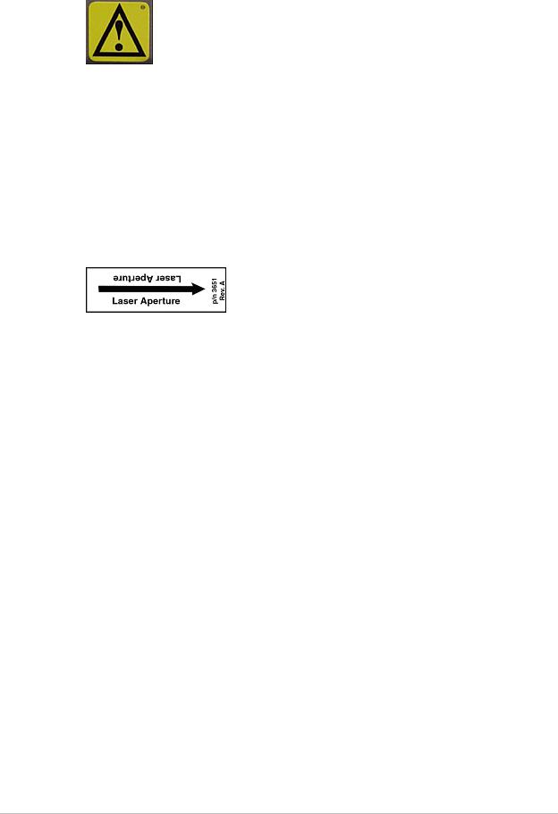
Laser Safety
DO NOT STARE INTO THE LASER BEAM during patient positioning and Quality Assurance procedures. This label is located under the scanner arm and shows the location of the laser aperture:
The laser aperture is located on the underside of the scanner arm, facing the patient. Keep the laser aperture away from the patient’s eyes during patient positioning.
Radiation Safety
X-ray exposure: The system makes radiation when electric voltage is supplied to, and current flows through, the x-ray tube. During a measurement, the shutter opens to let a beam of radiation pass through the scanner table and patient.
For iDXA systems, the nominal radiation field at the scanner table top is 18.4 mm x 3.3 mm.
For Prodigy systems, the nominal radiation field at the scanner table top is 19.5 mm x 3.4 mm.
For DPX systems, the nominal radiation field at the scanner table top is 2 mm.
Lead oxide shielding surrounds the x-ray tube insert inside the tube housing assembly and reduces radiation levels around the scanner table.
Leakage radiation: < 0.4 mR/hr at 1 meter.
28 |
X-ray Bone Densitometer with enCORE v17 software - User Manual |

Safety
Skin Entrance Dose
Refer to Current and Typical Dose Tables (307) for irradiation times and skin entrance doses. A Victoreen model 530 Precision Electrometer/Dosemeter with a Model 660-5 Ion Chamber was used to measure the X-ray entrance dose.
Mechanical Safety
The scanner arm moves down the entire length of the scanner table. Make sure the patient does not interfere with the movement of the scanner arm to prevent possible injury. In addition, make sure that there are no objects behind the scanner table that might obstruct movement of the scanner arm.
iDXA scanners: Weight applied to the scan table bed must not exceed 204 kg (450 lb).
DPX NT/MD+ scanners: Weight applied to the scan table bed must not exceed 136 kg (300 lb).
DPX Duo/Bravo and Prodigy scanners: Weight applied to the scan table bed or footstep (DPX Duo only) must not exceed 159 kg (350 lb).
Symbols
Symbol |
Name |
Description |
|
|
|
|
Electronic Instructions |
Symbol indicating that the Instructions for Use |
|
for Use |
are supplied in electronic form |
|
|
|
|
Emergency Stop |
Shows the location of the emergency stop |
|
|
button |
or |
|
|
|
|
|
|
Focal Point |
Symbol from EN60417-1, 5327 |
|
|
|
|
Functional Earth |
Shows location of Functional Earth terminal |
|
|
|
|
Laser On |
Shows the location of the Laser On indicator . |
|
|
|
|
Permanent Filtration |
Symbol from EN60417-1, 5381 |
|
|
|
29 |
X-ray Bone Densitometer with enCORE v17 software - User Manual |

Safety
Symbol |
Name |
Description |
|
|
|
|
Power Off |
Shows the switch position for Power Off |
|
|
|
|
Power On |
Shows the location of the Power On indicator |
|
|
and the switch position for Power On |
|
|
|
|
Protective Earth |
Shows location of Protective Earth terminal |
|
|
|
|
Refer to Instruction |
Alerts the user that the User Manual contains |
|
Manual |
important safety information |
|
|
|
|
Shutter Open |
Shows the location of the Shutter Open |
|
|
indicator |
|
|
|
|
Tube Insert |
Symbol from EN60417-1, 5337 |
|
|
|
|
Type B Equipment |
Shows that the scanner has Type B protection |
|
|
against electrical shock |
|
|
|
|
Warning |
Shows important safety warnings, such as the |
|
|
location of pinch points |
|
|
|
|
X-ray On |
Shows the location of the X-ray On indicator |
|
|
|
|
X-ray Source |
Symbol from EN60417-1, 5338 |
|
|
|
Sample Labels
Actual label appearance may vary from the samples displayed in this section.
For labels showing certification to 21 USCFR Subchapter J , the month in the Manufactured field is translated below.
English |
Translation |
English |
Translation |
|
|
|
|
|
January |
|
July |
|
|
|
|
|
February |
|
August |
|
|
|
|
30 |
X-ray Bone Densitometer with enCORE v17 software - User Manual |
Loading...