

Page 1
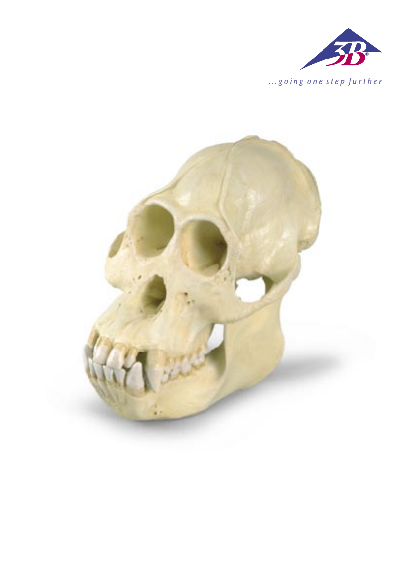
VP761/1
Page 2

1
EnglishOrang-Utan
Species Pongo pygmaeus (Hoppius)
Subfamily Ponginae Allen, 1925
Family Pongidae Elliot, 1913
Superfamily Hominoidea Simpson, 1931
Infraorder Catarrhina Hemprich, 1820
Suborder Simiae Haeckel, 1866
Order Primates Linnaeus, 1758
The skull of the mature, very large male orang-utan presents a very marked relief. The cerebral cranium
and the facial bones (viscerocranium) have a rough appearance and are marked with bumps, ridges, etc.,
which were formed in response to the requirements of the masticatory and neck musculature. They are in
no way comparable with those of the human.
In the orang-utan’s skull too, the disproportionate size of the face/jaw part (the facial bones, i.e. the
splanchnocranium or viscerocranium) in relation to the cerebral cranium is noticeable. This relationship,
however, only develops in the course of postnatal growth – particularly at the time of the second dentition.
On the sagittal suture, that is down the middle of the skull, a bony sagittal crest (crista sagittalis) develops,
becoming more pronounced towards the back. It is formed from the parietal bones and serves as the origin of the temporal muscle3, which increases in size as it approaches the crest. At the back of the head
(the occiput) the sagittal crest joins the occipital crest, which develops as the neck musculature becomes
stronger.
The occipital condyles of the head joint and the great occipital foramen they enclose (foramen occipitale
magnum) are located in the posterior region of the skull. Here too, the obvious contrast with the newborn
or infantile animal is evident.
The sexual dimorphism of the skull appears at first sight more pronounced in the orang-utan than in, for
example, chimpanzees, but less so than in the gorilla. As is the case with all primates that have been investigated in this respect, male orang-utans display on average a larger brain volume, larger and differently
shaped canine teeth, and a significantly more pronounced skull relief. In any case, all mature males, but
almost no mature females, have a sagittal crest. All adult animals develop occipital crests, due to their
“front-heavy” heads. In orang-utans, however, these crests are noticeably smaller than in the African
Ponginae.
2
®
Generally only supra-orbital arches develop. Related to this, there are no frontal sinuses. This development
takes place in connection with the formation of the permanent teeth and then with the wear of the teeth
with continuing abrasion from chewing4.
The upward branch of the lower jaw (ramus mandibulae) is relatively low. Typical of the Ponginae is the
more or less parallel arrangement of the premolar and molar teeth. In front of them are the incisor teeth.
The sexually differentiated, dagger-like canine teeth extend distinctly beyond the occlusion plane. For this
reason, in the upper jaw between the canine tooth and the first premolar, there is a gap, or diastema, into
which the lower canine tooth engages.
The anterior teeth engage one over the other like shears, which makes it easier to bite off a piece of food,
while the posterior teeth have broad, shallow crowns, suited to grinding; this constitutes-in the case of
the molars–what is known as the dryopithecine pattern, which is also displayed by humans. In the orangutan too, the enamel shows typical furrows on the occlusion plane next to the fissures. These are set close
together, and their arrangement is so characteristic that even a lay person can identify an orang-utan tooth
from them alone.
Since the pioneering studies of E. Selenka around 1900, orang-utans are no longer considered the closest
living relatives of humans.
Page 3

English Orang-Utan
Some dimensions of the original orang-utan skull5.
max. length of cranium (inc. occipital crest) 134 mm
max. breadth of cranium 131 mm
skull breadth in region of max. postorbital narrowing 65 mm
volume of cranial cavity = “brain size” 440 ccm
length of face 182 mm
breadth of upper face (external biorbital breadth) 107 mm
breadth of zygomatic arch 160 mm
max. separation of zygomatic arch from skull wall 45 mm
length of palate 94 mm
breadth of palate 41 mm
bicondylar breadth of mandible 100 mm
bigonial breadth of mandible 135 mm
height of corpus mandibulae 42 mm
ramus height of mandible 107 mm
ramus breadth 61 mm
total mass of skull 873 g
mass of cranium 551 g
mass of mandible 322 g
®
1
Author: Dr Dr Olav Röhrer-Ertl, Primates Section, SNSB, Munich
1
This model was cast from a replica of the original skull from the Senckenberg Research Institute and Natural History
Museum in Frankfurt/Main. For educational reasons the abraded teeth of the original were reconstructed following
younger male specimens in Munich, so as to be able to give a better representation of the tooth pattern. In this process,
some adaptations to the jaws had to be made.
2
The scientific name of the orang-utan has been under dispute for a good 40 years. This does not just concern the correct
attribution, as in the case of the chimpanzee, but also the specific name. Variant forms, known as synonyms, are found
in the literature.
3
Muscles cannot attach to one another, but require hard tissue for this purpose.
4
With increasing flattening of the tooth biting surfaces, the chewing force must be increased, which leads to increased
growth of the masticatory muscles, which in turn leads to more pronounced moulding of muscle attachment surfaces.
Here too, the distribution of the ever-increasing chewing force over the facial skeleton results in more pronounced
structures. Here we see the effect of the spatial relationship between the largest organ in the head, the brain, and the
others, particularly the eyes. In the African Ponginae this relationship is mainly horizontal (one behind the other),
whereas by contrast in the orang-utan they are arranged more vertically (one above the other).
5
All measurements were taken, from an original, by Dr sc. A. Windelband, Berlin. In general, model dimensions will vary
slightly from these.
Page 4

Orang-Utan
1
Deutsch
Spezies Pongo pygmaeus (Hoppius)
Unterfamilie Ponginae Allen, 1925
Familie Pongidae Elliot, 1913
Überfamilie Hominoidea Simpson, 1931
Teilordnung Catarrhina Hemprich, 1820
Unterordnung Simiae Haeckel, 1866
Ordnung Primates Linnaeus, 1758
Der Schädel des spät-erwachsenen (maturen) und recht großen, männlichen Orang-Utans zeigt ein kräftig
ausgeprägtes Schädelrelief. Dabei wirken Hirnschädel (Neurocranium) und Gesichtsschädel (Viscerocranium)
rauh und von Höckern, Leisten etc. besetzt, welche sich auf Anforderung der Kau- und Nackenmuskulatur
bildeten. Sie können in keinem Fall mit der des Menschen verglichen werden.
Auch beim Schädel (Cranium) des Orang-Utans fällt der übergroße Anteil der Gesichts-Kiefer-Partie (der
Gesichtsschädel bzw. das Splanchno- oder Viscerocranium) gegenüber dem Hirnschädel (Neurocranium)
auf. Dieses Verhältnis bildet sich aber erst im Verlauf des nachgeburtlichen (postnatalen) Wachstums heraus – insbesondere in der Zeit des Zahnwechsels.
Auf der Pfeilnaht (Sutura sagittalis), also auf der Schädelmitte erhebt sich der nach hinten verstärkende
knöcherne Scheitelkamm (Crista sagittalis). Er wird von den Scheitelbeinen (Ossa parietalia) aus gebildet
und dient als Ursprung des sich bis dorthin vergrößernden Schläfenmuskels (Musculus temporalis)
Hinterhaupt (occipital) trifft der Scheitelkamm (Crista sagittalis) auf den Nackenkamm (Crista occipitalis),
welcher sich im Zusammenhang mit der Verstärkung der Nackenmuskulatur ausbildet.
Die Höcker (Condyli occipitales) des Kopfgelenkes und das von ihnen umrahmte Hinterhauptsloch
(Foramen occipitale magnum) befinden sich im hinteren Schädelbereich. Auch hierin zeigt sich der augenfällige Unterschied zum neugeborenen oder kindlichen (infantilen) Tier.
Die geschlechtstypische Formausprägung (Geschlechtsdimorphismus) des Schädels erscheint beim OrangUtan auf den ersten Blick stärker ausgeprägt, als z.B. bei Schimpansen, aber geringer als bei Gorillas.
Wie bei allen dahingehend untersuchten Primates zeigen auch männliche Orang-Utans im Mittel höhere Hirnvolumina, größere und anders geformte Eckzähne (Canini) und deutlich stärker ausgeprägte
Schädelreliefs. Zumindest spät-erwachsene (mature) Männer verfügen immer, erwachsene Frauen aber so
gut wie nie über einen Scheitelkamm (Crista sagittalis). Nackenkämme (Cristae occipitales) bilden sich bei
allen erwachsenen Tieren aus und finden ihre Begründung in ihren „vorlastigen“ Köpfen. Bei Orang-Utans
aber fallen sie kleiner aus, als bei den afrikanischen Ponginae.
2
3
. Am
®
Es sind grundsätzlich lediglich Überaugenbögen (Arcus supraorbitales) ausgebildet. Im Zusammenhang
damit gibt es keine Stirnhöhlen (Sinus frontales). Diese Bildungen erfolgen im Zusammenhang mit
der Ausbildung des Dauergebisses (Dentes permanentes) und dann im Zusammenhang mit dem
Gebissgebrauch bei fortschreitender Abkauung (Abrasion)
Der aufsteigende Ast des Unterkiefers (Ramus mandibulae) ist relativ niedrig. Charakteristisch für das
Gebiss der Ponginae erscheint eine eher parallele Zahnanordnung der Vormahl- (Dentes praemolares) und
Mahlzähne (Dentes molares). Schneidezähne (Dentes incisivi) stehen dazu in Front. Die geschlechtstypisch
geformten, dolchförmigen Eckzähne überragen die Kauebene (Occlusionseben) deutlich. Von daher gibt
es im Oberkiefer (Maxilla) zwischen Eckzahn und 1. Praemolaren eine Lücke, das Diastema, in welches der
Eckzahn des Unterkiefers (Mandibula) bei Gebissschluss eingreift.
Die Zähne des Vordergebisses (Dentes anteriores) greifen scherenartig übereinander, was das Abbeißen
erleichtert, die des Hintergebisses (Dentes posteriores) zeigen breite, stumpfe Kronen, wie sie zum
Zermahlen günstig sind, dabei liegt – bei den Molares – das sog. Dryopithecinen-Muster vor, wie es auch
der Mensch zeigt. Auch beim Orang-Utan zeigt der Zahnschmelz (Enamelum) auf der Kauebene neben
4
.
Page 5

Deutsch
Orang-Utan
1
den Furchen (Fissurae) charakteristische Schmelzrunzeln. Diese erscheinen dichtgelagert und in ihrer
Anordnung so charakteristisch, dass jedermann einen Orang-Utan-Zahn allein danach sicher zuweisen
kann.
Orang-Utans gelten seit den hierfür bahnbrechenden Arbeiten E. Selenkas um 1900 nicht mehr als die
nächsten lebenden Verwandten des Menschen.
5
Einige Maße des originalen Orang-Utan-Schädels
.
größte Hirnschädellänge (mit Crista occipitalis) 134 mm
größte Hirnschädelbreite 131 mm
Schädelbreite im Bereich der stärksten postorbitalen Einschnürung 65 mm
Volumen der Schädelhöhle (Cavum cranii) = „Hirnvolumen“ 440 ccm
Gesichtslänge 182 mm
Obergesichtsbreite (äußere Biorbitalbreite) 107 mm
Jochbogenbreite 160 mm
Größter Abstand der Jochbögen zur Schädelwand 45 mm
Gaumenlänge 94 mm
Gaumenbreite 41 mm
Condylenbreite des Unterkiefers 100 mm
Winkelbreite des Unterkiefers 135 mm
Höhe des Corpus mandibulae 42 mm
Asthöhe des Unterkiefers 107 mm
Astbreite 61 mm
®
Gesamtmasse des Craniums 873 g
Masse des Calvariums 551 g
Masse der Mandibula 322 g
Verfasser: Dr. Dr. Olav Röhrer-Ertl, Sectio Primates der SNSB, München
1
Als Vorlage für den Abguss dieses Modells diente eine Nachbildung des Original-Schädels des Senckenberg Forschungs-
institutes und Naturmuseums in Frankfurt am Main. Aus didaktischen Gründen wurden die abgekauten (abradierten)
Zähne des Originals nach Originalbefunden jüngerer männlicher Tiere in München neu aufgebaut, um auch die
Zahnmuster besser darstellen zu können. Im Zusammenhang damit ergaben sich auch einige Kieferanpassungen.
2
Der wissenschaftliche Name – das Nomen – des Orang-Utan ist seit gut 40 Jahren in der Diskussion. Dabei geht es nicht,
wie bei dem Schimpansen, nur um den korrekten Autor, sondern auch um den Art-Namen, das Species-Nomen. In der
Literatur finden sich also auch von hier abweichende Formen, sog. Synonyma.
3
Muskeln können nicht aneinander ansetzen, sondern benötigen dafür biologisches Hartgewebe.
4
Bei zunehmender Einebnung der Zahn-Kauflächen muss der Kaudruck erhöht werden, was ein verstärktes Kaumuskel wachstum bewirkt, welches wiederum die Muskelansatzflächen bzw. –ursprünge verstärkt modelliert. Die Ableitung sich
ständig verstärkenden Kaudrucks über das Gesichtsskelett verstärkt auch hier die Strukturen. Wesentlich im Unterschied
afrikanischer Ponginae zum Orang-Utan wirkt sich hier aus, dass die Lagebeziehungen zwischen dem Gehirn als größtem
Kopforgan und den übrigen – insbesondere der Augen – eher horizontal (also hintereinander) und beim Orang-Utan
eher vertikal (also übereinander) angeordnet sind.
5
Alle Maße wurden durch Dr. sc. A. Windelband, Berlin an einem Original erhoben. Die Maße des Modells weichen in der
Regel davon geringfügig ab.
Page 6

Orangután
1
Español
Especie Pongo pygmaeus (Hoppius)
Subfamilia Ponginae Allen, 1925
Familia Pongidae Elliot, 1913
Superfamilia Hominoidea Simpson, 1931
Infraorden Catharrina Hemprich, 1820
Suborden Simiae Haeckel, 1866
Orden Primates Linnaeus, 1758
El cráneo del orangután macho adulto en edad avanzada (maturen), de complexión gruesa, presenta un
relieve craneal muy marcado. El cráneo cerebral (Neurocranium) y el cráneo facial (Viscerocranium) son
de aspecto bronco y están cubiertos de prominencias, molduras óseas etc. formadas por exigencia de las
musculaturas masticatoria y cervical. De ninguna manera pueden compararse con los del hombre.
Tambien en el cráneo (cranium) del orangután llama la atención la gran extensión que ocupa la parte
maxi-facial (cráneo facial/Splanchnocráneo o Viscerocranium) en comparación con el cráneo cerebral
(Neurocranium). Esta relación en las proporciones se configura en el transcurso del crecimiento postnatal
(postnatalen), sobre todo en el periodo de la segunda dentición.
Sobre la sutura sagital (Sutura sagittalis), es decir, en la mitad del cráneo, se alza una cresta ósea (Crista
sagittalis) que se refuerza hacia atrás. Se forma a partir del hueso parietal (Ossa parietalia) y sirve de base
al músculo temporal (Musculus temporalis)
cabeza (occipital) se encuentran la cresta sagital (Crista sagittalis) y la cresta occipital (Crista occipitalis), que
se desarrolla en relación con el fortale-cimiento de la musculatura cervical.
Las prominencias (Condyli occipitalis) de la articulación de la cabeza y el foramen occipital (Foramen occipitale magnum) rodeado por ellas, se encuentran en la zona posterior del cráneo. Igualmente se observa
en este punto la ostensiva diferencia con el animal recien nacido o con el de edad infantil (infantilen).
La típica forma del cráneo, determinada por el sexo (Dimorfismo sexual), se presenta en el orangután a
primera vista más acentuada que p.e. en los chimpancés, pero menos que en los gorilas. Como en todos los
primates estudiados, presentan tambien los orangutanes machos por término medio, un volumen craneal
más grande, dientes caninos (canini) de otra forma y un relieve craneal claramente más pronunciado. Por
lo menos poseen siempre los machos adultos en edad avanzada (mature) una cresta sagital, lo que las
hembras adultas poseen rara vez. Las crestas occipitales (Cristae occipitales) se forman en todos los animales adultos y tienen su razón de ser en sus “pesadas” cabezas . En los orangutanes son más pequeñas que
en los Ponginae africanos.
3
, que se agranda hasta dicho punto. En la parte posterior de la
2
®
En principio se han formado sólo arcos supraorbitales (Arcus supraorbitales). En conexión con esto, no existen los senos frontales (Sinus frontales). Estas configuraciones se forman en relación con el desarrollo de la
denticion permanente (dentes permanentes) y luego en conexión con el uso de la dentadura cuando hay
desgaste avanzado (Abrasion)
La rama ascendente de la mandíbula inferior (Ramus mandibulae) es relativamente baja. En la dentadura
de los Ponginae es característica la aparición de un orden más bien paralelo de los premolares (Dentes
praemolares) y molares (Dentes molares). Los incisivos (Dentes incisivi) están, por el contrario, de frente.
Los incisivos afilados , cuya forma típica proviene del dimorfismo sexual, dominan claramente la superficie
masticatoria (Occlusioneben). Por ello hay en la mandibula superior (Maxilla) entre el canino y el primer
premolar un hueco llamado diastema,en el cual encaja el canino de la mandibula inferior (Mandibula) al
cerrar la dentadura.
Los dientes anteriores (Dentes anteriores) encajan unos sobre otros como unas tijeras, lo que simplifica la
acción de morder; los de la dentadura posterior (Dentes posteriores) presentan coronas anchas y romas,
apropiadas para la masticación, sin olvidar que los molares presentan la llamada “muestra Dryopithecina”,
la misma que tambien presenta el hombre. Igualmente en el orangután presenta el esmalte (Enamelum)
4
.
Page 7

Español
Orangután
en la superficie masticatoria, aparte de surcos (Fissurae), arrugas características, que aparecen muy densas,
y es su ordenación tan característica, que cualquiera puede identificar un diente de orangután por ellas.
A partir de los trabajos revolucionarios sobre el tema de E. Selenkas alrededor de 1900, ya no se pueden
consideran los orangutanes como los más próximos parientes vivos del hombre.
5
Algunas medidas del cráneo original del orangután
:
Largo máximo del cráneo cerebral (con Crista occipitalis) 134 mm
Ancho máximo del cráneo cerebral 131 mm
Ancho del cráneo en la zona de máximo estrechamiento postorbital 65 mm
Volumen de la cavidad craneal (Cavum cranii)=Volumen cerebral 440 ccm
Largo facial 182 mm
Ancho facial superior (Ancho biorbital máximo ) 107 mm
Ancho del arco zigomático 160 mm
Distancia máxima entre los arcos zigomáticos y la pared craneana 45 mm
Largo del paladar 94 mm
Ancho del paladar 41 mm
Ancho condilar de la mandibula inferior 100 mm
Ancho del ángulo de la mandíbula inferior 135 mm
Altura del “Corpus mandibulae” 42 mm
Altura de la rama de la mandíbula inferior 107 mm
Ancho de la rama 61 mm
®
Masa total del Cranium 873 gr.
Masa del Calvarium 551 gr.
Masa de las mandíbulas 322 gr.
1
Autor: Dr. Dr. Olav Röhrer-Ertl. Sectio Primates del SNSB, Munich
1
Como molde para el vaciado de este modelo se empleó de muestra una réplica de un cráneo original del Instituto de
Investigaciones y Museo de Historia Natural Senckenberg de Frankfurt sobre el Meno. Por motivos didácticos se sustituyeron los dientes ya raspados del original por otros hechos en Munich según hallazgos originales de animales machos
más jovenes, tambien para poder exponer mejor la muestra dental. Por esta causa, hubo tambien algunas adaptaciones
de las mandíbulas
2
El nombre científico- Nomen – del orangután se viene discutiendo desde hace más de 40 años. Aquí no se trata solo,
como en el chimpancé,del autor correcto, sino tambien del nombre del género, del nombre de la especie. En la
Literatura se encuentran tambien otros que divergen de los de aquí, los llamados Synonyma.
3
Los músculos no pueden unirse unos a otros sino que necesitan para ello un estratificado de tejido biológico.
4
Cuando crece el aplanamiento de la superficie masticatoria de los dientes tiene por fuerza que aumentar la presión
masticatoria, lo que trae como consecuencia un crecimiento más fuerte de la musculatura correspondiente, y ésto a su
vez, refuerza el modelaje de las superficies de inserción/puntos de partida musculares. La presion mascatoria cada vez
más fuerte ejercida sobre el esqueleto de la región facial, refuerza por tanto tambien las estructuras. La diferencia entre
los Ponginae africanos con el Orangután se traduce aquí en que la relación de la situación que se da entre el cerebro,
como mas grande órgano de la cabeza y los demás (sobre todo los ojos), es más bien horizontal, es decir, uno tras otro, y
en el orangután más bien vertical, es decir, uno sobre otro.
5
Todas las medidas fueron tomadas de un original por el Dr. sc. A. Windelband, Berlín. Las medidas del modelo de
Page 8

Orang-oután
1
Français
Species Pongo pygmaeus (Hoppius)
Subfamilia Ponginae Allen, 1925
Familia Pongidae Elliot, 1913
Superfamilia Hominoidea Simpson, 1931
Infraordo Catarrhina Hemprich, 1820
Subordo Simiae Haeckel, 1866
Ordo Primates Linnaeus, 1758
Le crâne de l’orang-outan mâle mature de très grande taille présente un relief crânien très prononcé. Le
crâne cérébral (Neurocranium) et le crâne viscéral (Viscerocranium) ont un aspect rugueux et sont parsemés de bosses, de replis, etc. qui se sont formés en fonction du muscle masticateur et de la musculature de
la nuque. Ils ne peuvent en aucun cas être comparés à ceux de l’homme.
Egalement au niveau du crâne (Cranium) de l’orang-outan, la partie du visage/des mâchoires de taille
imposante (le crâne viscéral, respectivement le Splanchnocranium ou Viscerocranium) par rapport au crâne
cérébral (Neurocranium), est frappante. Ce phénomène ne se forme cependant qu’au cours de la croissance
postnatale – en particulier durant la phase de l’échange des dents.
La crête sagittale externe (Crista sagittalis) osseuse se développant vers l’arrière s’érige sur la suture sagittale (Sutura sagittalis), c’est-à-dire au centre du crâne. Elle est formée à partir des os pariétaux (Ossa parietalia) et sert d’origine au muscle temporal (Musculus temporalis)
(Crista occipitalis) rencontre la crête occipitale (Crista occipitalis) au niveau de l’occiput (occipital), la crête
occipitale se formant avec l’amplification de la musculature de la nuque.
Les tubérosités occipitales (Condyli occipitales) de l’articulation atlanto-occipitale et le trou occipital
(Foramen occipitale magnum) entouré par celles-ci sont situés dans la région occipitale du crâne. Ici également, on remarque la différence frappante par rapport à l’animal nouveau-né ou infantile.
La prononciation de la forme typique du crâne en fonction du sexe (dimorphisme sexuel) semble de prime
abord plus fortement marquée chez l’orang-outan que chez les chimpanzés, mais moins marquée que
chez les gorilles. Comme chez tous les primates examinés, les orangs-outans mâles montrent également en
moyenne un volume du cerveau plus élevé, des canines plus grandes et de forme différente et un relief du
crâne considérablement plus prononcé. Les hommes matures du moins présentent toujours, et les femmes
matures pratiquement toujours, une crête sagittale (Crista sagittalis). Les crêtes occipitales (Cristae occipitales) se forment chez tous les animaux adultes et trouvent leur justification dans leur tête „portant la charge
à l’avant“. Chez les orangs-outans, celles-ci sont plus petites que chez les Ponginae africains.
2
3
s’agrandissant jusque-là. La crête sagittale
®
En principe, des bourrelets supra-orbitaires (Tori supraorbitales) sont formés qui sont reliés au milieu
(médian) par un bourrelet glabellaire (Torus glabellaris) en un bourrelet frontal (Torus frontalis) uniforme.
En rapport avec ceci, leurs sinus frontaux (Sinus frontales) sont très prononcés. Cette formation se produit
en relation avec la formation des dents permanentes (Dentes permanentes) et ensuite en relation avec
l’abrasion
Le rameau ascendant de la mandibule (Ramus mandibulae) est relativement bas. Une disposition plutôt
parallèle des prémolaires (Dentes praemolares) et des molaires (Dentes molares) semble être caractéristique de la dentition des Ponginae. Les incisives (Dentes incisivi) occupent une position frontale. Les troisièmes incisives en forme de poignard et spécifiques au sexe dépassent considérablement le niveau de la
mastication (niveau de l’occlusion). Par conséquent, il existe une lacune dans le maxillaire (Maxilla) entre
l’incisive et les 1ères prémolaires, le diastème, dans lequel l’incisive de la mandibule (Mandibula) intervient dans l’occlusion des dents.
Les dents antérieures (Dentes anteriores) s’enchevêtrent les unes sur les autres comme des ciseaux, ce qui
facilite la coupure ; les dents postérieures (Dentes posteriores) présentent de larges couronnes émoussées
4
.
Page 9

Français
Orang-oután
comme elles sont nécessaires pour le broyage. En ce qui concerne les molaires, on observe l’échantillon
Ddryopithecinen, comme on le rencontre également chez l’homme. Chez le gorille également, l’émail
(Enamelum) présente en plus des fissures (Fissurae) quelques arêtes, en l’occurrence caractéristiques, sur la
surface de mastication.
Depuis les travaux révolutionnaires de E. Selenkas aux alentours de 1900, les orangs-outans ne sont plus
considérés comme les parents vivants les plus proches de l’homme.
5
Quelques dimensions du crâne original de l’orang-outan
.
Longueur maximale du crâne cérébral (avec Crista occipitalis) 134 mm
Largeur maximale du crâne cérébral 131 mm
Largeur du crâne dans la région de la constriction post-orbitaire la plus forte 65 mm
Volume de la cavité crânienne (Cavum cranii) = „volume du cerveau“ 440 ccm
Longueur du visage 182 mm
Largeur du visage supérieur (largeur biorbitaire externe) 107 mm
Largeur de l’arcade zygomatique 160 mm
Distance maximale des arcades zygomatiques jusqu’à la paroi crânienne 45 mm
Longueur du palais 94 mm
Largeur du palais 41 mm
Largeur du condyle de la mandibule 100 mm
Largeur de l’angle de la mandibule 135 mm
Hauteur du corps mandibulaire 42 mm
Hauteur du rameau de la mandibule 107 mm
®
Largeur du rameau 61 mm
Masse totale du crâne 873 g
Masse de la voûte crânienne 551 g
Masse de la mandibule 322 g
1
Auteur : Dr Dr Olav Röhrer-Ertl, section primates de la SNSB, Munich
1
Le moulage de ce modèle a été tiré d’une reproduction du crâne original de l’Institut de Recherche de Senckenberg et
Musée d’Histoire naturelle de Francfort-sur-le-Main. Pour des raisons didactiques, les dents abrasées de l’original ont
été reconstituées à Munich selon les résultats originaux de jeunes animaux mâles afin de mieux pouvoir représenter la
dentition. Dans ce contexte, quelques adaptations de la mâchoire ont également été effectuées.
2
Le nom scientifique de l’orang-outan est discuté depuis une bonne quarantaine d’années. Ce faisant, il ne s’agit pas,
comme pour le chimpanzé, uniquement de l’auteur correct, mais également du nom de l’espèce. Dans la littérature, on
rencontre par conséquent des variantes, c’est-à-dire des synonymes.
3
Les muscles ne peuvent pas s’insérer les uns dans les autres, mais nécessitent pour ce faire du tissu biologique dur.
4
Lors du nivellement croissant des surfaces de mastication des dents, la pression de mastication doit être augmentée,
ce qui provoque une croissance potentialisée du muscle masticateur qui à son tour modèle les surfaces d’insertion et les
origines du muscle de façon potentialisée. La dérivation de la pression de mastication se renforçant constamment sur le
squelette du visage renforce ici également les structures. La différence considérable des Ponginae africains par rapport à
l’orang-outan se traduit en l’occurrence par le fait que les relations de présentation entre le cerveau en tant qu’organe
de la tête le plus grand et les autres organes – en particulier les yeux – sont disposées horizontalement (par conséquent
les unes après les autres) et chez l’orang-outan plutôt verticalement (par conséquent les unes sur les autres).
5
Toutes les mesures ont été relevées sur un original par le Dr sc A. Windelband, Berlin. Les mesures du modèle ne
présentent en général que de faibles écarts.
Page 10

Orangotango
1
Português
Espécie Pongo pygmaeus (Hoppius)
Subfamília Ponginae Allen, 1925
Família Pongidae Elliot, 1913
Superfamília Hominoidea Simpson, 1931
Infraordem Catarrhina Hemprich, 1820
Subordem Simiae Haeckel, 1866
Ordem Primates Linnaeus, 1758
O crânio do orangotango macho adulto tardio (maduro), e bastante grande, apresenta um relevo craniano
fortemente marcado. Incluindo a forte esquama frontal (Torus frontalis), o crânio neural (Neurocranium)
e o crânio visceral (Viscerocranium) têm aparência rude e estão cobertos de protuberâncias, bordas, etc.,
que são criadas por necessidades da musculatura de mastigação e do pescoço. Em nenhum caso podem ser
comparados com os dos humanos.
Também, no caso do crânio (Cranium) do orangotango, é notória a maior importância das partes faciais e
mandibulares (crânio facial ou Viscerocranium) em relação ao crânio neural (Neurocranium). Esta relação
só vem a se estabelecer durante a fase de crescimento pós-natal, e em particular, durante a fase de substituição dental.
Sobre a sutura sagital (Sutura sagittalis), ou seja, no meio do crânio, eleva-se a crista sagital (Crista sagittalis) a qual apresenta um reforço ósseo para trás. Ela se forma a partir dos ossos parietais (Ossa parietalia)
e serve de origem para músculo temporal (Musculus temporalis)3 que ali aumenta de tamanho. Na parte
occipital da cabeça, a crista sagital (Crista sagittalis) encontra-se com a crista occipital (Crista occipitalis), a
qual se desenvolve proporcionalmente ao desenvolvimento da musculatura do pescoço.
As protuberâncias (Condyli occipitales) da articulação da cabeça e o forame occipital craniano (Foramen
occipitale magnum) por elas enquadrado encontram-se na parte posterior do crânio. Também neste caso
observa-se uma clara diferença em comparação ao animal recém-nascido ou infantil.
As características específicas de gênero sexual (dimorfismo sexual) no crânio aparecem a primeira vista de
forma mais nítida no orangotango do que no chimpanzé, mas menos nítida do que no gorila. Como em
todos os primatas estudados, os orangotangos machos também apresentam um volume cerebral superior
na parte mediana, caninos maiores de forma diferente, assim como um relevo craniano nitidamente mais
forte. Pelo menos os adultos tardios (maduros) machos, e as fêmeas adultas tardias freqüentemente também, sempre apresentam uma forte crista sagital (Crista sagittalis). A crista occipital se desenvolve em todos
os exemplares adultos e sua presença explica-se pelo maior volume das suas cabeças na parte frontal. No
orangotango, estas são menores do que no Ponginae africano.
2
®
De um modo geral encontram-se arcadas supraorbitais (Arcus supraorbitales) desenvolvidas. Em função
disto, não se encontram senos frontais (Sinus frontales). Esta formação ocorre em associação com o desenvolvimento da dentição definitiva (Dentes permanentes) e logo em relação à utilização da mandíbula e ao
subseqüente avanço do desgaste dos dentes (abrasão)4.
O ramo ascendente da mandíbula (Ramus mandibulae) é relativamente baixo. A ordem mais bem paralela
dos dentes molares e pré-molares encontrada é característica da dentadura dos Ponginae. Os dentes incisivos encontram-se à frente em relação àqueles. Os dentes caninos em forma de punhal, típicos do gênero,
ultrapassam claramente a linha de oclusão. Por isso existe na maxila superior uma falha entre o canino e o
primeiro pré-molar, o diastema, no qual se insere o canino da mandíbula quando a dentadura é fechada.
Os dentes da dentadura anterior fecham-se como tesouras uns sobre os outros, o que favorece o corte no
ato de morder. Os dentes da dentadura posterior apresentam coroas amplas e arredondadas, de modo
a favorecer a mastigação, sendo que no caso dos molares encontra-se uma estrutura do tipo da do driopiteco, como é também é encontrada no homem. Também no orangotango, o esmalte dental (Enamelum)
Page 11

Português
apresenta na linha de oclusão, ao lado das fissuras (Fissurae), as características ranhuras no esmalte. Estas
últimas são ordenadas estreitamente e de modo tão característico, que qualquer um pode identificar um
dente de orangotango de forma segura a partir delas.
Os orangotangos já não são mais considerados os parentes vivos mais próximos do homem desde as pioneiras pesquisas de E. Selenkas por volta de 1900.
Algumas medidas do crânio original de
Comprimento máximo do crânio neural (com crista occipital) 134 mm
Largura máxima do crânio neural 131 mm
Largura máxima do crânio na região da constrição pós-orbital maior 65 mm
Volume da cavidade craniana (Cavum cranii) = „volume do cérebro“ 440 ccm
Comprimento da face 182 mm
Largura da parte superior da face (largura biorbital externa) 107 mm
Largura do arco zigomático 160 mm
Maior distância dos arcos zigomáticos à parede craniana 45 mm
Comprimento do paladar 94 mm
Largura do paladar 41 mm
Largura condilar da mandíbula 100 mm
Largura angular da mandíbula 135 mm
Altura do corpo mandibular (Corpus mandibulae) 42 mm
Altura do ramo da mandíbula 107 mm
Largura do ramo 61 mm
Massa total do crânio 873 g
Massa da caixa craniana 551 g
Massa da mandíbula 322 g
orangotango5.
Orangotango
®
1
Autor: Dr. Dr. Olav Röhrer-Ertl, Sectio Primates da SNSB, Munique
1
Como matriz para a moldagem deste modelo foi utilizada uma reprodução do crânio original do Instituto de Pesquisa e
Museu da Natureza Senckenberg, em Frankfurt am Main. Por razões didáticas, os dentes desgastados (abrasados) do original foram reconstituídos a partir de originais de animais jovens encontrados em Munique, de modo a também poder
representar melhor estrutura dental. Em função disto, foram efetuadas várias adaptações na mandíbula.
2
O nome científico (o Nomem) do orangotango é sujeito a discussão já fazem 40 uns anos. Neste caso, não se trata
somente de saber qual o autor correto, como no caso do chimpanzé, mas também do nome da espécie em si. Na
literatura também encontram-se nomes divergentes, ou seja, sinônimos.
3
Os músculos não podem inserir-se uns nos outros, para tal, necessitam de tecidos biológicos duros.
4
Com o aumento do aplainamento da superfície de mastigação dos dentes a pressão mastigatória deve ser aumentada,
o que provoca um aumento da musculatura de mastigação, o que por sua vez intensifica a modelação dos pontos
de inserção ou as origens dos músculos. O desvio provocado na face do esqueleto por causa da pressão de mastigação
em constante aumento reforça aqui também as estruturas. Uma diferença fundamental entre o Ponginae africano e
o orangotango caracteriza-se aqui pelo fato que a posição relativa do cérebro como maior órgão da cabeça aos outros
órgãos, em particular aos olhos, obedece a uma ordem mais bem horizontal (ou seja, um atrás do outro), sendo que no
caso do orangotango, esta é mais bem vertical (ou seja, um acima do outro).
5
Todas as medidas foram levantadas de um original pelo Dr. sc. A. Windelband, Berlin. As medidas do modelo divergem
em geral ligeiramente do original.
Page 12

Orangutan
1
Italiano
Specie Pongo pygmaeus (Hoppius)
Sottofamiglia Ponginae Allen, 1925
Famiglia Pongidae Elliot, 1913
Superfamiglia Hominoidea Simpson, 1931
Infraordine Catarrhina Hemprich, 1820
Sottordine Simiae Haeckel, 1866
Ordine Primates Linnaeus, 1758
Il cranio di questo orangutan maschio, piuttosto grande e di età matura, mostra un rilievo craniale fortemente pronunciato. Il neurocranio e il viscerocranio hanno un aspetto scabro e ornato di gibbosità, sporgenze, ecc. originate dalle esigenze poste dalla muscolatura masticatoria e nucale. Tali caratteristiche non
sono comparabili con quelle umane.
Anche nel cranio dell’orangutan colpiscono le dimensioni sproporzionate della sezione viso-mascellare (del
viscerocranio ovvero del viscerocranio e dello splancnocranio) rispetto al neurocranio. Tale relazione tuttavia si sviluppa solo nel corso della crescita postnatale, in particolare nel periodo di cambio della dentatura.
Sulla sutura sagittale, ed anche al centro del cranio, si erge la cresta sagittale ossea, che aumenta di volume sul retro. Si forma a partire dalle ossa parietali ed è all’origine del muscolo temporale, che aumenta
il suo volume fino a quel punto3. Sull’occipite la cresta sagittale incontra la cresta occipitale, la quale si
sviluppa in concomitanza al rafforzamento della muscolatura nucale.
I condili occipitali dell’articolazione del capo che circondano il foro occipitale grande si trovano nell’area
posteriore del cranio. Anche qui appare evidente la differenza rispetto all’animale neonato o infantile.
Il dimorfismo sessuale del cranio nell’orangutan appare al primo sguardo più fortemente pronunciato
che per esempio negli scimpanzé, ma meno che nei gorilla. Come tutti gli altri primati analizzati, anche
gli orangutan maschi al centro dei volumi cerebrali superiori, denti canini più grandi e di forma diversa e
rilievi cranici notevolmente più pronunciati. I maschi di età matura presentano sempre la cresta sagittale,
mentre le femmine adulte quasi mai. Le creste occipitali si formano in tutti gli animali adulti e trovano la
loro ragione d’essere nella prominenza anteriore del capo. Negli orangutan sono tuttavia più piccoli che
nei Pongidi africani.
2
®
In linea di massima solo le arcate sopraorbitali sono sviluppate. Di conseguenza non ci sono seni frontali.
Queste formazioni nascono parallelamente allo sviluppo della dentatura permanente e proseguono poi
con l’uso della dentatura in conseguenza della progressiva abrasione dei denti4.
Il ramo ascendente della mandibola (ramo mandibolare) è relativamente basso. Caratteristico della dentatura dei Pongidi appare l’allineamento piuttosto parallelo dei denti premolari e molari. I denti incisivi
invece sono in posizione frontale. I denti canini a forma di pugnale, tipici di questa famiglia, sporgono chiaramente rispetto al piano di masticazione o occlusione. Per tale ragione nella mascella tra dente canino
e primo premolare c’è un vuoto, il diastema, nel quale a dentatura chiusa si inserisce il dente canino della
mandibola.
I denti anteriori si chiudono a forma di forbice, cosicché il morso risulta facilitato; i denti posteriori
mostrano delle corone larghe e smussate, idonee alla triturazione. I molari sono caratterizzati dal cosiddet-
Page 13

Italiano
Orangutan
to modello driopitecineo, come nell’uomo. Anche nell’orangutan lo smalto dentario mostra sulla superficie
di masticazione, accanto ai solchi, le tipiche grinze. Queste appaiono così compatte e con una disposizione
così caratteristica che qualsiasi persona sarebbe in grado di riconoscere un dente di orangutan.
A partire dal lavoro pionieristico di E. Selenkas attorno al 1900 gli orangutan non sono più considerati
come i parenti viventi più prossimi dell’uomo.
5
Qui di seguito indichiamo alcune misure del cranio originale dell’orangutan
.
Massima lunghezza del neurocranio (con cresta occipitale) 134 mm
Massima larghezza del neurocranio 131 mm
Larghezza del cranio nell’area del restringimento postorbitale maggiore 65 mm
Volume della cavità cranica = „volume del cervello“ 440 ccm
Lunghezza del viso 182 mm
Larghezza del viso superiore (larghezza biorbitale esterna) 107 mm
Larghezza dell’arcata zigomatica 160 mm
Massima distanza delle arcate zigomatiche rispetto alla parete cranica 45 mm
Lunghezza del palato 94 mm
Larghezza del palato 41 mm
Larghezza condilare della mascella inferiore 100 mm
Larghezza angolare della mascella inferiore 135 mm
Altezza del corpo mandibolare 42 mm
Altezza del ramo della mascella inferiore 107 mm
Larghezza del ramo 61 mm
®
Massa totale del cranio 873 g
Massa del cranio privo di mandibola 551 g
Massa della mandibola 322 g
1
Autore: Dr. Dr. Olav Röhrer-Ertl, Sezione Primati di SNSB, Monaco di Baviera
1
Como progetto per la colta di questo modello è stato necessario disporre di una riproduzione del cranio originale del
Istituto di ricerca e Museo di storia naturale Senckenberg di Francoforte sul Meno. Per ragioni didattiche i denti
dell’originale, che erano consumati, sono stati ricostruiti a Monaco di Baviera sulla base della dentatura di reperti
originali di giovani animali maschi, allo scopo di illustrare meglio il campione di dentatura. La ricostruzione ha reso
necessari anche degli adattamenti della mascella.
2
Il nome scientifico dell’orangutan viene discusso da oltre 40 anni. Non si tratta di chiarire solo quale sia l’autore
corretto, ma anche il nome della specie. Nella letteratura scientifica quindi si trovano forme diverse, cosiddetti sinomini.
3
I muscoli non si possono accostare gli uni agli altri, ma hanno bisogno di un tessuto-resina biologico.
4
Al progressivo livellamento delle superfici masticatorie dei denti consegue un aumento della pressione di masticazione,
il che provoca una crescita maggiore dei muscoli masticatori, che a sua volta produce un modellamento maggiore delle
superfici di inserzione e delle origini dei muscoli. La deviazione progressiva della pressione di masticazione rafforza
anche le strutture dello scheletro del viso. La differenza sostanziale tra Pongidi africani rispetto all’orangutan è che
nei primi il rapporto di localizzazione tra il cervello come organo maggiore del capo e il resto, in particolare gli occhi, è
piuttosto orizzontale (quindi uno dietro l’altro) mentre nell’orangutan è piuttosto verticale (quindi uno sopra l’altro).
5
Tutte le misure sono state rilevate da un originale dal Dr. sc. A. Windelband di Berlino. Le misure del modello sono di
solito leggermente diverse.
Page 14

オランウータン
種 ()
亜科 オランウータン亜科(
科 ショウジョウ科(
上科 ヒト上科(
下目 狭鼻猿類(
亜目 真猿亜目(
目 霊長目(
本模型は成熟したかなり大きいサイズの雄チンパンジーの頭骨模型であり,頭骨の隆起もかなり大きい。ヒト
の頭骨とは異なり,脳頭蓋と顔面骨(内臓頭蓋)の表面には凹凸が多く,咀嚼筋,頚筋の付着部の突起や隆起
などがよく発達している。これは咀嚼筋や頚部の筋肉の発達によるものである。
オランウータンの頭骨でもチンパンジーなどと同様に,顎顔面(顔面骨,例えば顔面頭蓋もしくは内臓頭蓋)
のサイズは脳頭蓋のサイズに対して不釣合いなくらい大きい。この特徴は生後の成長過程,とくに永久歯の発
生時期に獲得される。
頭頂骨の中央にある矢状縫合に沿って形成されている矢状稜は,後頭部の方への突出が目立つ。矢状稜は頭頂
骨から形成され,側頭筋の起始としての役割を担っている。矢状稜は,後頭部で後頭稜とつながっており,頚
部の筋が強くなるほど発達する。
この模型の後頭顆と大後頭孔は頭骨の後方にあるため,新生児期や乳幼児期のオランウータンではないことが
わかる。
オランウータンの頭骨の性差はチンパンジーのものに比べれば大きいが,ゴリラのものよりは小さい。しかし
他の霊長目の動物と同様に,雄オランウータンは雌オランウータンより頭蓋容量が大きく,犬歯も大きく形が
異なっており,頭骨の隆起もより発達している。矢状稜は,成熟した雄のオランウータンでは必ず見られる
が,雌のオランウータンではほとんど見られない。成熟したオランウータンは前頭部が重いため後頭稜が発達
している。しかし,オランウータンの後頭稜はチンパンジーと比べると明らかに小さい。
)
)
)
)
)
)
日本語
一般的にオランウータンは,永久歯の形成や成長時の咀嚼による歯の磨耗とも関連して眼窩上隆起は発達して
いるが,前頭洞は有さない。
オランウータンの下顎枝は比較的低い。前臼歯と臼歯が平行に近い状態で並んでいるのはオランウータン亜科
の特徴であり,それらの前には上顎側切歯がある。性差異のある鋭くとがった犬歯は,咬合平面を明らかに超
えて伸びている。このため,上顎の犬歯と第一前臼歯の間には,犬歯を収めるための空隙もしくは正中離開が
ある。
前歯は,はさみのように重なり合っており,食物を噛み切りやすくなっている。反対に奥歯は幅が広く歯冠は
浅いため,細かく噛み砕くのに適している。この臼歯はいわゆるドリオピテクス型のものとして知られている
もので,ヒトにも同様に見られる。またオランウータンの咬合面でもエナメル質の溝が見られる。オランウー
タンの歯の並びは密で,特徴的なため,専門家でなくても一見しただけでオランウータンの歯だと容易に判別
できるほどである。
オランウータンがヒトと最も近縁な種であるとされていたときもあったが,
草分け的研究以来,チンパンジーがヒトに最も近い種とされている。
年頃からの による
Page 15

日本語
実物標本の計測値
オランウータン
脳頭蓋最大長(後頭稜を含む)
脳頭蓋最大幅
眼窩後部幅が最大の点における頭骨幅
頭蓋腔容量=「脳の大きさ」
顔長
上顔幅(外眼窩顔幅)
頬骨弓幅
頬骨弓と側頭壁の最大距離
口蓋長
口蓋幅
下顎関節突起幅
下顎角幅
下顎体高
下顎枝高
下顎枝幅
頭骨総重量
脳頭蓋重量(下顎骨を除く頭骨の重量)
下顎骨重量
筆者
このモデルは,フランクフルトのゼンケンベルグ自然史研究博物館が所有する標本のレプリカを原形としている。元となった標
本の歯は磨り減っていたため,より良い状態で見られるよう
に数箇所修正を加えた。
オランウータンの学名については年もの間議論が続いている。これは分類学上の正確な位置づけが決まらないというチンパン
ジーと同様の問題に加え,種小名さえ確定していないためである。シノニム(同物異名)として知られている異型のオランウー
タンがいくつか文献には記されている。
筋だけではお互いに付着できないので,硬組織が必要とされる。
歯の咬合面が平らになるほど,咀嚼力は強化される。それが咀嚼筋の発達を促し,結果,筋付着表面の構造を変化させる。この
標本の顔面骨格においても,咀嚼力が増加し続けた結果とおもわれる構造上の特徴が明確にあらわれている。その効果を脳とそ
の他の器官,特に眼との位置関係で見ることができる。チンパンジーでは,脳と眼は横にならんでおり(脳は眼の後ろ),反対
にオラウータンではより縦の位置関係にある(脳は眼の上)。
が所有する若いオスの標本を参考に歯を復元,この過程で顎
Page 16
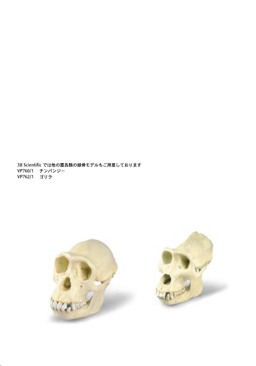
The following Primate skulls are also available from 3B Scientific:
VP760/1 Chimpanzee
VP762/1 Gorilla
Bei 3B Scientific erhalten Sie auch die folgenden Schädel von Primaten:
VP760/1 Schimpanse
VP762/1 Gorilla
En 3B Scientific consigue también los cráneos de primates siguientes:
VP760/1 Chimpancé
VP762/1 Gorila
A 3B Scientific, vous pouvez aussi commander les crânes de primates suivants:
VP760/1 Chimpanzé
VP762/1 Gorille
Na 3B Scientific você também poderá obter os seguintes crâneos de primatas:
VP760/1 Chimpanzé
VP762/1 Gorila
I seguenti crani primati sono disponibili anche presso 3B Scientific:
VP760/1 Scimpanzè
VP762/1 Gorilla
VP760/1
VP762/1
Page 17

Page 18

Page 19

Page 20

VP761/1-07/05-2
®
3B SCIENTIFIC
3B Scientific GmbH
Rudorffweg 8 • 21031 Hamburg • Germany
Tel.: + 49-40-73966-0 • Fax: + 49-40-73966-100
www.3bscientific.com • 3b@3bscientific.com
© Copyright 2005 for instruction manual and design of product:
3B Scientific GmbH, Germany
PRODUC TS
 Loading...