

Page 1
For over 130 years, Toshiba has
led the world in developing technology to improve the quality of life. This Made for Life
™
commitment is reflected
in our family of leading-edge imaging systems for MRI, CT, ultrasound, cath labs, X-ray and nuclear medicine.
From creating our first X-ray tube in 1915 to introducing the first Dynamic Volume CT Scanner in 2007, Toshiba
continues to build upon our legacy with technological innovation that improves patient care while providing lasting
quality for a lifetime of value.
INNOVATION BY DESIGN
12" × 12"/12" × 12" Flat Panel Detector
©Toshiba Medical Systems Corporation 2009 all rights reserved.
Design and specifications subject to change without notice.
MCAXR0197EA 2009-06 TME/D/D
http://www.toshibamedicalsystems.com
Printed in Japan
Toshiba Medical Systems Corporation meets internationally recognized
standards for Quality Management System ISO 9001, ISO 13485.
Toshiba Medical Systems Corporation Nasu Operations meets the
Environmental Management System standard, ISO 14001.
Made for Life and Infinix are trademarks of Toshiba Medical Systems
Corporation.
VF-i/BP
- Type S
-
INFX-8000V
nel D
X-80V
Toshiba — A History
of Leadership
1875 • Founding of Toshiba
1915 • First X-ray Tube
1973 • First Real-time Ultrasound Scanner
1989 • First Helical CT Scanner
1990 • First Tissue Doppler Imaging System
1993 • First One-million-pixel CCD
1997 • First Open, Superconducting Magnet
2000 • First All-digital Multipurpose X-ray System
2003 • First 64-slice CT Scanner
2005 • First Compact Dual Plane Cath Lab with
Flat Panel Detectors
2007 • First Dynamic Volume CT Scanner
Page 2
Major improvements in image quality, patient access
and ease of use
Comprehensive 12" x 12" biplane imaging without
compromising patient access
High-resolution, flat panel images with uniform
brightness and no distortion
Quiet instant-on, liquid-metal bearing X-ray tube for
efficient exams
Unique lateral arm adjustment to quickly optimize
imaging angles
Valuable dose-saving features:
- Various pulse rate controlled with grid
- X-ray beam filtration
- Variable frame rates in fluoroscopy and digital angiography
- Last image hold with virtual collimation
Major DICOM service classes included, which provide open access
to patient information
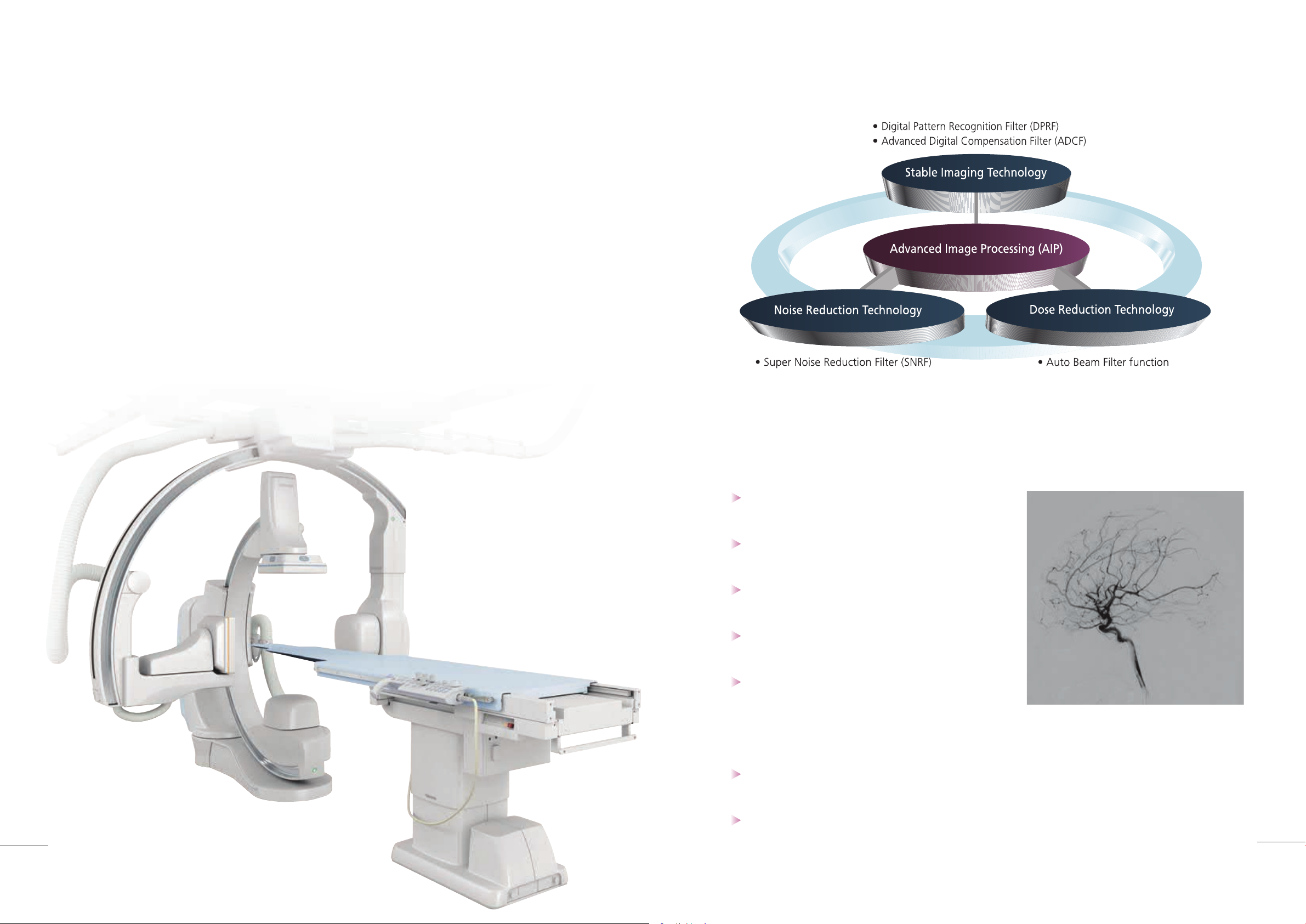
New generation filter made it possible the reduction of noise with high spatial resolution
and less lag. The new filter enhances high-definition images of small devides and structures
(Super Noise Reduction Filter: SNRF).
Advanced technologies deliver
optimized biplane imaging
Designed in concert with leading pediatric physicians, the VF-i/BP provides
advanced, versatile patient access to meet the demands of today’s multi-discipline
imaging environments. The system’s revolutionary multi-tasking computer and intuitive
user interface deliver optimum image quality, time-saving ease of use and improved
workflow. Ideal for diagnostic, interventional and hybrid procedures, the
VF-i/BP is
a completely new approach to biplane imaging designed to take advantage of its
revolutionary multi-axis C-arm.
2
3
Advanced Image Processing (AIP) provides superb
image quality for visualization of vessels and device.
Page 3
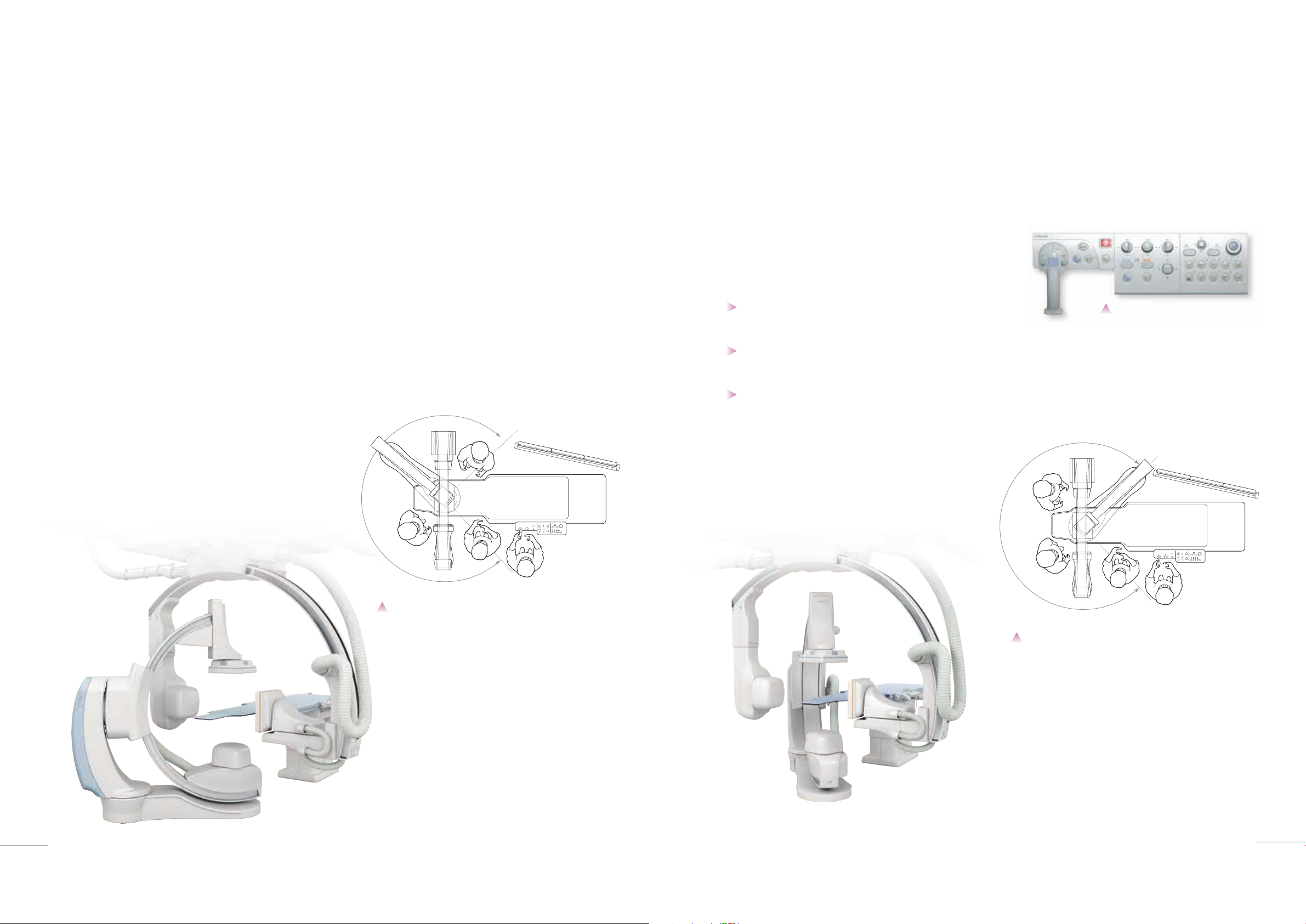
The five-axis design provides a new level of access to the patient.
The head end of the table has a full 180 degrees of space allowing
the necessary physicians to conveniently access the patient and still
provide biplane viewing.
Compact tableside control provides
ergonomic and tactile control of all exam
functions for rapid component positioning,
and control of digital processing functions.
I n this confi guration, the head end of the table has 145 degrees of
space allowing easy access for your anesthesiologist.
Efficient tableside control
The hyperhandle design and tableside console layout allow
clinicians to more effectively concentrate on the patient and the
image data providing a more patient focused examination.
Workflow is enhanced by tableside access to key functions
through a specially designed graphical user interface
During image review, a single keystroke enables system
setup from any selected image
Automatic archiving provides immediate recall of images
at tableside without interruption
Unparalleled patient access:
meeting the needs of all physicians
The VF-i/BP is designed to provide superior access to the patient — an
important point of distinction in the imaging landscape that now often requires
the attention of a wide range of specialists. In hybrid procedures that may require
a full complement of specialists including surgeons, neuroradiologists and
anesthesiologists, the
VF-i/BP is at its best.
4
5
Page 4

Quick and easy flat panel
detector positioning
Vertical movement of the flat panel detector of the
Ω-arm can be linked with vertical movement of the
X-ray tube, enabling quick and easy positioning.
PA/Cranial projection acquired in 8" FOV.
Multiple frames integrated for PEAK image.
Lateral projection simultaneously acquired
on 8" FOV. Multiple frame integrated for
PEAK image.
Optimum abdominal coverage can be
achieved with the 12" x 12" FPD, as
evidence by this SMA injection for GI
bleeding.
12" x 12" FPD can be utilized for a frontal
or lateral projection, in this case providing
coverage of cervical carotid and cerebral
carotid circulation.
Real-time processing capabilities produce high-
resolution flat panel images with uniform brightness
and no distortion in both single plane and biplane mode
Biplane acquisition at 15 pps with 1024 x 1024
resolution stops rapid motion and allows simultaneous
display of both AP and lateral images in real time
Advanced processing capability delivers
high-quality biplane imaging from the smallest
pediatric patients to the heaviest adults
The 12" x 12" FPDs provided on both planes can be set
close to the head for neuro imaging.
Distortion-free
flat panel biplane imaging
Toshiba’s high-definition flat panel detectors deliver superior contrast
and dynamic resolution. Whether processing biplane fluoroscopy or
biplane digital angiography, the images demonstrate a fine balance of low
noise and easy visualization of contrast flow, with a sharp display of small
details of interventional devices.
6
7
Page 5

3D-Angio
Easy setup and execution of mask and arterial phase are used to create bone or device fusion.
Peripheral DSA
Oriented for wide coverage, the 12" x 12" flat panel provides full imaging of the lower extremities. After
programmed setup, the table is stepped by manual activation while watching the bolus flow for accurate
reliable results. Typically, a single injection can cover the total peripheral anatomy.
+ =
Guide View provides a clinical “roadmap”
Toshiba’s “Guide View” provides a superimposed roadmap over live fluoroscopy images, facilitating
accurate device placement within a targeted vascular anatomy.
Unique technology enhances visualization (in black or white) of the catheters or guide wires
Advanced image processing technology
The use of Toshiba's unique Advanced Digital Compensation Filter (ADCF) and Digital
Pattern Recognition Filter (DPRF) in combination produces images of unparalleled clarity.
ADCF is a background processing technique that is useful for reducing halation in the lung
field and for correcting dark areas such as the mediastinum. DPRF is useful for depicting
devices and blood vessels. It enhances the contrast of devices and blood vessels and at the
same time recognizes all other areas as noise, reducing the amplitude of the signals from
these areas.
SNRF significantly reduces image noise in 14-bit gray scale images without requiring the
X-ray dose to be increased. It achieves this by recognizing and reducing the noise components
in each individual image frame.
Advancing biplane imaging
from head-to-toe
Signal suppression
Signal enhancement
With its comprehensive positioning and image review capabilities, the VF-i/BP
accommodates a wide range of procedures. Advanced conventional and 3D imaging
technologies provide unprecedented imaging with unique tools to enhance both
diagnostic and interventional procedures. These powerful imaging and processing
tools enhance clinicians overall treatment planning capabilities.
8
9
Page 6

Variable dose mode
With the touch of a tableside button, the operator can choose
from four pre-programmed fluoroscopy modes. Different
combinations of pulse rates, dose level, and image processing
parameters optimize various study protocols.
Dose display
Radiation dose can be monitored in real time. The operator can
observe dose levels on a digital display in the examination room.
Virtual collimation
After fluoroscopy, virtual collimation uses software to simulate
collimator and beam filter positions. This lets operators adjust
collimation without additional fluoroscopy, further reducing
radiation dose.
Electronic zoom
Electronic zoom digitally enlarges images in real time during
fluoroscopy, without increasing dose. This eliminates the need to use
smaller fields of view on the detector for magnification purposes,
which would increase the dose required.
Fluoroscopic acquisition
Operators can capture still and dynamic images for future reference during
fluoroscopy. These archived images represent an alternative to fluorography
and a major reduction in dose exposure.
F-STORE: Fluoroscopic images for up to the last 10 seconds can be recorded
on the image disk after fluoroscopy is completed.
Clinicians enjoy the added advantage of
increased productivity and patient care with
complete tableside control.
X-ray beam filter
Toshiba's beam filtration can dramatically reduce absorbed patient
dose and radiation scatter. At tableside, clinicians can select the
mode of choice to limit dose and optimize image quality.
Dose-reduction technologies
for patient and operator
10
11
Page 7

More efficient exams with parallel processing and true multitasking
Simultaneously processing and transferring image data during acquisition yields quick, efficient exams. For
example, during fluoroscopy and fluorography, operators can prepare for the next scheduled patient, process and
save images from a previous (or current) study, and transfer or archive images to an associated network.
The advantages of parallel processing
Representative reference images can be displayed on
the reference monitor as a thumbnail. The images can be
easily selected by mouse operation.
Live monitor Reference monitor
Main console take over the same design with the table
side hyper handle for user friendliness.
Customizable exam parameters include:
• C/Ω-arm position and angulation
• Table height
• Source-to-image distance
• Compensation filter settings
• Acquisition rate
• Image size
• Field of view
• Generator settings
• Digital processing
Advanced system design
drives higher productivity
VF-i/BP can store virtually any number
of customized exam types for any number
of operators. This unique Toshiba feature
dramatically boosts productivity.
VF-i/BP
is equipped with Sequential Navigation for physicians to quickly “navigate” through
an exam (e.g., carotid, renal or runoff).
VF-i/BP executes the preferred angles, projections,
and acquisition parameters, all from memory. One touch of a button enables navigation
through the routine settings for each exam type. Operators have the freedom to change any
parameter throughout the procedure without disrupting Sequential Navigation.
12
13
Page 8

Compact design for easy siting
Access to patient information
with seamless network integration
PACS/network storage: Provides online
dynamic review of patient images. Storage
and transfer of multi-modality images are
handled at high speed.
Presentations: Clinical data can be
exported as PC format files for use in
presentations.
DICOM CD-R/DVD-RAM: Serve as
long-term and portable storage media for
valuable image data.
A typical system layout
The VF-i/BP comes standard with the six major DICOM Service Classes enabling
efficient network integration. These DICOM features allow open access to patient
information while reducing examination time and enhancing overall department workflow.
Infinix-i: Dynamic viewing and flexible
network integration permits rapid export and
retrieval of images. Open communications
with HIS /RIS provides rapid transfer of
patient information.
14
15
6,000
3,000
(236.2)
(118.1)
2,000
(78.7)
Machine
room
7,500
(295.3)
Angiography room
Control room
Floor-mounted C-arm suppor t (CAS-880A)
Ceiling-suspended Ω-arm suppor t (CAS-820B)
Catheterization table (CAT-850B)
Ceiling-suspended monitor
CAS-820B control cabinet
CAS-880A control cabinet
XTBP-8100G power cabinet
XTP-8100G power cabinet
XTP-8100G system power cabinet
HEX-125 X-ray tube cooling unit
Digital radiography unit
System console
System cabinet for control room
Coolant circulator
Unit: mm (in)
 Loading...