

Page 1
Leica MM Multi Wavelength
Cell Scoring powered by MetaMorph
Analysis Software Drop-in for Leica MM AF
• Multi-Parameter analysis of up to seven wavelengths
• Identication of subpopulations of cells
• Adaptive background correction™ for improved segmentation
• Field and Cell-by-Cell measurements
The Leica MM Multi Wavelength Cell Scoring Application Module
for Leica MM AF software is designed for the scoring of cells with
up to seven uorescent stains.
This exible module is ideal for counting and logging measurements of cells in multiple wavelength experiments. Using a uorescent marker for the nucleus and additional markers for the cytoplasm or the entire cell, each wavelength is analyzed and cells
are assigned scoring proles for the presence or absence of each
marker.
The module makes a number of additional cell-by-cell measurements including per-wavelength number and percentage of cells
scored as positive or negative, scoring proles, area and intensity.
The module utilizes Adaptive Background Correction adapting the
cell detection algorithm to the local intensity ranges between and
within cells to provide the most robust segmentation available.
This technique enables probe detection even with highly variable
background uorescence within a single image.
The simple interface minimizes setup efforts and at the same time
enables users to customize the settings and measurements to obtain the best possible results specic to the type of cells used in
the experiments.
®
Page 2
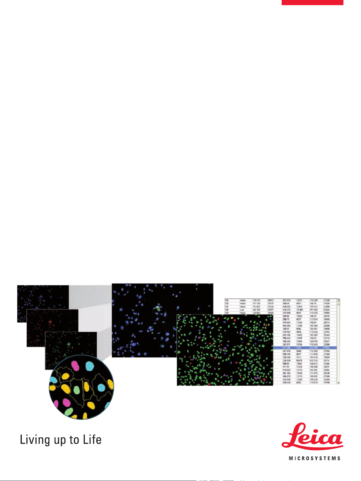
Multiple wavelengths
Leica MM AF provides a exible integrated
solution for multi wavelength image acquisition
and analysis. Up to seven wavelengths can be
analyzed simultaneously by the module. U2OS
rat β-arrestin 2-RrGFP cells treated with 1 μm
isoproterenol. Blue: Hoechst 33342, red: AntiPhospho-Histone H3 (Ser28), green: Transuor® vesicles.
Configuration for analysis
Set the number of wavelengths used and repeat
the following steps for each wavelength:
1. Select the image of interest
2. Choose the stained area (nucleus,
cytoplasm, or both)
3. Specify approximate minimum and maximum widths of the objects to be detected
4. Adjust detection sensitivity by specifying the
intensity above local background
5. Set positive scoring criterion (minimum stained area)
6. Optionally specify the reporting parameters
Multi-parameter analysis
Field measurements include:
• Wavelength-specic count and percentage of
negative and positive cells
• Scoring prole counts
Cell-by-cell measurements include:
• Wavelength-specic stained area, and integrated and average intensities
• Scoring prole
Powerful data export capabilities
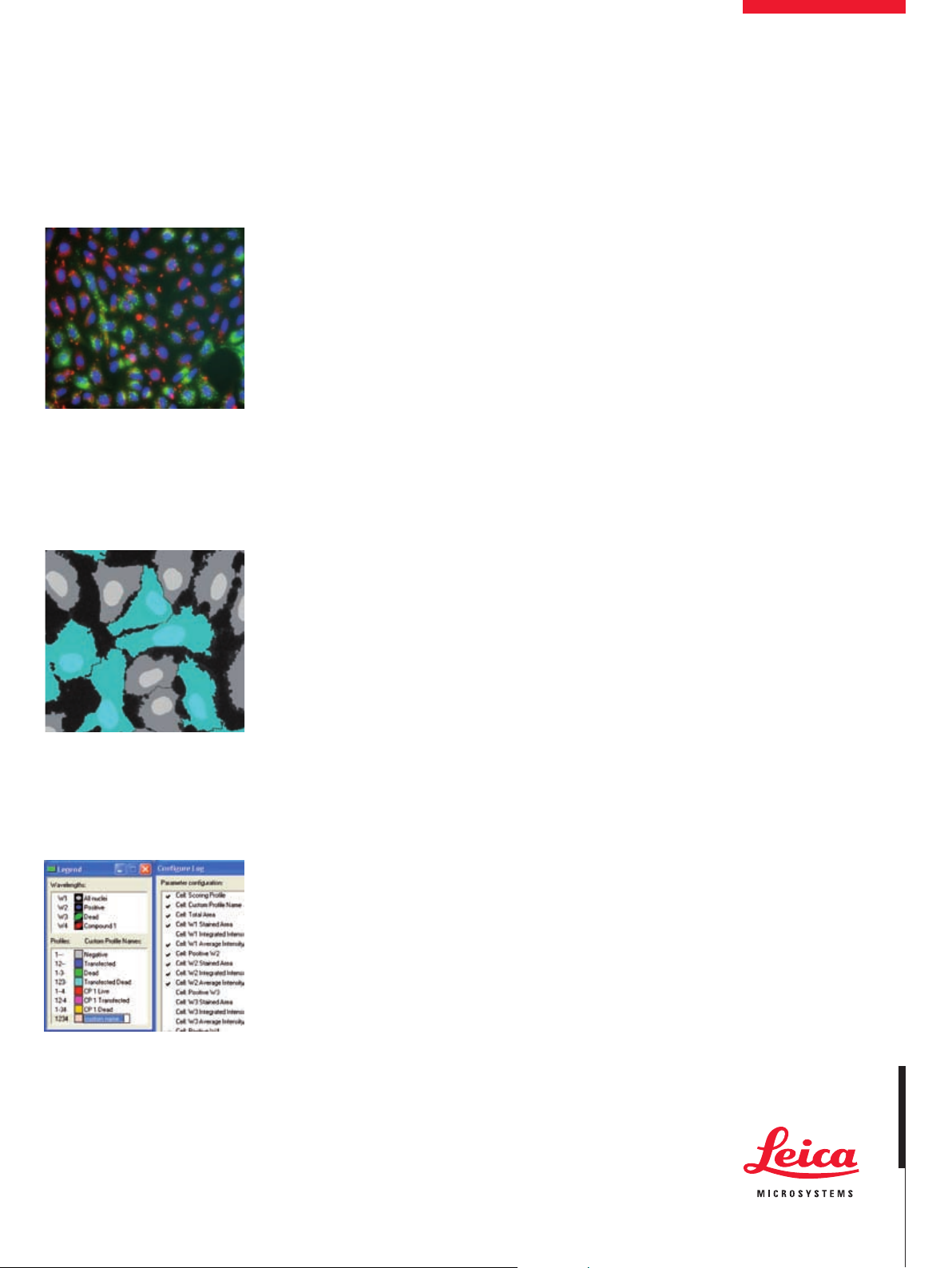
Accurate segmentation
Detection of cells uses Adaptive Background
Correction and displays interactive graphics on
the original images indicating scoring for each
wavelength for immediate verication of accuracy. The module also generates a segmentation image.
Customization
Wavelength and prole naming, graphics
colors and measurement parameters can all
be customized to match the biology of your
experiment. All settings can be saved for future
experiments.
Interactive data display
Once the analysis is run, the Cellular Results table allows you to interactively view individual
cells’ data. Clicking one or multiple cells in the
image highlights the data for the selected cell(s)
in the table.
Customization through journaling
Journals are sophisticated and powerful macros
that record and perform a series of tasks without
the need for a programming language. The modules can be incorporated into a Leica MM AF
journal to increase the customization and automation of your analysis.
All measurements can be directly exported to
a text le or Microsoft
®
Excel® for further ana-
lysis.
“MetaMorph® is a Registered Trademark of MDS Analyti-
cal Technologies”
www.leica-microsystems.com
 Loading...