

Page 1
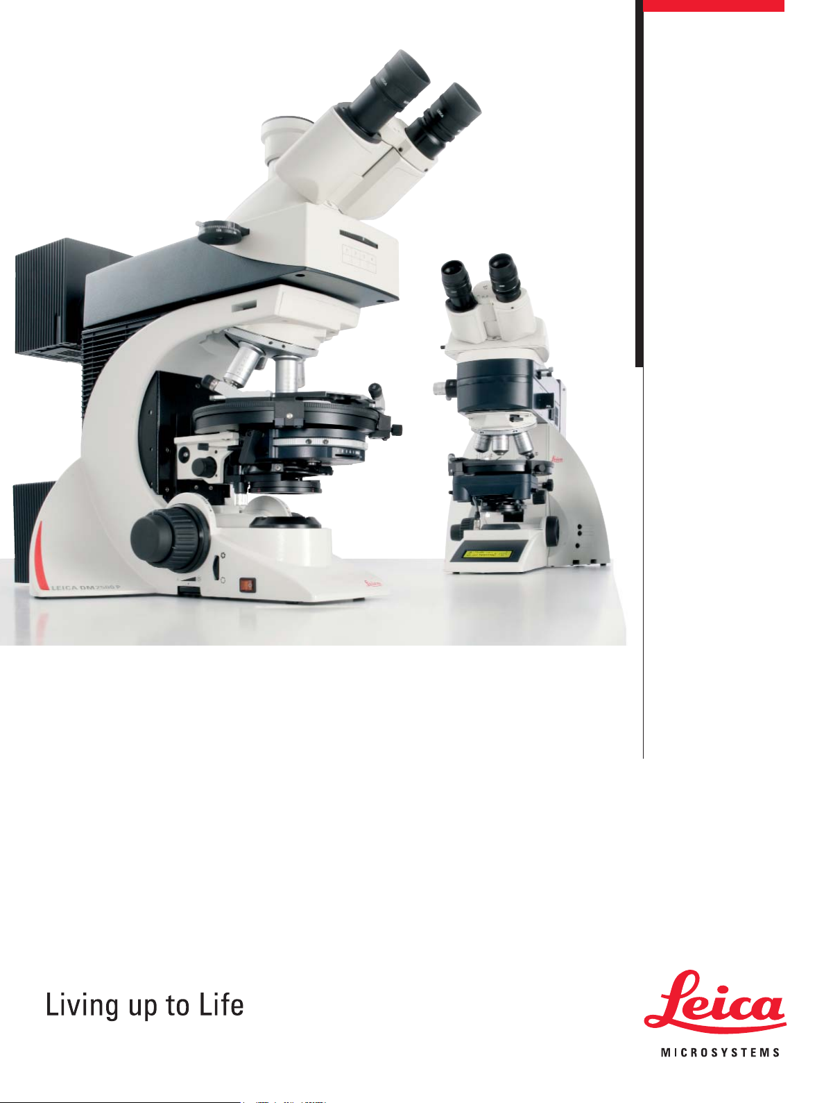
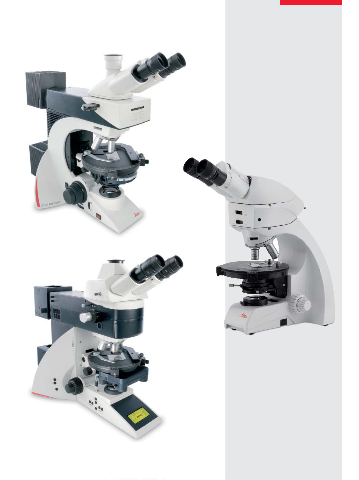
Leica DM4500 P,
DM2500 P, DM750 P
Breaking New Ground in Polarizing Microscopy
Page 2

Brilliance
Reliability
Flexibility
Documentation
Simply Precise
Polarizing microscopes
for geosciences and industry
The new Leica microscope series
is designed for all polarizing examinations: petrography, mineralogy, structure characterization and examination of liquid crystals.
Leica’s new polarizing microscopes are ideal for a wide range of
applications.
With versatile instrument options, Leica polarizing microscopes
are also an ideal match for industrial analysis and quality control,
such as analyzing glass, plastics, textiles and fibers or testing displays in the semiconductor industry. Leica microscopes always
provide the most accurate and reliable results.
Specifically designed for your application:
• Leica DM4500 P for research and development
• Leica DM2500 P for routine polarization applications
• Leica DM750 P for university and other instructional use
Accurate results:
The new Leica polarizing microscopes will show you how easy
and reliable microscopy can be. Leica’s convenient operating
concept allows you to improve your workflow and concentrate
entirely on the task at hand.
Advantages that speak for themselves:
• Improved polarization contrast to obtain more information
from a sample
• Easy operation for accurate sample evaluation in both
orthoscopy and conoscopy
• Ergonomic design for user comfort
• Camera and software modules can be integrated for fast, easy,
and reproducible documentation
Page 3
Leica Design by Christophe Apothéloz und Werner Hölbl
3
Page 4
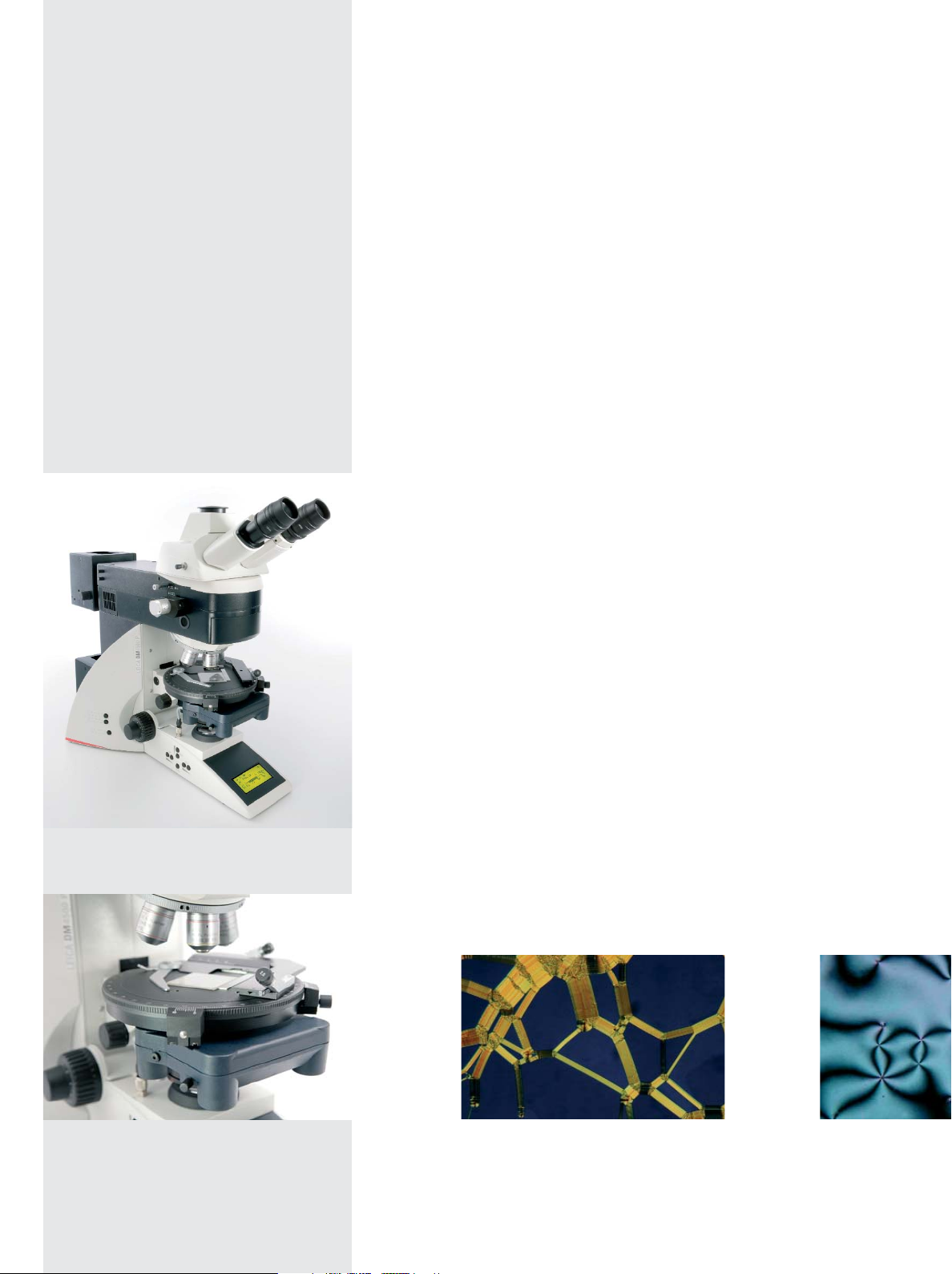
Leica DM4500 P
The Microscope that Guides You
• Automation that anticipates your next
work step:
– Automatic diaphragm setting and
light intensity
– Constant Color Intensity Control
for constant color temperature
– Condenser cap swings in and out
automatically
The right diaphragm – automatically
The Leica DM4500 P automatically detects which contrast method
and objective are being used. This provides valuable consistency
and reproducibility for your research. Manual diaphragm setting
is no longer required, either in the transmitted light or incident
light method. You can concentrate on your work – the Leica
DM4500 P takes care of the rest for you.
Always in the right light
Light intensity automatically adjusts to the objective. Image brightness remains constant when switching objectives, which eliminates glare. You can always adjust the light intensity manually as
well.
Constant color temperature
The Leica DM4500 P’s transmitted light axis is ideally suited for
mineralogical stone identification. Its Constant Color Intensity
Control automatically maintains a constant color temperature,
and you no longer need neutral density filters to compensate for
changes in light intensity.
Designed for use in research and development: the new
Leica DM4500 P – polarizing microscopy has never been
easier.
For the most precision:
the Leica DM4500 P’s rotating stage
4
The correct condenser setting – immediately
All condensers are designed with condenser heads that are
perfectly matched optically and automatically swing in and out
depending on the objective magnification. They are effective from
1.25x–100x magnification.
Oily strikes of a cholesteric liquid crystal mixture.
Crossed polarizers, magnification 10x.
Images courtesy of Dr. Toralf Scharf, Institute of Microtechnology (IMT), University of Neuchâtel, S
Defective texture in planar a
Crossed polarizers, magnific
Page 5

All settings at a glance
You can see all microscope settings at a glance on the easy-toread, integrated display: information such as contrast method,
orthoscopic or conoscopic mode, objective, diaphragm setting,
and light intensity are clearly indicated. With this feedback,
results can easily be reproduced.
• Conveniently arranged functions:
– New, convenient display
– Variable, programmable function buttons
• Great optical quality for crystal-clear results:
– Improved conoscopy module
– Precise orthoscopy
Easily assign function buttons
You can assign the function buttons to any function you want – no
programming skills are required. Six conveniently located buttons
behind the focus knobs provide fast and easy access to the functions you use most.
Perfect interaction of all functions
The interaction between the display and coding of the individual
modules allows the microscope to guide your work. With just one
look at the display, all relevant information is at your fingertips.
For example, the display indicates when to swing the conoscopy
module into or out of the beam path. You have the ability to adjust
the light and diaphragm values to obtain the best conoscopic
image at any time.
• State-of-the art functions:
– Microscope guides you to the next work step
– Displays current operation status
Everything seen on the display of the Leica DM4500 P
is saved automatically. This allows you to reproduce the
settings at any time.
Liquid crystal, defective texture in a hybrid aligned cell.
Crossed polarizers, magnification 5x.
The Leica DM4500 P anticipates your next work step.
Settings on the conoscopy module appear immediately
on the display. This shows the current operating
status of your instrument at all times.
5
Page 6

Leica DM2500 P
The Microscope that Adapts to Each User
• Ergonomic design adjusts to you:
– Height-adjustable focus knobs
• Convenient features let you work faster:
– Color-coded objectives and condenser
diaphragms match lenses
• Safety feature protects the sample and objective:
– Integrated focus stop prevents
objective/sample collisions
Comfortable and relaxed work
No two people are alike. The Leica DM2500 P ensures that every
user can work at the microscope in a relaxed manner. The height
of the microscope’s focus knobs can be individually adjusted to fit
each user’s exact hand size, which prevents hand, arm, and shoulder tension and ensures a comfortable and fatigue-free posture.
Efficient and reproducible microscopy
Color-coded lenses match the color-coded field and aperture
diaphragm adjustment (CDA), to ensure that the illumination conditions are always matched to the objective. Using a manual
microscope has never been easier. With CDA, the Leica DM2500
P offers a level of reproducibility that is one-of-a-kind in its class.
Reliably and accurately adjusts to your sample
The built-in focus stop protects your samples and the front lens of
the objective. For samples of equal height, the focus stop makes
the focusing plane easier to reconstruct so you can concentrate
entirely on your application.
The Leica DM2500 P will show you how easy and reliable
polarizing microscopy can be.
Ergonomically designed to the last detail: you can adjust
focus knob height to match your hand size.
6
Textile fibers, crossed polarizers with lambda plate,
magnification 100x.
Images courtesy of Michael Doppler, Leica Microsystems
Light augite with biotite repla
rims. Black magnetite grains
magnification 200x.
Page 7

Versatile and adaptable
You have a choice of two conoscopy modules to supplement the
Leica DM2500 P. The advanced conoscopy module with a centerable, focusable Bertrand lens and extended field of view has been
designed for advanced requirements in conoscopy. As an economical alternative, Leica offers the standard conoscopy module
with a pre-focused, centerable Bertrand lens, built-in analyzer,
and integrated pinhole for examining small grains.
The 4-position polarization incident light axis is ideally suited to
research applications. Reflected light contrast methods such as
brightfield according to Smith, quantitative polarization (POL) or
fluorescence (Fluo) – provide ideal imaging conditions for mineralogical or geological examinations. A centerable Bertrand lens
module is also available for conoscopy.
• Flexibility to meet your needs:
– Choice of Bertrand lens modules
– Orthoscopy
– 4-position Pol incident light axis
– 5-position, centerable objective turret
The 5-position objective nosepiece provides individual centration
for each objective, and two rotating stages are available. A 45°
stage rotation with click stop is optional.
Biotite hornblende granite myrmekite
(quartz-feldspar) with lambda plate.
Crossed polarizers, magnification 200x.
A first on the world market: correct diaphragm setting at
all times – the Color-coded Diaphragm Assistant helps set
the diaphragm values needed.
Developed for everyday use on the Leica DM2500 P –
the new POL rotating stage with 45° click stop to indicate
the illumination positions.
7
Page 8

Leica DM750 P
The Microscope for Teaching
• Advanced performance in a teaching
polarizing microscope:
– Standard and advanced conoscopy modules
– Polarizer with notch markings
– 4-position objective turret, centerable
– Sturdy, compact design
• Convenience that makes work easy:
– Easy-to-access control functions
– Ergonomic viewing angle
– Accurate angular measurement with
verniers on the rotating stage
Accurate and versatile for teaching
The Leica DM750 P is the ideal polarizing microscope for university and other instructional use, offering a standard and an
advanced Bertrand lens module for unsurpassed ease of operation. With a wide range of accessories and Leica’s renowned
optics, the Leica DM750 P is exceptional not only for its compact,
durable design, but also for its efficiency and ease of operation.
Designed for optical brilliance and long life illumination
The standard Köhler field diaphragm and magnetically fixed blue
filter provide vivid, pin-sharp images. The 2,000-hour, 35-watt
halogen lamp saves hundreds of dollars in replacement bulb cost
over the life of the microscope. Based on the same optical platform as Leica Microsystems’ research microscope line, students
enjoy outstanding optical performance and full access to virtually
all accessories from the Leica Microsystems microscope product
line.
Developed for college teaching and research use:
the Leica DM750 P.
8
Maximum ease of use and high optical brilliance are
the outstanding features of the Leica DM750 P.
Page 9

Camera and Software Modules
Complete the System
Ready to expand at any time
To seamlessly interface with the new Leica polarizing microscopes, Leica offers a comprehensive camera and software solution for fast, convenient documentation of your work. You can
expand your system at any time using Leica’s cameras and application-specific software modules. All future software and hardware components from Leica will operate on a uniform interface.
Archiving and documentation is easy
The basic core functionality of the Leica Application Suite (LAS) is
provided with every Leica microscope and digital camera as part
of an integrated system solution. Together, the combined system
provides an intelligent, automated microimaging environment.
LAS is the basic software for microscope configuration and control, and provides a platform for acquiring, analyzing, and processing the highest quality digital images.
LAS Reticule for comparison and measurement
The LAS Reticule application provides electronic
tools for displaying live images and overlaying
different types of measuring reticules. LAS Reticule
provides visual feedback about the approximate
size of the field of view. In this way, object size
comparisons and distribution measurements can be carried out quickly and
effortlessly.
• Leica’s complete polarizing microscope
systems integrate the following components:
– Leica polarizing microscope
– Leica Digital FireWire Camera (DFC)
– Leica Application Suite (LAS) software
Advanced interactive measurement
The Interactive Measurement module of
the Leica Application Suite has been
designed for particularly difficult measurements. Using this module, samples can be individually counted and
assigned to an identified
class as well.
9
Page 10

Modular, Customized Configurations –
Microscopes Designed for You
• Flexibility that gives the freedom you need:
– Wide selection of POL objectives
• Compatibility that knows no bounds:
– Fully compatible components across Leica’s
polarizing microscope product line
– Wide selection of analyzers, polarizers,
and compensators
– Full wave & quarter wave plates are available
– Wide selection of POL observation tubes
The result of combining maximum precision and optimum
ergonomic design – the 360° analyzer.
Flexibility – Designed for you
Flexible to the last detail. All Leica polarizing microscope components can be configured for all microscopes in the polarizing line.
For example, you can choose from over twenty POL objectives for
the Leica DM4500 P, DM2500 P or DM750 P. The optical possibilities are unlimited. You will enjoy the benefits provided by this
complete system when using the new 360° analyzer, the 360°
polarizer or even with full wave plates. All components can be
used for classroom teaching, everyday routine work, and
research.
Leica’s entire line of DIN standard compensators can be used in
all Leica polarizing microscopes, as can the attachable mechanical
stage for accurate sample positioning. This always ensures flexible
interchange and replacement of parts.
Flexibility is key. All of Leica’s rotating stage polarizing
microscopes feature attachable, interchangeable
mechanical stages.
10
Page 11

Technical Data
• Objective turret
• Objectives
• Usable field
of view
• Contrast method
Changeover
Color reproduction
Transmitted light
Incident light
Leica DM750 P
4x (M25), centerable
HI Plan POL
N Plan POL
Immersion objectives
20 mm
Manual
Polarization contrast
Orthoscopy
Conoscopy
Brightfield
Phase contrast
Darkfield
Polarization contrast
Brightfield
Leica DM2500 P
5x (M25), centerable
HI Plan POL
N Plan POL
PL Fluotar POL
Immersion objectives
25 mm
Manual
Polarization contrast
Orthoscopy
Conoscopy
Brightfield
Phase contrast
DIC
Darkfield
Polarization contrast
Brightfield
Darkfield*
DIC
Fluorescence
Leica DM4500 P
6x (M25), centerable, absolute encoded
HI Plan POL
N Plan POL
PL Fluotar POL
Immersion objectives
25 mm
Motorized
CCIC: Constant Color Intensity Control
Polarization contrast
Orthoscopy
Conoscopy
Brightfield
Phase contrast
DIC
Darkfield
Polarization contrast
Brightfield
Darkfield*
DIC
Fluorescence
• Conoscopy
• Transmitted
light axis
Illumination
Operation
• Incident light axis
• Condensers
• Focus drive
* on request
Bertrand lens cube
in new IL axis
Bertrand lens module (AB module)
Advanced conoscopy module
12 V 35 W halogen lamp
Manual
User guidance with CDA
Manual
User guidance with CDA
Manual changeover
User guidance with CDA
Manual, 2-gear gearbox
Bertrand lens cube
Bertrand lens module (AB module)
Advanced conoscopy module
12 V 100 W halogen lamp
Manual
User guidance with CDA
Manual
User guidance with CDA
Manual changeover
User guidance with CDA
Manual, height-adjustable,
Focus stop, 2 or 3-gear gearbox
Fully integrated conoscopy beam path
User guidance with display feedback
12 V 100 W halogen lamp
Motorized
Integrated illumination manager
Motorized
Integrated illumination manager, round and
rectangular field diaphragms for ocular or
camera observation
Motorized changeover of condenser
head, 7x condenser disc, polarizer
Manual, 2-gear gearbox
11
Page 12

“With the user, for the user”
Leica Microsystems
Leica Microsystems operates globally in four divisions,
where we rank with the market leaders.
Life Science Division
•
The Leica Microsystems Life Science Division supports the
imaging needs of the scientific community with advanced
innovation and technical expertise for the visualization,
measurement, and analysis of microstructures. Our strong
focus on understanding scientific applications puts Leica
Microsystems’ customers at the leading edge of science.
Industry Division
•
The Leica Microsystems Industry Division’s focus is to
support customers’ pursuit of the highest quality end result.
Leica Microsystems provide the best and most innovative
imaging systems to see, measure, and analyze the microstructures in routine and research industrial applications,
materials science, quality control, forensic science investigation, and educational applications.
Biosystems Division
•
The Leica Microsystems Biosystems Division brings histopathology labs and researchers the highest-quality,
most comprehensive product range. From patient to pathologist, the range includes the ideal product for each
histology step and high-productivity workflow solutions
for the entire lab. With complete histology systems featuring innovative automation and Novocastra™ reagents,
Leica Microsystems creates better patient care through
rapid turnaround, diagnostic confidence, and close customer collaboration.
Surgical Division
•
The Leica Microsystems Surgical Division’s focus is to
partner with and support surgeons and their care of patients with the highest-quality, most innovative surgical
microscope technology today and into the future.
The statement by Ernst Leitz in 1907, “with the user, for the user,” describes the fruitful collaboration
with end users and driving force of innovation at Leica Microsystems. We have developed five
brand values to live up to this tradition: Pioneering, High-end Quality, Team Spirit, Dedication to
Science, and Continuous Improvement. For us, living up to these values means: Living up to Life.
Active worldwide
Australia: North Ryde Tel. +61 2 8870 3500 Fax +61 2 9878 1055
Austria: Vienna Tel. +43 1 486 80 50 0 Fax +43 1 486 80 50 30
Belgium: Groot Bijgaarden Tel. +32 2 790 98 50 Fax +32 2 790 98 68
Canada: Richmond Hill/Ontario Tel. +1 905 762 2000 Fax +1 905 762 8937
Denmark: Herlev Tel. +45 4454 0101 Fax +45 4454 0111
France: Nanterre Cedex Tel. +33 811 000 664 Fax +33 1 56 05 23 23
Germany: Wetzlar Tel. +49 64 41 29 40 00 Fax +49 64 41 29 41 55
Italy: Milan Tel. +39 02 574 861 Fax +39 02 574 03392
Japan: Tokyo Tel. +81 3 5421 2800 Fax +81 3 5421 2896
Korea: Seoul Tel. +82 2 514 65 43 Fax +82 2 514 65 48
Netherlands: Rijswijk Tel. +31 70 4132 100 Fax +31 70 4132 109
People’s Rep. of China: Hong Kong Tel. +852 2564 6699 Fax +852 2564 4163
Portugal: Lisbon Tel. +351 21 388 9112 Fax +351 21 385 4668
Singapore Tel. +65 6779 7823 Fax +65 6773 0628
Spain: Barcelona Tel. +34 93 494 95 30 Fax +34 93 494 95 32
Sweden: Kista Tel. +46 8 625 45 45 Fax +46 8 625 45 10
Switzerland: Heerbrugg Tel. +41 71 726 34 34 Fax +41 71 726 34 44
United Kingdom: Milton Keynes Tel. +44 1908 246 246 Fax +44 1908 609 992
USA: Bannockburn/lllinois Tel. +1 847 405 0123 Fax +1 847 405 0164
and representatives in more than 100 countries
11/09/???/????
•
Order No.: English 914 549
LEICA and the Leica Logo are registered trademarks of Leica Microsystems IR GmbH.
www.leica-microsystems.com
 Loading...