How it Works
Log In / Sign Up
Buy Points
How it Works
FAQ
Contact Us
Questions and Suggestions
Users
Hitachi
Loading...
A
air purifier with humidifying function
AJ-1200CE
AJ-S55FX
AJ-S55GZ
AJ-S55GZP
AJ-S55KR
AJ-S55KX
AJ-S55KXP
AJ-S60TX
AJ-S60TXP
AJ-S65KXP
AJ-S70F
AJ-S70FP
AJ-S70GZP
AJ-S70KX
AJ-S70KXP
AJ-S70TX
AJ-S70TXP
AJ-S70WX
AJ-S70WXP
AJ-S75TX
AJ-S75TXP
AJ-S80FX
AJ-S80MX
AKG-88
All Digital Models 2H
All models
2
AllPlay Adaptor
AMS1000
2
AMS200
2
AMS 2000
2
AMS 2100
4
AMS 2300
4
AMS 2500
3
AMS500
AN021003-1
AN032404-1
AN0604041
AN070827-1
AN072302-1
AN072803-1
AN081804-1
AN091802-1
AOB
AP1
AP3600
AP52
AP 720S
2
AP 720STM
ARIETTA 50LE
ARIETTA-70
Arietta S70
ATT-20
ATW-RTU-02
ATW-RTU-06
Audio HMA-7500
Audio HTADD1E
Audio HTDK150E
Audio HTDK160
Auto 480
AV3000E
AVC01U
2
AVC08U
3
AVC20
2
AVC50
AW 100
4
AW 130
2
AW 150
3
AW18DBL
AX-6
AX-67
AX-C10
AX-F100-E
3
AX-F100-W
3
AX-F300-E
2
AX-F300-EBS
2
AX-F300-WUN
2
AX-M131U
AX-M133
AX-M136I
2
AX-M138
AX-M140
3
AX-M20E
AX-M20-EBS
2
AX-M40MP3
AX-M5
AX-M5-E
AX-M5-EBS
2
AX-M5-UC
AX-M5-W
AX-M5-WUN
AX-M649-BT
2
AX-M68D
AX-M7
AX-M70MP3
AX-M717
AX-M7-EBS
3
AX-M7-UC
2
AX-M7-W
2
AX-M7-WUN
2
Loading...
Loading...
Nothing found
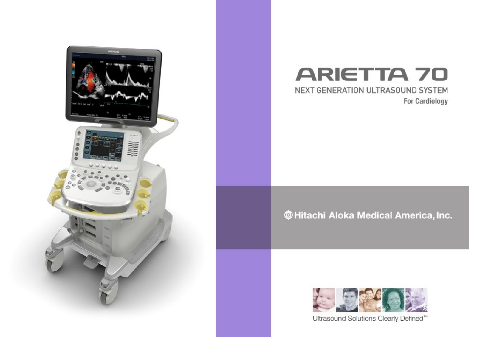
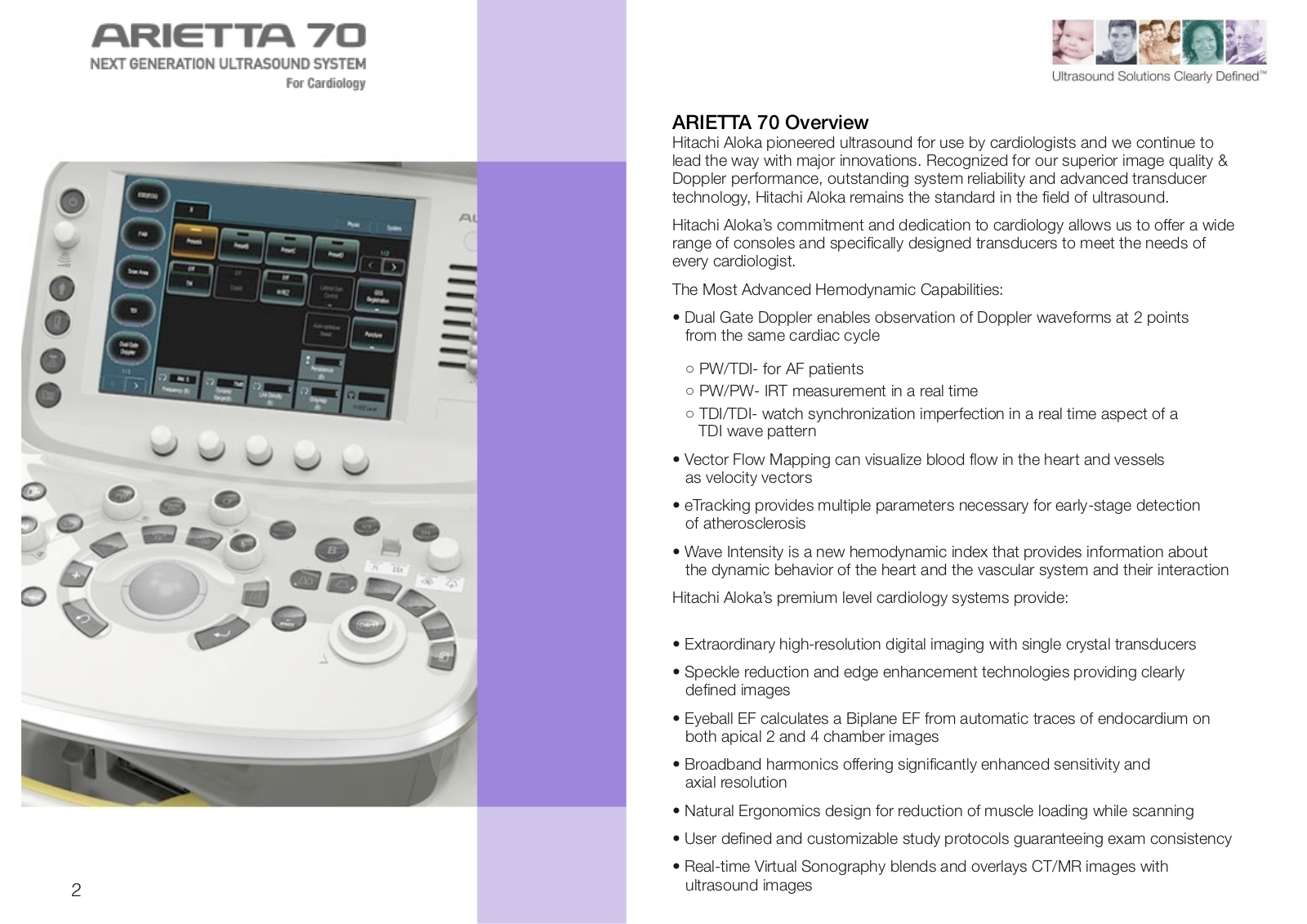
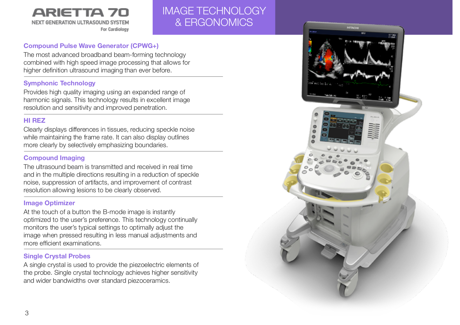
Arietta S70
Brochure
15 pgs
3.11 Mb
0
Table of contents
Loading...
Hitachi Arietta S70 Brochure
...
Hitachi Brochure
Download
Specifications and Main Features
Frequently Asked Questions
User Manual
Download
Loading...
+
10
hidden pages
Unhide
You need points to download manuals.
1 point = 1 manual.
You can buy points or you can get point for every manual you upload.
Buy points
Upload your manuals
 Loading...