

Page 1
Oriole™ Fluorescent
Gel Stain
Instruction Manual
Catalog #
161-0495, 1x solution, 200 ml
161-0496, 1x solution, 1 L
161-0497, kit for 5 L
For technical suppor t call yo ur local Bio-Rad offi ce.
In the U.S. ca ll 1-800-424-6723.
Page 2

Table of Contents
Section 1: Introduction an d General Informat ion 1
1.1 Introduction 1
1.2 Product D escription 2
1.3 Storage 2
1.4 Materials and Equipment Required
but Not Supplied 3
1.5 Reagents Required but Not Supplied 3
1.6 Safety Considerations 4
1.7 Disposal Considerations 4
1.8 Fluorescence Characteristics 4
Section 2: Instru ctions 6
2.1 General Considerations 6
2.2 Stain Solution Preparation 8
2.3 Gel Staining 9
2.4 Gel Imaging 11
Page 3

Section 3: Technical Information 14
3.1 Sensitivity of Staining — 1-D Gels 14
3.2 Compatibilit y with Mass Spectrometr y 15
3.3 Protein-to-Protein Variability 15
3.4 Dynamic Range 16
3.5 2-D Gel Staining 17
Section 4: Troubleshooting 18
Section 5: Product Information 21
™
5.1 Oriole
Fluorescent Gel Stain 21
5.2 Related Products 21
Page 4

Page 5

Section 1
Introduction and General Information
1.1. Introduction
™
fluorescent gel stain is an easy to use, rapid, and
Oriole
sensitive stain for visualization and quantitation of proteins
separated by SDS-PAGE. The product is available in
three configurations. The 200 ml and 1 L sizes are
provided ready to use. The product is also available as a
kit containing components to make 5 L of ready to use
staining solution.
The staining procedure is a simple one-step protocol
that can be completed in as little as 9 0 minutes.
Gels stained with Oriole fluore scent gel stain may
be visualized with a variety of dif ferent UV-based
fluorescence imaging systems.
Oriole fluorescent gel stain gives exceptional sensitivity
and dynamic range (see pages 14–16) and is compatible
with subsequent analysis by enzymatic digestion and
mass spectrometry. It is thus particularly well suited to
proteomics applications.
1
Page 6

1.2. Product Description
Oriole fluorescent gel stain comes in three package
configurations.
The 200 ml size — fully diluted and ready to use;
provide s enough stain for four mini format
Mini-PROTEAN
Criterion
®
gels (~8.6 x 6.8 cm), or two midi format
™
gels (13.3 x 8.7 cm).
The 1 L size — fully diluted and ready to use; provides
enough stain for 20 Mini-PROTEAN gels (~8.6 x 6.8
cm), ten Criter ion gels (13.3 x 8.7 cm), four large format
PROTE AN
®
II gels (16 x 16 cm or 16 x 20 cm), or two large
format PROTEAN Plus gels (25 x 20.5 cm).
The 5 L kit — contains concentrated compone nts to
prepare 5 L of staining solution and can be diluted to
1x according to demand.
1.3. Storage
The product is sta ble for at least 18 months from the date
of manufacture or until the expiration date on the label
when stored at 24°C or below. Consult the expiration date
2
Page 7

before using. Avoid prolonged exposure to temperatures
greater than 37°C and protect from light.
1.4. Materials a nd Equipment Required but
Not Sup plied
n
Staining containers — Any glass or plastic tray capable
of holding the recommended volume of solution may
be used
n
Imaging equipment — Gels are best imaged using a
UV-based fluorescence imager capable of excitation
near 270 nm and detection near 604 nm such as the
Molecular Imager
ChemiDoc
®
Gel Doc™ XR+, Molecular Imager®
™
XRS+, VersaDoc™ MP 4000, ExQuest™
spot cutter, and VersaDoc MP 500 0 systems. For a
more complete list of compatible imaging systems,
see pages 12–13
n
L aboratory shaker or rocker
n
Powder-free latex, vinyl, or nitrile gloves
1.5. Reag ents Required but Not Supplied
Methanol, reage nt grade (for 5 L kit only)
3
Page 8

1.6. Safet y Consideration s
Oriole fluorescent gel stain is a dilute solution of a
fluorescent dye. T he working solution is flammable and
should be handled in a manner that prevents exposure to
open flame or sparks. The complete proper ties of the dye
component have not been investigated. Eye protection
and gloves should be worn and general laboratory safety
precautions followed while handling both the diluted and
undiluted product.
1.7. Disposal Considerations
Laws governing the disposal of laboratory chemicals
vary by re gion. Consult the MSDS (available online at
www.bio-rad.com) and check local laws for proper
disposal guidelines.
1.8. Fluorescence Characteristics
Oriole fluorescent gel stain has a fluorescence excitation
maximum of 270 nm and a fluorescence emission
maximum of 604 nm.
4
Page 9
120
100
80
60
40
20
Fluore scence, norm alized
0
200 300 40 0 500 600 700 800
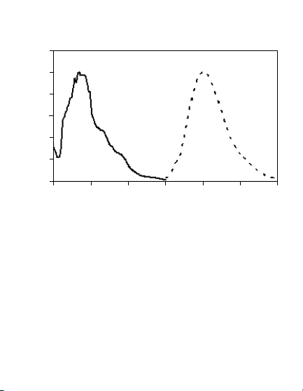
Fig. 1. Fluo resce nce exci tatio n and emissio n spec tra of
Oriol e stai n. Orio le stai n has its exc itatio n maxi mum at 270 nm
and emi ssion m axim um at 604 n m, making it compatible with
UV-base d image rs. —, Excitation sp ectru m;
Wavelen gth, nm
....
, emiss ion spe ctrum.
5
Page 10

Section 2
Instructions
2.1. General Considerat ions
Best results are obtained by using clean technique.
Any dust or dirt transferred to the surface of the gel
may appear in the fluorescence image as smudges or
speckles. Oriole
sensitive. Contaminant proteins such as keratin will
appear in the gel image if care is not taken to minimize
such contamination.
All glassware used should be cleaned with laboratory
glassware cleaner and rinsed with distilled or deionized
water. Use dust-free gloves and limit dust exposure by
keeping reagent vessels and gel trays covered as much
as possible. If gels are cast in the laborator y, the glass
plates used should be thoroughly cleaned with lint-free
laboratory wipes.
Oriole fluorescent gel stain is very se nsitive, and less
protein can be visualized than what is possible using
a visible stain like Coomassie Blue. Sensitivity is of the
™
fluorescent gel stain is exceptionally
6
Page 11

same general order as silver stain or other fluorescent
protein stains. Oriole fluoresce nt gel stain has a wide
dynamic range, and variability in the amount of protein to
be visualized can be accommodated simply by varying
the exposure settings during imaging. As a general rule,
the max imum quantity of protein re comme nded for
visualization with Oriole fluorescent gel stain is 1–2 µg for
individual proteins and 10–20 µg for complex mixtures on
1-D gels. The limit of sensitivity for individual proteins is
1 ng or less.
Oriole fluorescent gel stain is moderately light sensitive.
If gels are left in stain for more than 90 min, the gel tray
should be covered with aluminum foil or an opaque lid.
Oriole fluorescent gel stain is intended only for staining
1-D and 2-D SDS-PAGE gels. Native gels and IEF gels
cannot be stained with Or iole stain. Oriole stain is not
recommended for staining protein blots.
Instructions given are for standard 1 mm thick SDS-PAGE
gels. Thicker gels may benefit from longer stain times and
larger volumes of solution.
7
Page 12

Molecular weight standards that have bee n prestained
with a visible dye such as Precision Plus Protein
Dual Color or Kaleidoscope
™
prestained standards do
™
All Blue,
not stain with Oriole fluorescent gel stain and cannot be
imaged by fluorescence in gels stained with Oriole stain.
We recommend the use of unstained protein standards on
gels to be stained with Oriole stain.
2.2 . Stain Solut ion Preparation
The 200 ml and 1 L configurations are provided ready
to use.
The 5 L kit comprises 5 individual 1 L bottles, each
containing 590 ml of Oriole fluorescent gel stain diluent,
and a single bottle containing 50 ml of Oriole gel stain
concentrate. Staining solution (1x) is prepared as follows.
n
To a 1 L bottle holding 590 ml of diluent,
add (in sequence):
– 400 ml methanol (reagent grade)
– 10 ml of Oriole fluorescent gel stain concentrate
n
Mix well by shaking
n
Stain is now ready to be used
8
Page 13

NOTE: Use only methanol in preparing staining
solution from the 5 L kit. The use of water, ethanol,
or other solvents will re sult in poor staining
performance.
Reco mmen ded St ain Vol ume
Volume of S tain
Gel Siz e Solut ion per G el
Ready G el® or Mini- PROTEA N gel
(8.6 cm × 6.8 cm) 50 ml
Criter ion gel (13.3 cm × 8.7 cm) 100 ml
PROTEA N II gel (16 cm × 16 cm or 16 cm x 20 cm) 250 ml
PROTEA N Plus gel (25.6 cm × 23 cm) 500 ml
2.3. G el Staining
NOTE: Do not fix or was h gel prior to staining.
This will make staining less sensitive.
1. Place gel directly into a clean tray containing the
recommended volume of Oriole fluorescent gel stain.
Cover the tray, place on a rocker or shaker, and agitate
as vigorously as possible without splashing liquid or
damaging the gel.
9
Page 14

2. Stain for 90 min for maximum sensitivity.
3. Cover the tray to exclude light if the gel is stained
longer than 90 min.
NOTE: Best results are obtained if the gels are left in
staining solution no longer than 2 hr.
4. Transfer gel to water prior to imaging. This step
prevents ex posure of the imaging equipment
to moderately corrosive staining solution.
NOTE: Destaining is not necessar y. Staining intensity
persists when the gel is stored in water.
Stained gels can be stored in water for up to 6 months
and imaged without significant loss of sensitivity if
protected from light and stored at 2–8°C.
10
Page 15

2.4. G el Imaging
Gels stained with Oriole stain are visualized using UV
light excitation. Bio-Rad Molecular Imager Gel Doc XR+,
Molecular Imager Che miDoc X RS+, EXQuest spot cutter,
VersaDoc MP 4000, or VersaD oc MP 5000 systems are
recommended for imaging gels stained with Oriole stain.
If the imaging equipment has no preprogrammed imaging
function for Oriole fluorescent gel stain, the imaging
setting for SYPRO Ruby stain or ethidium bromide that
uses UV transillumination is recommended.
Any imaging system using UV light excitation may be
used to image Oriole fluore scent gel stain. Such imaging
systems almost always have midrange (300 nm, 30 6 nm,
or 312 nm) UV excitation and red or orange emission filters
as standard options for imaging ethidium bromide –
stained gels. The excitation and emission properties of
Oriole stain are very compatible with ethidium bromide,
therefore imager settings for ethidium bromide can be
used when imaging gels stained with Oriole stain.
Imaging systems capable of imaging gels stained with
Oriole fluorescent gel stain include the following:
11
Page 16

Manuf actur er I magin g system Recom mende d settings
Bio-Rad Gel Doc, ChemiDo c, For Gel Doc and ChemiDoc
Labor atorie s VersaD oc, EXQu est systems, use S tanda rd
spot cutter systems (302 nm) UV lamp with
Stand ard Fil ter (580 nm
bandp ass for et hidiu m
bromide). For Versa Doc
system s use Sta ndard
(302 nm) UV l amp and
either the 520 nm
longpass filter or the
605 nm bandpass filter
(either one or the other is
inclu ded wit h the
instrument)
GE Healthcare ImageQuant UV 302 illuminatio n with
ethidium bromide (orange)
emission filter
Fuji LAS 30 00, LA S 4000 312 nm illuminati on with
ethidium bromide
emission filter
Alpha Innotech AlphaImager, UV 302 with standard
FluorChem (orange) emission filter
12
Page 17

Manuf actur er I magin g system Recom mende d settings
UVP BioDo c-It, Midrange (302 nm)
VisiD oc-It, UV exci tation w ith ethi dium
DigiD oc-It, bromide re d emiss ion filte r
MultiDoc-It,
Photo Doc-It,
BioSpectrum,
EC3
Carest ream Gel Logic UV trans e xcitati on
with 590 nm bandpass
(ethidium bromide)
emission filter
Kodak 2200, 4000MM, 306 nm UV excitation
4000MM Pro, with ethidium bromide
4000R, 400 0R Pro standard orange (600 nm)
emission filter
Syngene G:Box, InGenius, Mid range UV excitation
U:Geniu s with ethidium bromide
emission filter
Biometra BioDocAn alyze Midrange (312 nm) UV
excitation with ethidiu m
bromide emis sion fil ter
Imaging systems using laser light excitation or other visible light
source excitation are not reco mmended for imag ing gels stained
with Or iole flu orescent gel stain. Th ese inc lude PharosF X™ system
(Bio-Rad), Typhoon, Storm, and Ettan DIG E Imager (GE Healthcare),
Odyss ey (Li-Cor), FLA (Fuji), an d FM Bio (Hi tachi ) systems.
13
Page 18

Section 3
Technical Information
3.1. Sensitivity of S taining — 1-D Gel s
™
The dye in Oriole
tightly to proteins. Background staining is low, and the
limit of se nsitivity is generally below 1 ng.
Fig. 2. U nadju sted im age of a ge l stai ned wit h Orio le sta in.
A diluti on series of Bio -Rad SDS-PAGE standards wa s run on a
4–20% Cr iterio n Tris-HCl li near gradien t gel, sta ined wi th Orio le
stain, a nd imaged using a Molec ular Im ager VersaDoc M P 4000
imagin g system w ith imag e setti ngs for SYP RO Ruby stain.
The res ulting i mage fil e was not adjusted.
stain is highly fluorescent and binds
14
Page 19

4 2 1 0.5 0.25 0.125 ng/ba nd
Fig. 3. I mage of t he gel
stain ed wit h Oriol e stai n,
adjus ted to sho w the limit
of sens itivi ty. The im age
from the p reviou s figure
was inver ted, cropped to
show prote in load s ≤ 4 ng,
and adjusted to show the
limit of s ensitivity.
3.2. Compatibility With Mas s Spec trometry
Oriole fluorescent protein gel stain is fully compatible with
downstream proteolysis and mass spectrometric analysis.
3.3. Pr otein-to-Protein Variab ility
Oriole fluorescent gel stain will stain most proteins
and exhibits lit tle protein-to-protein variabilit y in
staining intensity.
15
Page 20

3.4. Dynamic Range
Oriole fluorescent gel stain has a dynamic range of up to
three orders of magnitude. This allows protein quantitation
in samples of var ying concentration over a wide range of
relative abundance.
4.5
4
3.5
3
arbitrary u nits
2.5
Log (fluo resce nce),
2
0 0.5 1 1.5 2 2.5 3
Fig. 4. Li near ity of O riole stain . A dilution ser ies of ca rboni c
anhydrase was ru n on a Crite rion g el, stai ned with Oriol e stain,
and imaged on the M olecular Imager Vers aDoc M P 4000 imaging
system. Fl uores cence a ssociated with the car bonic a nhydra se
band was p lotted f ollowi ng backg round su btracti on.
Log car bonic a nhydrase, ng
16
Page 21
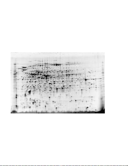
3.5. 2- D Gel Staining
Oriole fluorescent gel stain is ideal for staining 2-D
polyacryla mide ge ls. Clear background-fre e image s
are obtained without interference from CHAPS, carrier
ampholytes, or other components of the first-dimension
separation.
Fig. 5. 2- D gel st ained with Or iole st ain. E. c oli prote in
(40 μg) was run on an 11 cm pH 5– 8 Ready Strip
first dimension and Cr iterio n Tris-HCl 8 –16% gel for the se cond
dimen sion. Th e gel was s taine d with Or iole stain and wa s image d
on the Mol ecular Image r VersaDoc MP 40 00 imag ing system. The
result ing image was inve rted.
™
IPG stri p for the
17
Page 22

Section 4
Troubleshooting
Probl em Pos sible C ause Remedy
Bands o r spots N o protein o n gel Verify th at there i s actua lly
not visible protein on the gel by staining
with another method such as
Bio-Safe™ Coomassie stain.
Malfunctioning Check instrument manual
imaging system for troubleshooting, or
contact the imaging
instrument manufacturer.
Poor sta ining Insuffic ient Stain sensitiv ity ma ximi zes
sensitivi ty staini ng time after 90 min.
Dirty stai ning Make sure that the st ainin g
trays trays and other equipment
have been th oroug hly
cleaned with laboratory
glassware cleaner.
Insufficient Follow the recommendations
stain volume for sta in volume appropriate
to the gel size (Section 2.2).
continued
18
Page 23

Probl em Pos sible C ause Remedy
Poor sta ining Reuse of t he stain Reuse of Oriol e stain i s
sensitivi ty not recommended.
Use of Use only methanol and
nonre comme nded provided di luent.
solve nt for the 5 L k it
Fixing or washing Do not fix or wash gel prior
the gel p rior to to stai ning.
staining
High or uneven Dirty equ ipment Make s ure that th e stain ing
backgro und or st ainin g trays trays and ot her equipme nt
staini ng have been thoroughly
cleaned with laboratory
glassware cleaner.
Too much time in Restrict time of
staining solution staining solution
treatmen t to 90–120 min.
Backgrou nd resulting from
overstai ning ca n be redu ced
by washing the gel in water
for 30 min or m ore.
continued
19
Page 24

Probl em Possib le Caus e Reme dy
High or uneven Reagent impu rities Use high quality reag ents.
backgro und when prepari ng stain
staini ng s olution from th e
5 L kit
Speck les or Par ticul ate mater ial Make sure that the
blotches in the from reagents, staining trays are
gel ima ge staining tray, dus t, thoroughly c leane d.
or gloves
Limit the time that the gels
and staining so lutio n are
exposed to open air.
Use dust-free gloves and
handle gels only by
the edges.
Uneven s taining Insuffici ent sha king Ensure that the gel
durin g staini ng is well agitated
during staining.
Gel shr inkag e Some ge l shrin kage The gel will reswe ll
occur s durin g staining following transfe r to water.
20
Page 25

Section 5
Product Information
5.1. Oriole Fluorescent Gel Stain
Catalo g # D escription
161-0495 Oriol e
161-0496 Oriol e Fluor escen t Gel Sta in, 1x soluti on, 1 L
161-0497 Oriol e Fluor escen t Gel Sta in, kit fo r 5 L
5.2. Relate d Products
Catalo g # D escription
163-2091 Ready Prep
170-8640 Molec ular Im ager Ver saDoc M P 4000 S ystem
170-8650 Molec ular Im ager Ver saDoc M P 5000 S ystem
170-8251 Molec ular Im ager Ch emiDo c XRS+ Sy stem
170-8170 Molecular Im ager G el Doc XR+ S ystem
345-9920 Criterion Ge l/Blo tting Trays, p ack of 12
161-0786 Bio-S afe Coomassi e stain, 1 L
161-0787 Bio-S afe Coomassi e stain, 5 L
161-0363 Precis ion Plu s Protein Unstain ed Stan dards
161-0378 Precis ion Plu s Protein S tanda rds Plug s
161-0303 SDS-PAGE St andards, high r ange
161-0304 SDS-PAGE St andards, low ran ge
161-0317 SDS-PAGE St andar ds, broad r ange
™
Fluore scent G el Stain, 1x solution, 200 ml
™
Proteomics Grade Water, 500 ml
21
Page 26

Bio-Ra d Labora tories, In c. is licensed by Invi trogen Corpora tion to sell SY PRO
product s for resea rch use only, under U.S. pa tent 5,616,502.
AlphaI mager an d FluorC hem are trademar ks of Alpha Innotech C orporation.
BioDoc -It, BioSp ectrum, DigiDo c-It, Doc-It, and Mul tiDoc-It are trade marks
of UVP, LLC. Coomass ie is a trademark of BA SF Akti engese llschaft. Etta n,
ImageQ uant, Typhoon, and Storm a re trademarks of GE H ealthcare Grou p
Compan ies.Odys sey is a trademark of L I-COR, I nc.
22
Page 27

Life Science
Group
10-0361 0510 Sig 1109
10017295 Rev B US/EG
Bio-Rad
Laboratories, Inc.
Web site www.bio-rad.com USA 800 424 6723 Australia 61 2 9914 2800
Austria 01 877 89 01 Belg ium 09 385 55 11 Brazil 55 31 3689 6600
Canada 905 364 3435 China 86 20 8732 2339
Czech Republic 420 241 430 532 Denmark 44 52 10 00
Finland 09 804 22 00 Fran ce 01 47 95 69 65 Germany 089 31 884 0
Greece 30 210 777 4396 Ho ng Kong 852 2789 3300
Hungary 36 1 459 6100 Ind ia 91 124 4029300 Israel 03 963 6050
Italy 39 02 216091 Japan 03 6361 7000 Korea 82 2 3473 4460
Mexico 52 555 488 7670 Th e Netherlands 0318 540666
New Zealand 0508 805 500 Norway 23 38 41 30 Poland 48 22 331 99 99
Portugal 351 21 472 7700 Russia 7 495 721 14 04
Singapore 65 6415 3188 S outh Africa 27 861 246 723
Spain 34 91 590 5200 Sweden 08 555 12700 Switzerland 061 717 95 55
Taiwan 886 2 2578 7189 U nited Kingdom 020 8328 2000
 Loading...