

Page 1
Works-in-Progress package
Version 2.0
For the
SIEMENS Magnetom under VA21B
Installation and User’s Guide
NUMARIS/4A21
April 4th, 2003
Section of Medical Physics, University Hospital Freiburg, Germany
Contact:
Oliver Speck PhD, Hugstetterstr. 55, D 79106 Freiburg,
email: oliver.speck@uniklinik-freiburg.de
, phone: +49 761 2703832
Page 2

Multi-Echo EPI
Table of Contents
1 Basic principles................................................................................................. 3
1.1 Principle of Multi-Echo EPI........................................................................... 3
1.2 Image contrast of Multi-Echo EPI................................................................. 4
1.3 Processing options....................................................................................... 4
1.4 References................................................................................................... 4
2 Software Installation Procedure....................................................................... 5
2.1 Installation....................................................................................................5
3 Sequences and Protocols................................................................................. 7
3.1 General Remarks ......................................................................................... 7
3.2 New Parameters........................................................................................... 7
3.3 Remarks on reconstruction performance...................................................... 9
3.4 Protocols....................................................................................................... 9
Page 2 of 9 Numaris/4 VA21B
Page 3
Multi-Echo EPI
1 Basic principles
1.1 Principle of Multi-Echo EPI
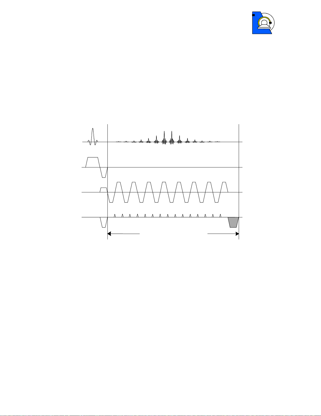
The basic timing scheme of Multi-Echo EPI is shown in Figure 1. It is based on the
original EPI sequence proposed by Mansfield (1). After a single RF-excitation, n
complete EPI images are acquired in a single shot. The phase gradient is rewound to
the original starting position after each echo-image to ensure identical k-space
trajectories for all echo-images (2).
RF
G
S
G
R
G
P
echo-image loop
n
Figure 1 – Basic timing of the Multi-Echo EPI sequence. The shaded gradient in
phase encoding direction ensures identical k-space trajectories for all echo-images.
The resulting echo-images have different increasing echo times and thus T2*weighting. The single echo-images correspond to standard gradient echo EPI images
with the same echo time.
Page 3 of 9 Numaris/4 VA21B
Page 4

Multi-Echo EPI
1.2 Image contrast of Multi-Echo EPI
The signal intensity of the single echo images of the Multi-Echo EPI sequence is
simply given by:
*)2/exp(0 TTESS −=
where S0 is the signal intensity at echo time 0, which corresponds to the proton
density for long repetition times TR, and TE is the echo time. Therefore, the echo
times of the different echo-images should be on the order or less than the tissue T2*.
Parametric images of the proton density and T2* can be obtained by fitting a model
function to the signal intensities at the different echo times (3). Please see the
processing options described below.
Figure 2 – Head images acquired on Magnetom TRIO with the Multi-Echo EPI
sequence with 4 different echo times in a single shot.
1.3 Processing options
Several processing options are given in the method. The different echo images are
always saved separately as the original images and optionally parametric maps can
be calculated in the reconstruction. In addition, online 3D-motion correction (even
prospective – PACE) and t-test can be performed. Please see sequence and
protocols for details.
1.4 References
1. P. Mansfield, A.A. Maudsley. Planar Spin Imaging. J. Magn. Reson. 27, 101
(1977)
2. O. Speck, J. Hennig. Functional Imaging by I
using Multi-Image-EPI. Magn. Reson. Med. 40(2), 243-248 (1998)
- and T2*-Parameter Mapping
0
Page 4 of 9 Numaris/4 VA21B
Page 5

Multi-Echo EPI
3. O. Speck, T. Ernst, L. Chang. Bi-exponential modeling of multi gradient echo
data of the brain. Magn. Reson. Med. 45(6), 1116-1121 (2001)
4. O. Speck, J. Hennig. Motion Correction of Parametric fMRI Data from MultiSlice Single-Shot Multi-Echo Acquisitions, Magn. Reson. Med. 46(5): 10231027 (2001)
2 Software Installation Procedure
2.1 Installation
The sequence consists of 5 files. The sequence file for the host, the sequence file for
the MPCU and the ICE (EVA) program.
Copy the sequence .dll-file for the host to the customer sequence directory:
copy os_mepi2d_VA21.dll %CustomerSeq% (\n4\x86\prod\bin\)
Copy the sequence file for the MPCU to the corresponding sequence directory:
copy os_mepi2d_VA21.m750 \n4\ppc750\prod\bin
!!Attention!! This is only correct for systems with Reco-PC and m750 MPCU!
Otherwise, copy the relevant MPCU file to the corresponding directory. Software
does not run on Alpha reconstruction mac hi n es.
CoilSelectManipulator.h ist ein noch fehlendes File in
C.\idea\n4\comp\Measurement\CCSpuSer\CoilInterface.
Copy the EVA program .dll-files to the customer EVA directory:
copy MotionCorrDecorator.dll q:\bin
copy MosaicDecorator.dll q:\bin
Copy the EVA protocol :
copy t-test_10B10A_moco_mepi.evp
C:\MedCom\MRICustomer\EvaDefProt\Bold
Create a new default protocol:
Open the Exam Explorer, select a Protocol location, select ‘Insert … Sequence’,
select ‘Folder: USER’, select ‘os_mepi2d_VA21’, click ‘Insert’.
Double click the new protocol and adjust the parameters as desired.
The first two and the last steps have to be repeated for the os_mepi2d_pace_VA21
sequence for prospective motion correction.
Page 5 of 9 Numaris/4 VA21B
Page 6

Multi-Echo EPI
Page 6 of 9 Numaris/4 VA21B
Page 7

Multi-Echo EPI
3 Sequences and Prot ocols
3.1 General Remarks
Since the sequence is derived from the standard EPI sequence, most of the user
interface and features are identical to this method. Due to the large number of images
created in a short time, the images are saved to the database in the mosaic format.
One mosaic-image contains all slices of one echo time for one repetition. Repetitions
can be saved in multiple series if desired (Contrast card or BOLD card).
Prior to the first run of the sequence, the 3D standard shim procedure is performed to
ensure best field homogeneity. As with standard EPI this option cannot be disabled.
Due to the high switching rates used in EPI imaging, the peripheral nerve stimulation
monitor will sometimes not allow the measurement to be performed with the
parameters selected. In general, a lower bandwidth will decrease the stimulation and
a different selection of the readout gradient may also help (read direction along L/Raxis shows generally the lowest stimulation).
3.2 New Parameters
Number of echoes: The number of echo-images per excitation can be set on the
sequence card (part1) as the parameter contrasts. The number of echo-images can
be selected between 1 and 8. The number of TE-times is changes accordingly.
Please note that only the first echo time can be changed by the user. All other echo
times are set to the minimum value according to the remaining selected parameters.
!!Attention!!: Even echo time 1 is adjusted to longer times without warning if required
by another parameter! It is not set back to minimum if the parameter is changed back!
Please check echo time 1 after changing other timing relevant parameters of the
sequence.
Page 7 of 9 Numaris/4 VA21B
Page 8

Multi-Echo EPI
Separate Correction: In the source code of the sequence card a new parameter
‘Separate Correction’ has been introduced. If this parameter is enabled (during
compilation) each echo image is preceded by a new set of navigators used for the
Nyquist ghost correction by the reconstruction. If the feature is disabled the same
correction is used for all echo-images. The echo times are somewhat shorter when
this feature is disabled, however, in general the image quality is better with the
separate correction enabled (especially for high imaging bandwidth). In rare cases
the signal in the later echoes is too low and the reconstruction introduces artifacts in
the images (small noisy stripes in the image) if this feature is selected. Please contact
the developer if changes are needed.
T2*-Fit (parameter not shown): This new feature allows online calculation of I0- and
T2*-maps from the echo images according to the model function shown in 1.2. I0 and
T2* are determined for each voxel who’s intensity exceeds 8% of the maximum
intensity in the first echo. I0 and T2* of voxels with a signal intensity below this
threshold are set to 0. This option is automatically and only enabled when
motion correction is selected. The resulting T2*-maps are scaled such that an
image intensity of 1000 corresponds to a T2* of 100ms. T2* is forced to lie within
physiological limits (0<T2*<333ms). Values higher than 333ms are set to 334ms to
avoid very large signal intensity differences in case of fitting errors and allow a T2*resolution of 0.1ms without overflows. Therefore, T2* maps of phantoms with very
long T2 and good homogeneity may be calculated falsely. The T2*-fit is only
performed when Mosaic-images are created (for more than 1 slice)!
Motion Correction: Similar to the motion correction in the a_ep2d_mosaic sequence
a full 3D motion correction can be performed online. In the Multi-Echo EPI sequence
this option also enables the T2*-Fit since the parameter maps are needed for motion
correction. For most accurate results the motion correction is based on the I0-maps,
which are used to determine the motion parameters. Subsequently these parameters
are applied to reorient the T2*-maps. Only the motion corrected T2*-maps are written
to the database together with the original images.
The PACE-variant of the sequence (separate sequence: os_mepi2d_pace_VA21)
allows prospective motion correction. Thus, the volume is rotated and shifted after
each measurement to ensure acquisition in the same patient based reference frame.
Residual motion is corrected for in the processing. This option is only enabled when
the motion correction is selected, which in turn enables the T2*-fit (which is
necessary for motion correction (see above). Be aware that the prospective motion
correction stops if the motion between two scans is too large (as in the product EPIPACE sequence).
Interpolation: The interpolation should be set to ‘k-Space’! (see 3D Filter)
3D Filter: The filter has the same function as in standard EPI (remember: smaller
values cause stronger spatial filtering!).
Page 8 of 9 Numaris/4 VA21B
Page 9

Multi-Echo EPI
t-Test: The online t-test and display work as in the original EPI sequence. However,
the t-test only works properly when motion correction is selected! Alternatively,
T2*-Maps can be processed within the BOLD-card.
3.3 Remarks on reconstructi on performa nce
The number of images that can be acquired with the Multi-Echo EPI method is very
high (up to 30 images per second for 64*64 matrix). The processing (especially T2*Fit and Motion Correction) requires computationally intensive calculations and may
required slower acquisition speeds (increased TR) to be performed online. If the
reconstruction system lags behind the acquisition, the raw data is saved to disk
during acquisition and reconstructed afterwards. Therefore, long reconstruction times
after the scan are possible. The scan is not aborted! However, no new scan can be
started during reconstruction! Do not move the patient table to the home position
during reconstruction, otherwise the reconstruction will stop and data is lost!
The number of images that can be processed online depends on the reconstruction
hardware. Up to 20 slices with 4 echoes and a TR of 3s can be acquired without
motion correction and without long reconstruction delays.
3.4 Protocols
When the default protocol is created, motion correction and t-test are selected!
Please adjust to your needs. Remember, t-test only works correctly when motion
correction is enabled (due to inflexibility in the BOLD task card t-test could not be
disabled for this condition).
As in the standard EPI sequence, the sequence automatically introduces ‘dummy
scans’ at the beginning if more than one measurement is specified! The number of
dummy scans depends on the TR.
Page 9 of 9 Numaris/4 VA21B
 Loading...