

Page 1
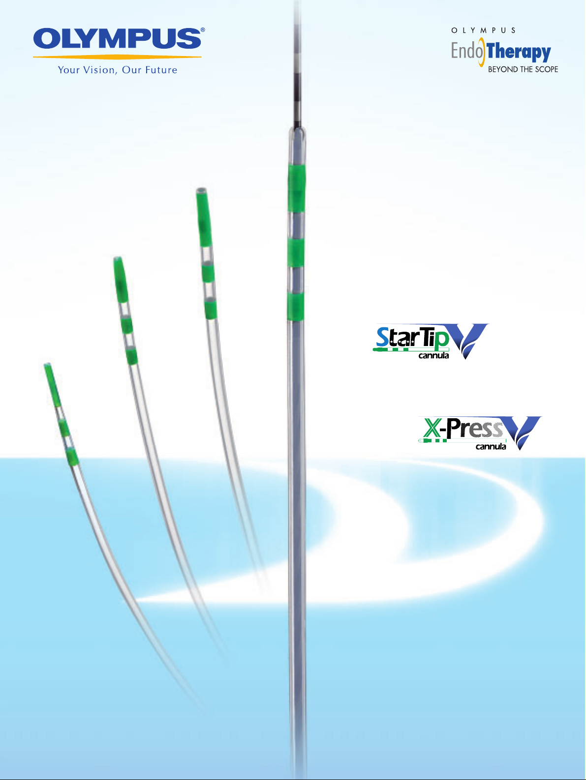
Single Use Cannula V
Single Use Cannula V
Page 2
Taper (PR-V435Q)
Standard (PR-V416Q)
Short taper (PR-V418Q)
Slit (PR-V427Q)
Ball Tip (PR-V223Q)
Taper (PR-V434Q)
Short taper (PR-V414Q)
Long taper (PR-V420Q)
The V-System is a complete system that integrates Olympus endoscopes and EndoTherapy devices. The revolutionary
V-System design offers the option of guidewire manipulation by the physician or the assistant, allows easier exchange
of catheters, and enhances cannulation capability.
The convenient C-Hook allows the device handle to be attached to the endoscope’s control section, putting
it within easy reach of the endoscopist. With the device handle right at hand, the endoscopist can maneuver
the guidewire, inject contrast media, and manipulate the handle — all while keeping a grip on the scope
control section.
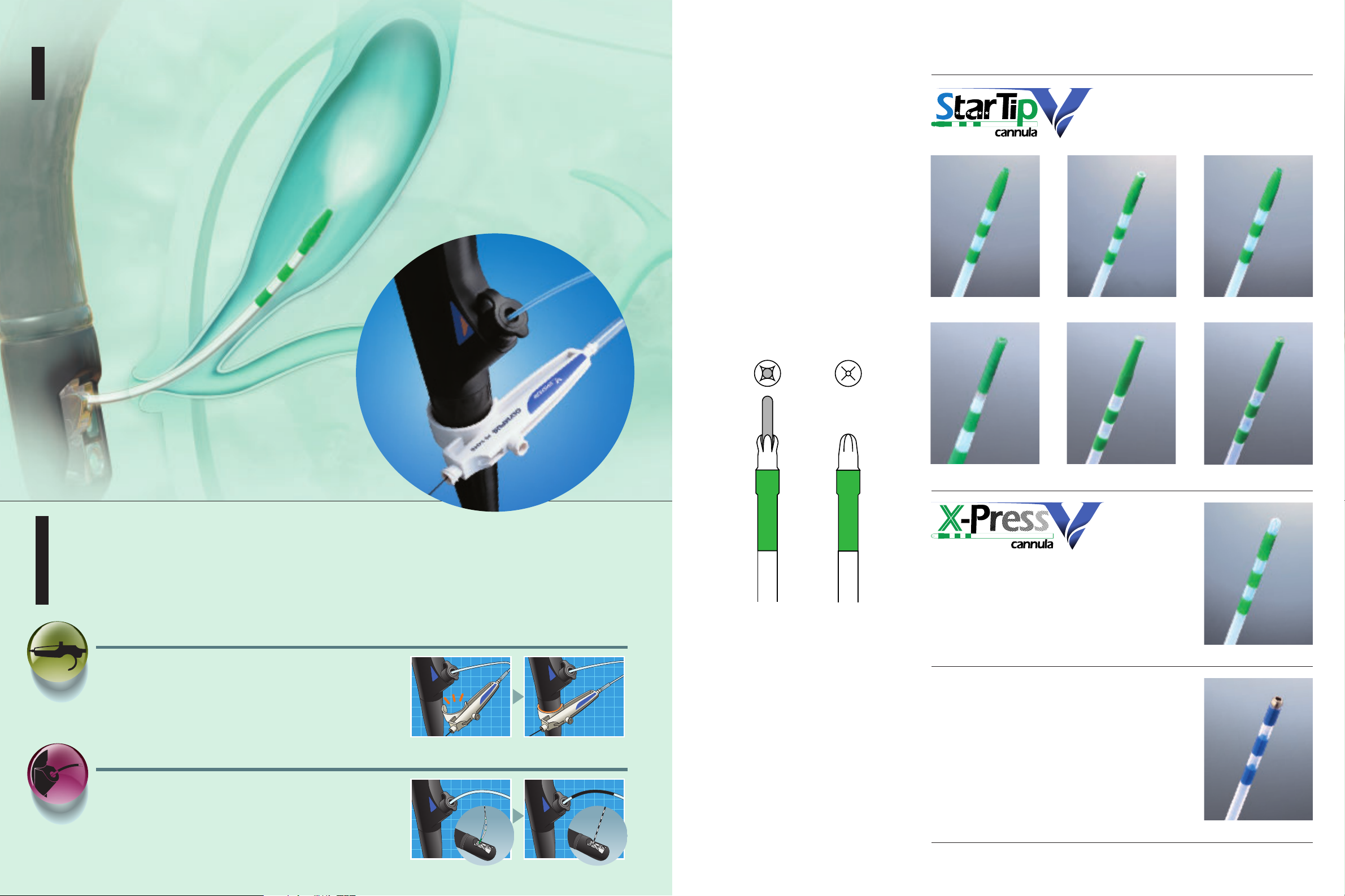
The exclusive V-Marking is located on the proximal side of the sheath. When this marking reaches the
channel port on the scope’s control section, it indicates that the device tip has reached the distal end of
the scope and the V-Groove forceps elevator may be lowered. When withdrawing the device from the
scope, the same marking indicates when to raise the elevator to lock the guidewire.
C-Hook
Now endoscopists have the option to
manipulate guidewires and devices.
V-Marking
Indicates when to raise and lower
the V-Groove forceps elevator.
The Innovative V-System Design Lets You Proceed
with Confidence and Efficiency
The Innovative V-System Exchange Capability Makes
Olympus' ERCP Cannulae Even More Efficient
Excellent visibility under
fluoroscopy
The platinum marking at the distal
end ensures that the tip is clearly
visible under fluoroscopy.
Wide selection of cannulae
A full line-up of 8 models of different
tip designs and diameters is available
to meet every clinical requirement.
Smooth cannulation of
even minor or
constricted papilla
The X-PressV’s cross-cut round
tip is not only ideally suited for
smooth cannulation of the papilla
of Vater, but is also equally effective
for cannulation of the minor or
constricted papilla.
Expanded guidewire
compatibility
Compatible with guidewires of
various sizes.
Comprehensive distal
marking system
Cannulation depth is indicated by
markings at 3mm intervals at the
distal end.
Single-use design for
convenience and reliability
All the cannulae in this line are
designed for single use only.
Standard position of
X-PressV cannula
During guidewire
insertion or fluid injection
Page 3

Specifications, design and accessories are subject to change without any notice or obligation on the part of the manufacturer.
Shinjuku Monolith, 3-1 Nishi-Shinjuku 2-chome, Shinjuku-ku, Tokyo 163-0914, Japan
Postfach 10 49 08, 20034 Hamburg / Wendenstrasse 14-18, 20097 Hamburg, Germany
6100 Blue Lagoon Drive, Suite 390 Miami, Florida 33126-2087, U.S.A
Keymed House, Stock Road, Southend-on-Sea, Essex SS2 5QH, England
491B River Valley Road #12-01/04,Valley Point Office Tower,Singapore 248373
Room 1520-1527, Ocean Centre, 5 Canton Road, Tsimshatsui, Kowloon, Hong Kong
Room.1401, East Tower, Gong Yuan No6 Royal Palace, No6 Gon YuanXijie, JianGuoMenNei, DongCheng District, Beijing, 100005, China
117071, Moscow, Malaya Kaluzhskaya 19, bld. 1, fl.2, Russia
31 Gilby Road, Mount Waverley, VIC., 3149, Australia
2 Corporate Center Drive, Melville, N.Y. 11747-3157, U.S.A.
Spacifications
Model Distal tip diameter Portion DiameterShape of the distal end
1700mm
Working length Compatible guidewire
PR-V414Q
PR-V416Q
PR-V418Q
PR-V420Q
PR-V427Q
PR-V434Q
PR-V435Q
PR-V223Q
2.1mm
2.0mm
1950mm
0.89mm(0.035inch)
0.89mm(0.035inch)
0.64mm(0.025inch)
0.64mm(0.025inch)
0.89mm(0.035inch)
0.89mm(0.035inch)
0.64mm(0.025inch)
0.89mm(0.035inch)
Short taper
Standard
Short taper
Long taper
Slit
Taper
Taper
Ball Tip
4.5Fr
4.0Fr
3.5Fr
3.5Fr
2.5Fr
4.0Fr
3.5Fr
6.0Fr
Printed in Japan F1086SB-3-0804
Page 4
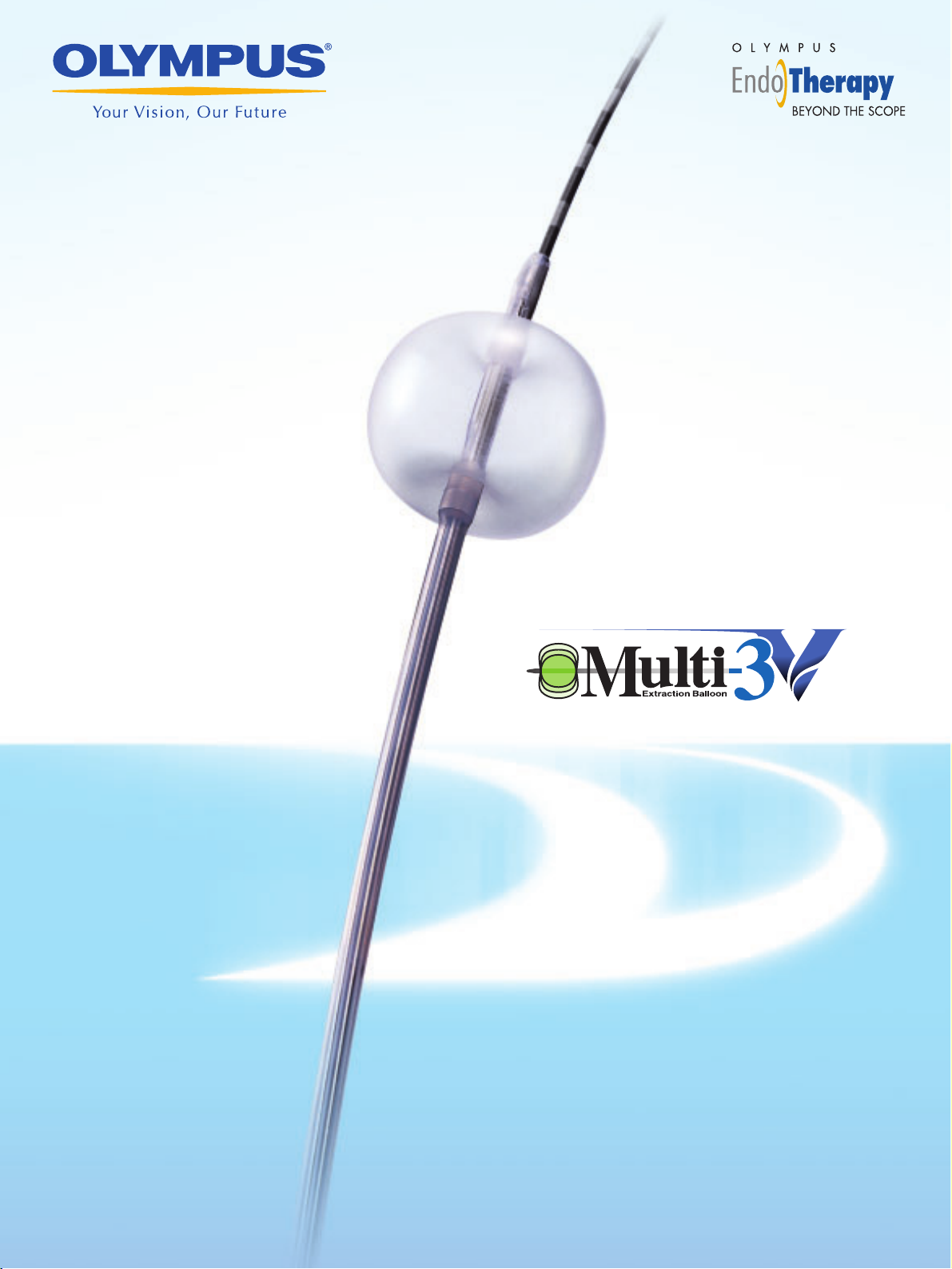
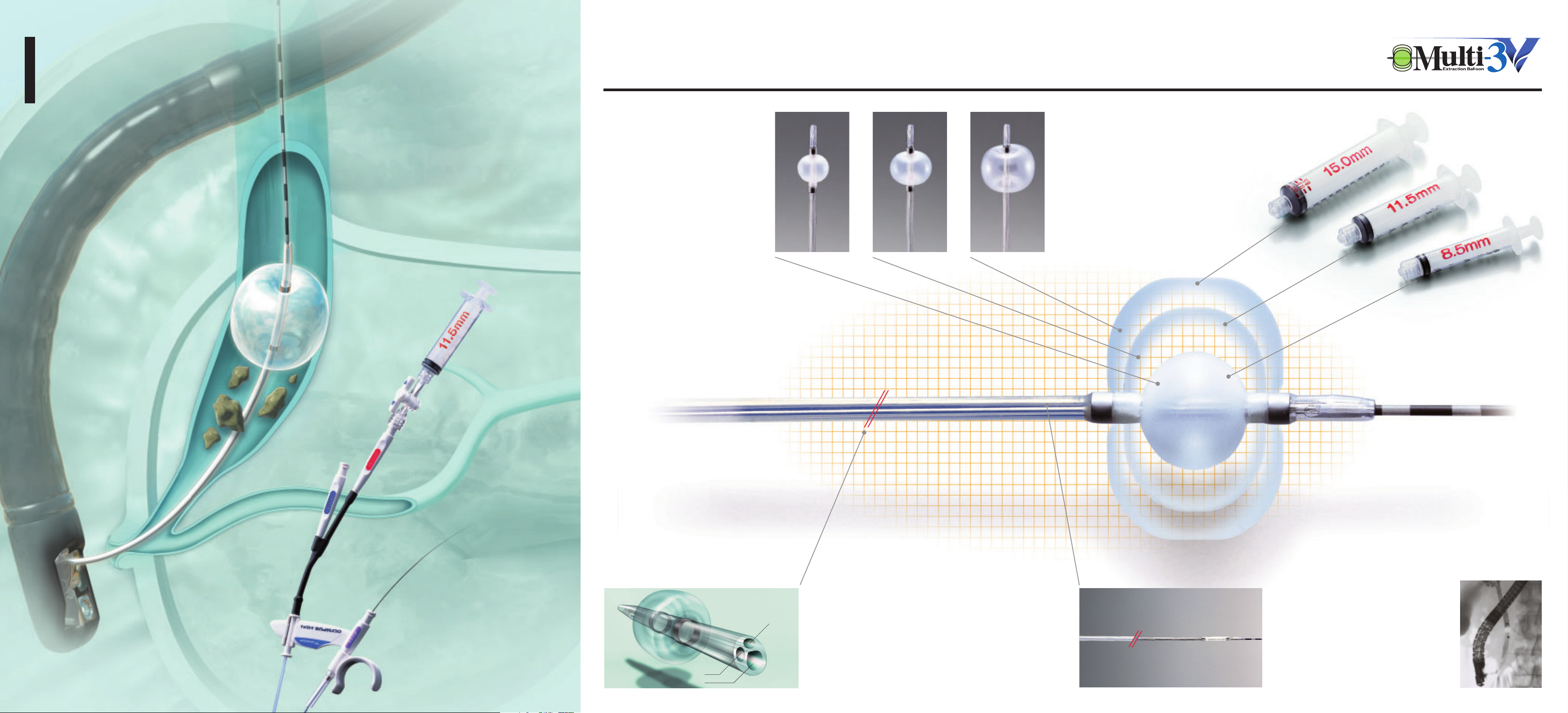
Single Use 3-Lumen Extraction Balloon V
Page 5
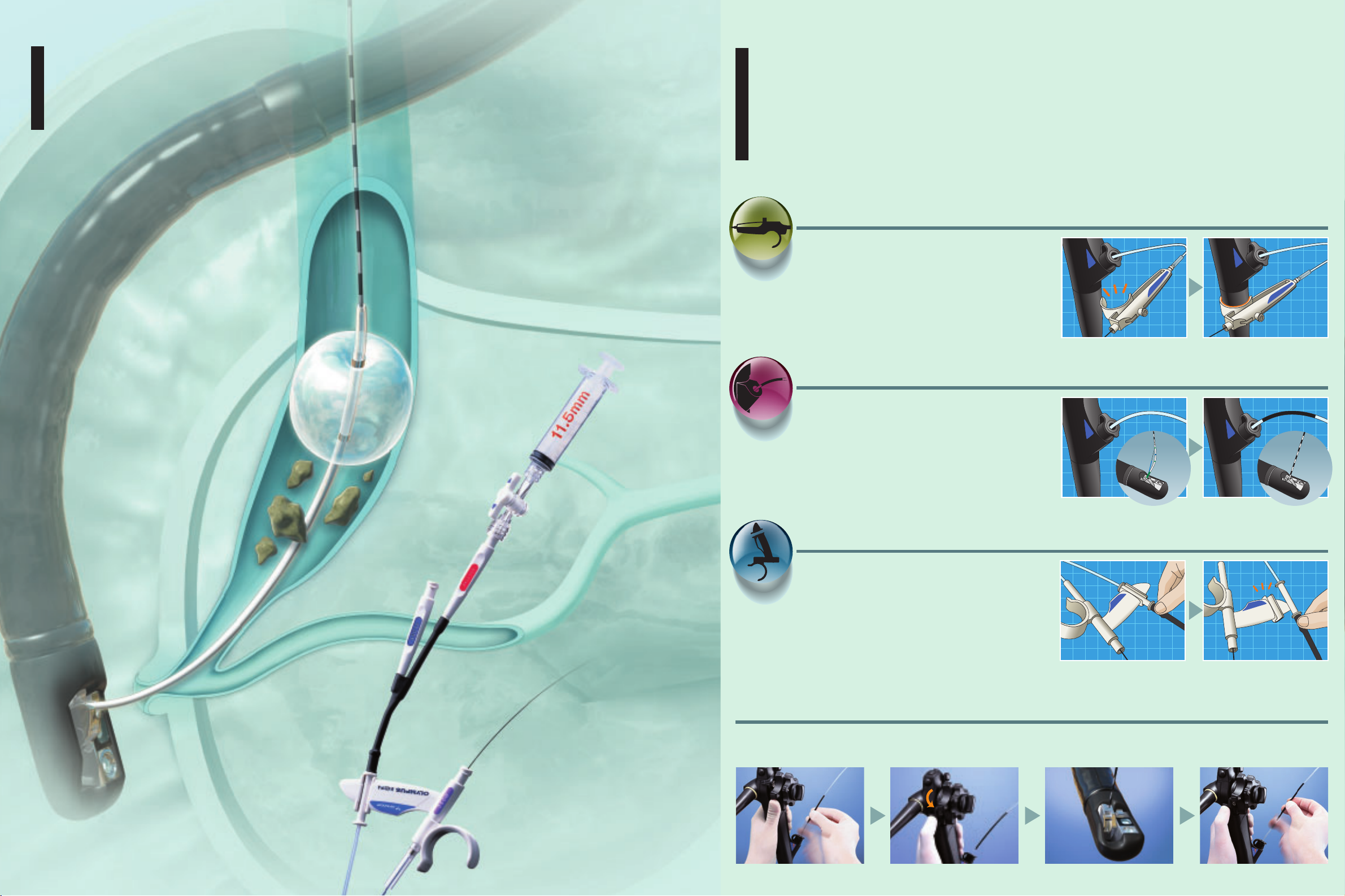
B
The guidewire and injection lumens are separated to
ensure smoother passage of the guidewire. Additionally,
contrast media can be injected without removing the
guidewire. Two models are available — one with the
injection port located above the balloon and the other
with the injection port located below the balloon.
Triple-lumen design allows for
easy passage of the guidewire
The balloon catheter sheath is manufactured from a
special material that allows it for improved insertion
into the papilla, ensuring smoother guidewire passage,
and facilitating contrast injection. The tapered design
— 7 Fr. catheter at the proximal end tapers to 5 Fr. at
the distal end — enables an easy approach to the bile
duct and accommodates a 0.035" guidewire.
Easier cannulation
and tapered sheath design
Multiple balloon sizing
Injection Lumen
0.035" Guidewire Lumen
Air Lumen
The balloon can be inflated to
one of three diameters— 8.5mm,
11.5mm, and 15mm. The balloon size can
be easily adjusted to suit the requirements
and conditions of each case so there is
no need to change catheters during
the procedure.
Reliable, Efficient Stone Extraction with the Multi-Size Balloon Design, Th
Manufactured from a Special Material That Allows for Easier Cannulation,
Tr iple Lumen, Multi Size Balloon with
Revolutionary V-System Exchange Capability for More
Precise Balloon Inflation and Efficient Stone Extraction
V-Marking
The exclusive V-Marking is located on the proximal side of the sheath. When
this marking reaches the channel port on the scope’s control section, it indicates
that the device tip has reached the distal end of the scope and the V-Groove
forceps elevator may be lowered. When withdrawing the device from the scope,
the same marking indicates when to raise the elevator to lock the guidewire.
The V-System is a complete system that integrates Olympus endoscopes and EndoTherapy devices. The revolutionary
V-System design offers the option of guidewire manipulation by the physician or the assistant, allows easier exchange
of catheters, and enhances cannulation capability.
The V-Sheath allows the endoscopist complete device control or, if preferred,
device control may be given to the assistant. The unique device design allows
the guidewire sheath and injection sheath/handle to be separated. This forked
sheath design allows either the endoscopist or the assistant to control the device.
V-Sheath
The Innovative V-System Design Lets You Proceed
with Confidence and Efficiency
V-System device replacement procedure
C-Hook
The convenient C-Hook allows the device handle to be attached to the
endoscope’s control section, putting it within easy reach of the endoscopist.
With the device handle right at hand, the endoscopist can maneuver the
guidewire, inject contrast media, and manipulate the handle — all while keeping
a grip on the scope control section.
Indicates when to raise and lower
the V-Groove forceps elevator.
Device control by the endoscopist
or the assistant.
Confirm the position of the V-Marking
on the V-System EndoTherapy accessory.
Now endoscopists have the option to
manipulate guidewires and devices.
When the V-Marking is completely visible
above the instrument channel port, lift the
forceps elevator to lock the guidewire.
The guidewire is now locked
into the V-Groove.
Completely remove the device.
Page 6
B
The guidewire and injection lumens are separated to
ensure smoother passage of the guidewire. Additionally,
contrast media can be injected without removing the
guidewire. Two models are available — one with the
injection port located above the balloon and the other
with the injection port located below the balloon.
Triple-lumen design allows for
easy passage of the guidewire
The balloon catheter sheath is manufactured from a
special material that allows it for improved insertion
into the papilla, ensuring smoother guidewire passage,
and facilitating contrast injection. The tapered design
— 7 Fr. catheter at the proximal end tapers to 5 Fr. at
the distal end — enables an easy approach to the bile
duct and accommodates a 0.035" guidewire.
Easier cannulation
and tapered sheath design
Dual radiopaque bands
for position confirmation
The 15mm syringe also has markings at 8.5mm and 11.5mm.
This makes it possible to inflate to three balloon sizes using
only one syringe.(This is not pre-measured)
Three pre-measured syringes for reliable inflation
Multiple balloon sizing
Injection Lumen
0.035" Guidewire Lumen
Air Lumen
For easy confirmation of balloon
position during fluoroscopy, two
radiopaque bands are
incorporated — one at the distal
end of the balloon and one at
the proximal end.
Three pre-measured, clearly marked syringes enable precise inflation
of each balloon to the desired size. Much easier and much more
reliable than using a single syringe with different markings,
the pre-measured syringes allow inflation of the balloon
quickly and accurately without having to visually
check the markings on the syringe.
The balloon can be inflated to
one of three diameters— 8.5mm,
11.5mm, and 15mm. The balloon size can
be easily adjusted to suit the requirements
and conditions of each case so there is
no need to change catheters during
the procedure.
Reliable, Efficient Stone Extraction with the Multi-Size Balloon Design, Three Pre-Measured Syringes, and The Sheath is
Manufactured from a Special Material That Allows for Easier Cannulation, Exchange and Injection of Contrast Media.
Tr iple Lumen, Multi Size Balloon with
Revolutionary V-System Exchange Capability for More
Precise Balloon Inflation and Efficient Stone Extraction
Page 7

Spacifications
Model B-V231P-BB-V231P-A
Injection port
Balloon diameter
Sheath
Working length
Minimum channel size
Compatible guidewire
Radiopaque band
Syringes
Below
8.5mm/11.5mm/15.0mm
Distal end 5.5Fr/Proximal end 7Fr
1900mm
2.8mm min
0.89mm(0.035inch)
One band at the distal end and one band
at the proximal end of balloon
Three(3)syringes in different sizes are
contained in the balloon package
Above
8.5mm/11.5mm/15.0mm
Distal end 5.5Fr/Proximal end 7Fr
1900mm
2.8mm min
0.89mm(0.035inch)
One band at the distal end and one band
at the proximal end of balloon
Three(3)syringes in different sizes are
contained in the balloon package
Specifications, design and accessories are subject to change without any notice or obligation on the part of the manufacturer.
Shinjuku Monolith, 3-1 Nishi-Shinjuku 2-chome, Shinjuku-ku, Tokyo 163-0914, Japan
Postfach 10 49 08, 20034 Hamburg / Wendenstrasse 14-18, 20097 Hamburg, Germany
6100 Blue Lagoon Drive, Suite 390 Miami, Florida 33126-2087, U.S.A
Keymed House, Stock Road, Southend-on-Sea, Essex SS2 5QH, England
491B River Valley Road #12-01/04,Valley Point Office Tower,Singapore 248373
Room 1520-1527, Ocean Centre, 5 Canton Road, Tsimshatsui, Kowloon, Hong Kong
Room.1401, East Tower, Gong Yuan No6 Royal Palace, No6 Gon YuanXijie, JianGuoMenNei, DongCheng District, Beijing, 100005, China
117071, Moscow, Malaya Kaluzhskaya 19, bld. 1, fl.2, Russia
31 Gilby Road, Mount Waverley, VIC., 3149, Australia
2 Corporate Center Drive, Melville, N.Y. 11747-3157, U.S.A.
Printed in Japan F1084SB-3-0804
Page 8

SingleUseRetrievalBasketVSingleUseRetrievalBasketV
Page 9

Injection port for contrast media facilitates fluoroscopic visualization.
Sterile single-use design for convenience both before and after procedures.
Eight-wire basket
designed for the
capture and removal
of small stones.
Four-wire basket
design for
versatile retrieval
Rotation mechanism and
bullet-shaped tip
Wire guided ability for
superior insertion capability
The FG-V411Q and FG-V412Q are designed to be wire
guided with the wire passing only through the distal end
of the basket. This makes it possible to insert the basket
into a small incision on the papilla, and allows selective
insertion into the intrahepatic bile duct. In addition,
these baskets have the same expansion capability as our
conventional models(FG-401Q/FG-22Q-1 etc) because
the guidewire is only attached to the basket’s distal tip.
For versatile retrieval of a
wide range of stones, the
TetraCatchV features a
standard four-wire retrieval
basket configuration.
The FlowerBasketV has
eight wires on the distal side
and four wires on the
proximal side. The eight-wire
section allows the precise
retrieval of small stones while
the four-wire section makes
it easier to release them.
V-Marking
The exclusive V-Marking is located on the proximal side of the sheath. When
this marking reaches the channel port on the scope’s control section, it indicates
that the device tip has reached the distal end of the scope and the V-Groove
forceps elevator may be lowered. When withdrawing the device from the scope,
the same marking indicates when to raise the elevator to lock the guidewire.
The Innovative V-System Design Lets You Proceed
with Confidence and Efficiency
Indicates when to raise and lower
the V-Groove forceps elevator.
Revolutionary V-System Device Exchange
is Now Combined with New Basket Designs,
a Rotation Mechanism, and Wire Guided Access
Unique Distal Eight-wire Basket is
Ideal for Retrieval of Small Stones
and with Four Proximal Wires to
Facilitate Release of Captured Stones
Standard Four-wire Basket
Meets Most Stone Retrieval
Requirements in the Bile and
Pancreatic Ducts
The rotatable basket (FG-V401QR / FG-V402QR)
allows the retrieval of small stones, especially
floating stones or stones located in the lower bile
duct above the papilla. The bullet-shaped tip
ensures smooth insertion into the bile duct.
The V-System is a complete system that integrates Olympus endoscopes and EndoTherapy devices. The revolutionary
V-System design offers the option of guidewire manipulation by the physician or the assistant, allows easier exchange
of catheters, and enhances cannulation capability.
Page 10

An emergency device, this is only to be used in
the unlikely event of basket impaction due to an
extremely hard calculus.
Specifications, design and accessories are subject to change without any notice or obligation on the part of the manufacturer.
Shinjuku Monolith, 3-1 Nishi-Shinjuku 2-chome, Shinjuku-ku, Tokyo 163-0914, Japan
Postfach 10 49 08, 20034 Hamburg / Wendenstrasse 14-18, 20097 Hamburg, Germany
6100 Blue Lagoon Drive, Suite 390 Miami, Florida 33126-2087, U.S.A
Keymed House, Stock Road, Southend-on-Sea, Essex SS2 5QH, England
491B River Valley Road #12-01/04,Valley Point Office Tower,Singapore 248373
Room 1520-1527, Ocean Centre, 5 Canton Road, Tsimshatsui, Kowloon, Hong Kong
Room.1401, East Tower, Gong Yuan No6 Royal Palace, No6 Gon YuanXijie, JianGuoMenNei, DongCheng District, Beijing, 100005, China
117071, Moscow, Malaya Kaluzhskaya 19, bld. 1, fl.2, Russia
31 Gilby Road, Mount Waverley, VIC., 3149, Australia
2 Corporate Center Drive, Melville, N.Y. 11747-3157, U.S.A.
BML-110A-1
Printed in Japan F1091SB-3-0804
Spacifications
Model
Rotatable/Guidewire type
Basket type
FG-V401QR
FG-V402QR
FG-V411Q
FG-V412Q
Rotatable type
Rotatable type
Guidewire type
Guidewire type
1950mm
1950mm
1950mm
1950mm
Opening widthWorking length
20mm
22mm
20mm
22mm
Minimum channel size
2.8mm
2.8mm
3.7mm
3.7mm
Compatible guidewire
-
-
0.89mm(0.035inch)
0.89mm(0.035inch)
8wire type
4wire type
8wire type
4wire type
Page 11

Single Use 2-Lumen Sphincterotome V
Single Use 3-Lumen Sphincterotome V
Page 12

Exceptional Cutting Performance and Easy,
Fast Exchange Capability for Enhanced Efficiency
in ERCP Sphincterotomy
Distal marking on the sheath for
improved view field visibility
The distal marking on the sheath
clearly indicates both the center and
cutting position of the knife.
Injection lumen
Guidewire lumen
Cutting wire lumen
Sheath design for stable and
reliable cannulation
Designed to optimize insertion into the
scope, this sheath is narrower at the distal
end and thicker at the proximal end.
This improves handling and ensures smoother
insertion, while also providing excellent
cannulation capability into the papilla.
Easy identification
of ports
The guidewire port and the
injection port are easily
identified by symbols.
Unique Device Design and Attention to Every Detail of
The CleverCut2V and CleverCut3V Sphincterotomes
CleverCut coating enhances safety
Olympus’s signature CleverCut coating on the
proximal end of the cutting wire minimizes damage
to the surrounding tissue. In addition,
CleverCut Coating reduces the risk of electrical
contact between the wire and the endoscope.
The CleverCut3V wire, injection lumen and guidewire
lumen are arranged to allow easier orientation of the
cutting wire for effective sphincterotomy. Since the
injection lumen and the guidewire lumen are completely
separate, contrast media can be smoothly injected with
a guidewire in place.
The CleverCut3V offers excellent
orientation and smooth injection
Features that display
the icon on the top
are available with
CleverCut2V
double-lumen
models. Those
displaying the
bottom icon are
available with
CleverCut3V
triple-lumen models.
V-Marking
The exclusive V-Marking is located on the proximal side of the sheath. When
this marking reaches the channel port on the scope’s control section, it indicates
that the device tip has reached the distal end of the scope and the V-Groove
forceps elevator may be lowered. When withdrawing the device from the scope,
the same marking indicates when to raise the elevator to lock the guidewire.
The V-Sheath allows the endoscopist complete device control or, if preferred,
device control may be given to the assistant. The unique device design allows
the guidewire sheath and injection sheath/handle to be separated. This forked
sheath design allows either the endoscopist or the assistant to control the device.
V-Sheath
The Innovative V-System Design Lets You Proceed
with Confidence and Efficiency
V-System device replacement procedure
C-Hook
The convenient C-Hook allows the device handle to be attached to the
endoscope’s control section, putting it within easy reach of the endoscopist.
With the device handle right at hand, the endoscopist can maneuver the
guidewire, inject contrast media, and manipulate the handle — all while keeping
a grip on the scope control section.
Indicates when to raise and lower
the V-Groove forceps elevator.
Device control by the endoscopist
or the assistant.
Confirm the position of the V-Marking
on the V-System EndoTherapy accessory.
Now endoscopists have the option to
manipulate guidewires and devices.
When the V-Marking is completely visible
above the instrument channel port, lift the
forceps elevator to lock the guidewire.
The guidewire is now locked
into the V-Groove.
Completely remove the device.
The V-System is a complete system that integrates Olympus endoscopes and EndoTherapy devices. The revolutionary
V-System design offers the option of guidewire manipulation by the physician or the assistant, allows easier exchange of
catheters, and enhances cannulation capability.
Page 13

Exceptional Cutting Performance and Easy,
Fast Exchange Capability for Enhanced Efficiency
in ERCP Sphincterotomy
Tapered tip design for smooth insertion
into strictures and the minor papilla
(KD-V431Q series only)
Pre-curved distal end
for easier knife positioning
Distal marking on the sheath for
improved view field visibility
The distal marking on the sheath
clearly indicates both the center and
cutting position of the knife.
Injection lumen
Guidewire lumen
Cutting wire lumen
Cutting wire
Guidewire/Injection lumen
Stiffening wire
The CleverCut2V provides
efficient cannulation capability
The CleverCut2V has two stiffening wires
to provide stable cannulation and orientation.
Sheath design for stable and
reliable cannulation
Designed to optimize insertion into the
scope, this sheath is narrower at the distal
end and thicker at the proximal end.
This improves handling and ensures smoother
insertion, while also providing excellent
cannulation capability into the papilla.
Single-use design for
use-and-dispose convenience
Radiopaque tip markings for
optimal visibility under fluoroscopy
Easy identification
of ports
The guidewire port and the
injection port are easily
identified by symbols.
The tapered tip design is ideally suited for cases in
which cannulation is difficult due to strictures or when
insertion into the minor papilla is required. The tapered
tip CleverCut3V is compatible with a 0.025"
diameter guidewire.
The distal ends of the CleverCut2V and CleverCut3V
are pre-curved to achieve stable cannulation capability.
This distal configuration also facilitates easy positioning
of the knife into the papilla.
The CleverCut2V and CleverCut3V
are designed for single use only.
The radiopaque tips of the CleverCut2V
and CleverCut3V provide excellent
visibility under fluoroscopy.
Unique Device Design and Attention to Every Detail of
The CleverCut2V and CleverCut3V Sphincterotomes
CleverCut coating enhances safety
Olympus’s signature CleverCut coating on the
proximal end of the cutting wire minimizes damage
to the surrounding tissue. In addition,
CleverCut Coating reduces the risk of electrical
contact between the wire and the endoscope.
The CleverCut3V wire, injection lumen and guidewire
lumen are arranged to allow easier orientation of the
cutting wire for effective sphincterotomy. Since the
injection lumen and the guidewire lumen are completely
separate, contrast media can be smoothly injected with
a guidewire in place.
The CleverCut3V offers excellent
orientation and smooth injection
Features that display
the icon on the top
are available with
CleverCut2V
double-lumen
models. Those
displaying the
bottom icon are
available with
CleverCut3V
triple-lumen models.
Page 14

Specifications, design and accessories are subject to change without any notice or obligation on the part of the manufacturer.
Shinjuku Monolith, 3-1 Nishi-Shinjuku 2-chome, Shinjuku-ku, Tokyo 163-0914, Japan
Postfach 10 49 08, 20034 Hamburg / Wendenstrasse 14-18, 20097 Hamburg, Germany
6100 Blue Lagoon Drive, Suite 390 Miami, Florida 33126-2087, U.S.A
Keymed House, Stock Road, Southend-on-Sea, Essex SS2 5QH, England
491B River Valley Road #12-01/04,Valley Point Office Tower,Singapore 248373
Room 1520-1527, Ocean Centre, 5 Canton Road, Tsimshatsui, Kowloon, Hong Kong
Room.1401, East Tower, Gong Yuan No6 Royal Palace, No6 Gon YuanXijie, JianGuoMenNei, DongCheng District, Beijing, 100005, China
117071, Moscow, Malaya Kaluzhskaya 19, bld. 1, fl.2, Russia
31 Gilby Road, Mount Waverley, VIC., 3149, Australia
2 Corporate Center Drive, Melville, N.Y. 11747-3157, U.S.A.
Spacifications
Model
Maximum insertion
portion diameter
Distal tip diameter
Lumen type
KD-V411M-0320
KD-V411M-0330
KD-V411M-0720
KD-V411M-0725
KD-V411M-0730
KD-V411M-1520
KD-V411M-1530
KD-V411M-3030
KD-V431M-0720
KD-V431M-0730
KD-V211M-0720
KD-V211M-0725
KD-V211M-0730
KD-V211M-1520
KD-V211M-1530
KD-V211M-3030
4.5Fr
4.0Fr
4.5Fr
2.5mm
2.1mm
Working length
1700mm
Compatible guidewire
0.89mm(0.035inch)
0.64mm(0.025inch)
0.89mm(0.035inch)
Tip length
3mm
3mm
7mm
7mm
7mm
15mm
15mm
30mm
7mm
7mm
7mm
7mm
7mm
15mm
15mm
30mm
Knife length
20mm
30mm
20mm
25mm
30mm
20mm
30mm
30mm
20mm
30mm
20mm
25mm
30mm
20mm
30mm
30mm
3-Lumen type
2-Lumen type
Printed in Japan F1082SB-3-0804
Page 15

DISPOSABLE GUIDEWIRE
Page 16

An Essential Tool to achieve a New Level of Efficiency
in Biliary-Pancreatic Procedures. Designed to Enhance
the Revolutionary Exchange Capability of the V-System.
130mm
Ring-marker section
Spiral-marker section
Ring-markers are visible from 50mm
to 120mm from the distal end to
help determine duct penetration.
50mm
120mm
400mm
2700mm/4500mm
PTFE Coating
50mm
120mm
400mm
2700mm/4500mm
PTFE Coating
Hydrophilic Coating Length:500mm
Hydrophilic Coating Length:500mm
All Designs are Optimized to Assure Smooth,
Efficient Guidance of Devices to the Bile And Pancreati
Two-step markers ensure visibility
in the endoscopic field of view
Two-step markers on the LinearGuideV make
it easy to confirm the movement and position
of the guidewire within the endoscopic field of view.
The LinearGuideV has been designed to provide
superior insertion capability, maximum endoscopic
visibility, and easy device exchange. When the
LinearGuideV’s spiral markings are visible from
the scope tip, the guidewire can be locked to
simplify device exchange.
Endoscopic visibility of the spiral-
markers indicates that
LinearGuideV may be locked in
the V-Groove.
The Innovative V-System Design Lets You Proceed
with Confidence and Efficiency
Schematic of the V-Groove locking mechanism
V-System device replacement procedure
Conventional scope (TJF-160) V-System scope (TJF-160V)
LinearGuideVGuidewire
V-G roove
The V-System is a complete system that integrates Olympus endoscopes and EndoTherapy devices.
The revolutionary V-System design offers the option of guidewire manipulation by the physician
or the assistant, allows easier exchange of catheters, and enhances cannulation capability.
Confirm the position of the V-Marking
on the V-System EndoTherapy accessory.
When the V-Marking is completely visible
above the instrument channel port, lift the
forceps elevator to lock the guidewire.
The guidewire is now locked
into the V-Groove.
Completely remove the device.
The V-Groove in the V-Scope forceps
elevator locks LinearGuideV in place
When a guidewire slips out of position, it can
be extremely frustrating. With the new
LinearGuideV, unwanted movement is
a thing of the past. Olympus’ V-System
scopes feature a revolutionary V-Groove
in the V-Scope foreceps elevator that
allows LinearGuideV to be securely
locked in place without any special
attachment when extended 130mm from
the distal end of the scope. The approach
to the bile or pancreatic ducts via the
papilla can be accomplished quickly and easily
without worrying about the guidewire slipping.
Page 17

An Essential Tool to achieve a New Level of Efficiency
in Biliary-Pancreatic Procedures. Designed to Enhance
the Revolutionary Exchange Capability of the V-System.
Specially coated to insure
smooth wire manipulation
To ensure smooth passage through tight strictures in the bile
duct, the distal 500mm from the tip is coated with a special
hydrophilic coating that provides exceptional lubricity.
The balance of the guidewire is coated with PTFE for
smoother exchange. These coatings allow the
guidewire to be manipulated easily
130mm
Ring-marker section
Spiral-marker section
Ring-markers are visible from 50mm
to 120mm from the distal end to
help determine duct penetration.
50mm
120mm
400mm
2700mm/4500mm
PTFE Coating
50mm
120mm
400mm
2700mm/4500mm
PTFE Coating
Hydrophilic Coating Length:500mm
Hydrophilic Coating Length:500mm
Note:
This instrument meets the recognized standard for high
frequency electrosurgical leakage current - ANSI/AAMI HF-18(1993),
4.2.5.2 and 5.2.5.2 - when used with an Olympus sphinctetorome.
Removal of the guidewire is not necessary during sphincterotomy.
All Designs are Optimized to Assure Smooth,
Efficient Guidance of Devices to the Bile And Pancreatic Ducts
Two-step markers ensure visibility
in the endoscopic field of view
Two-step markers on the LinearGuideV make
it easy to confirm the movement and position
of the guidewire within the endoscopic field of view.
Two distal tip configurations to suit
different techniques and preferences
The LinearGuideV comes in two tip
configurations — straight and angulated.
The straight tip can be used for a variety of purposes,
while the angulated tip is best suited for passing a
stricture and for selective insertion.
Choose the tip that best suits your techniques
and preferences.
The LinearGuideV has been designed to provide
superior insertion capability, maximum endoscopic
visibility, and easy device exchange. When the
LinearGuideV’s spiral markings are visible from
the scope tip, the guidewire can be locked to
simplify device exchange.
Endoscopic visibility of the spiral-
markers indicates that
LinearGuideV may be locked in
the V-Groove.
Page 18

Specifications, design and accessories are subject to change without any notice or obligation on the part of the manufacturer.
Shinjuku Monolith, 3-1 Nishi-Shinjuku 2-chome, Shinjuku-ku, Tokyo 163-0914, Japan
Postfach 10 49 08, 20034 Hamburg / Wendenstrasse 14-18, 20097 Hamburg, Germany
6100 Blue Lagoon Drive, Suite 390 Miami, Florida 33126-2087, U.S.A
Keymed House, Stock Road, Southend-on-Sea, Essex SS2 5QH, England
491B River Valley Road #12-01/04,Valley Point Office Tower,Singapore 248373
Room 1520-1527, Ocean Centre, 5 Canton Road, Tsimshatsui, Kowloon, Hong Kong
Room.1401, East Tower, Gong Yuan No6 Royal Palace, No6 Gon YuanXijie, JianGuoMenNei, DongCheng District, Beijing, 100005, China
117071, Moscow, Malaya Kaluzhskaya 19, bld. 1, fl.2, Russia
31 Gilby Road, Mount Waverley, VIC., 3149, Australia
2 Corporate Center Drive, Melville, N.Y. 11747-3157, U.S.A.
Printed in Japan F1080SB-3-0804
Spacifications
Model Shape of distal portionWorking lengthOuter diameter
G-V210-3527S
G-V210-3527A
G-V210-3545S
G-V210-3545A
0.89mm(0.035")
2,700mm
4,500mm
Straight
Angulated
Straight
Angulated
Hydrophilic
coating length
500mm
 Loading...