

Page 1
Virtual Microscopy Solutions
Leica Microsystems’ Complete Virtual Microscopy Product Range
Page 2
Of Course We Scan
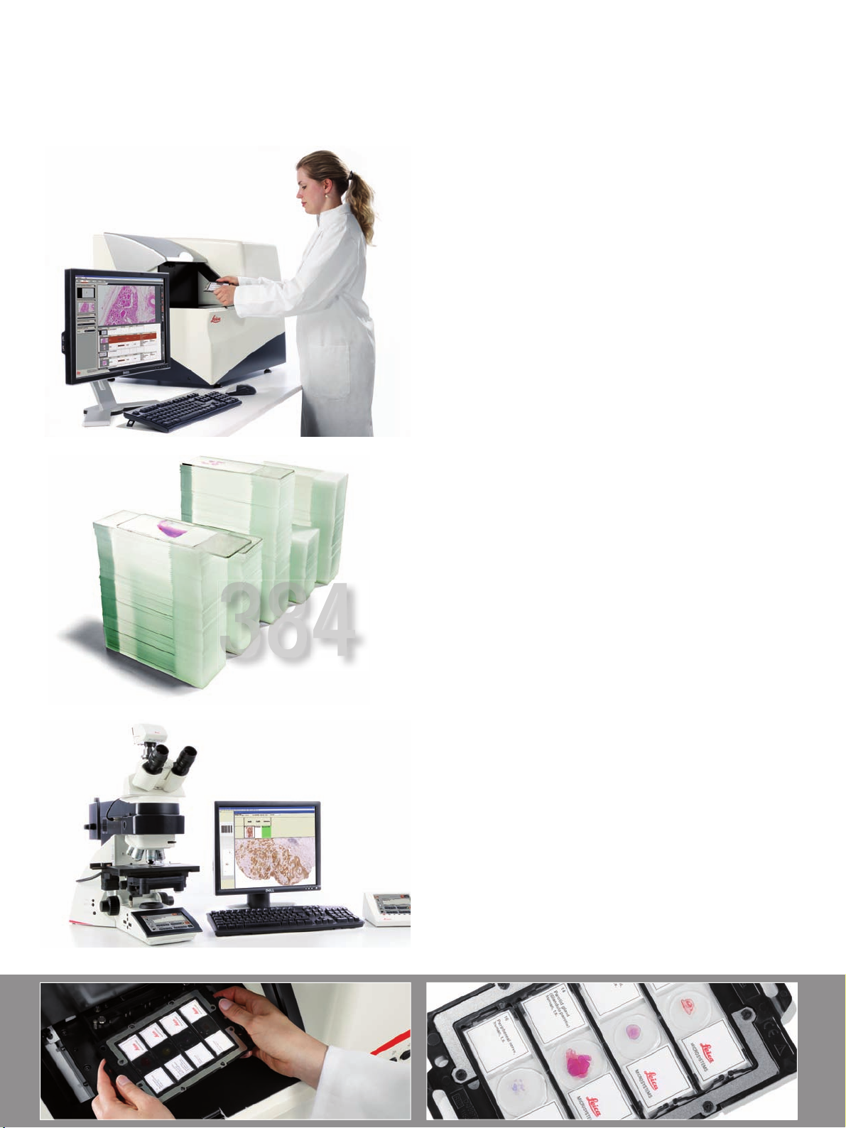
Leica SCN400
The fast, reliable, and fl exible way to scan and digitize your slides
• High resolution – high-quality, high-contrast images
• Always in focus – patented Dynamic Focus Tracking
• Reliable, precise scans – Minimize the risk of failed scans
• Unprecedented rapid scanning speed
Leica SL801
The fast, reliable, and fl exible way to handle large volumes of slides
• Automatically scan up to 384 slides without
• Easy upgrade – Leica SCN400 can be retrofi tted with
• Flexibility – use automatic scanning with user-defi ned
• Convenience – load slides as needed, even during the
with specially designed optics
reveals greater specimen detail
manual interaction
Leica SL801 at the user’s facility
settings or fully interactive mode for complete control
scanning process
Ariol Platform
Unmatched fl exibility in microscope-based slide scanning with
the Ariol system
• Highest quality brightfi eld and fl uorescent slide scanning
• Flexible hardware confi guration with user selected
objectives and fi lters
• Diagnostic image quality with advanced optional
analysis capabilities
• Professional integrated reports with gold standard
scoring protocols
Page 3
Scanning is Just the Beginning
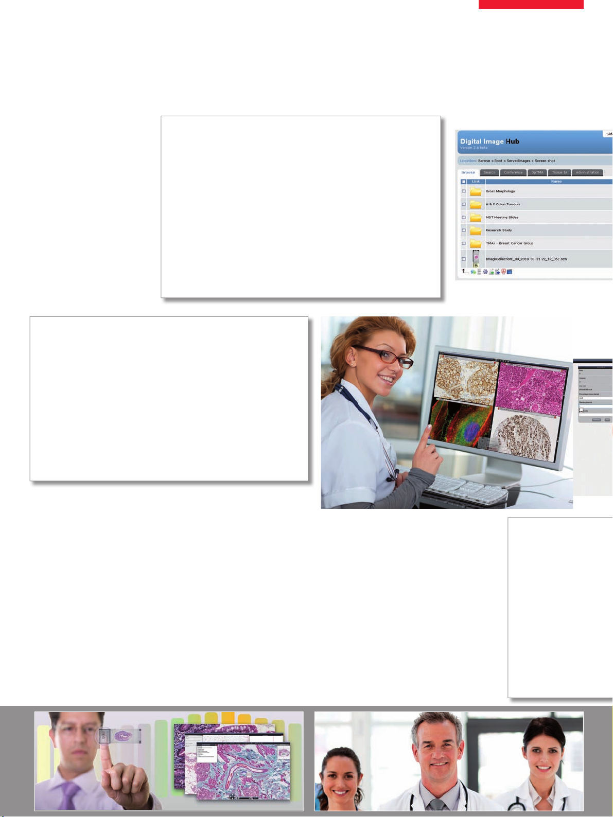
Manage
Use SlidePath’s Digital Image Hub for fl exible online access
• User defi ned image associated data for intuitive archiving,
search and retrieval
• Centralized multi-format image library including digital
slides, DICOM and standard image formats
• Easy to use folder structure for effi cient image management
• Communication protocols for seamless integration
with third party systems
• Flexible storage architecture ensures system scalability
and future proofi ng
Increase Effi ciency
Intuitive software workfl ows reduce time and cost & increase quality
• Rapidly review image cohorts with multi-parametric
searching and high throughput review
• User-defi ned glossaries and dictionaries standardize
nomenclature resulting in accurate data mining and analysis
• Use OpTMA for automatic core identifi cation and rapid core
review to increase TMA throughput and reduce risk of error
• Navigate images, record data and create annotations within
the slide viewer
Share Informat
Rapid access to slides any
• Support multi-users co
image library
• Reduce time taken for
remote slide reading
• Conferencing tool simu
environment for rapid s
• Review multiple image
viewing and improved s
• Online solution require
Page 4

Data Security
Ensure safety, control, and compliance of your complete image
management system
• Multi-level user control ensures correct access rights to
defi ned image and data collections
• Unique user names and encrypted passwords required
for system access
• Comprehensive audit trails and controls for 21 CFR part 11
• Secure system architecture and backup capabilities
e-Learning
Modernize slide-based learning with cutting edge technology
• Reduce costs and increase class size with SlidePath’s Digital
SlideBox
• Maximize teaching effi ciency and standardize course
material – same digital slide instead of similar glass slides
• Integrate rare cases and expose students to cases they
would not see with fragile glass slides
• Students have 24/7 access to course material
• Easy integration of course content, slides, and questionnaires
in a single online system
• Improve quality assurance and continuing professional
development schemes with standardized content and
no shipping time or costs
Page 5

The Image is Not the End
Flexible Image Analysis
for Your Research Laboratory
SlidePath’s Tissue IA provides high-throughput, web-enabled
analysis for digital slides
• Web-deployed software enables control and review of
image analysis from anywhere
• Flexible solution for whole slide, region of interest
or TMA analysis
• Standard algorithms (membrane, nuclear, and positive pixel)
can be adapted for multiple biomarker and tissue types
• Grid computing approach enables whole slide analysis
• Scalable solution to increase processing power
and decrease analysis time
Quantify Biomarkers
for Clinical Diagnostics
The Ariol platform couples high quality imaging with advanced
intelligent analysis
Clinical Her2/neu brightfi eld IHC, ER, PR in breast cancer analysis
• Gold-standard scoring protocols fully supported
(Allred scoring, Histoscoring)
• Unique region-of-interest analysis readily integrates
into existing pathology workfl ows
• Produce case reports with easy export into information
management systems
Clinical Her2/neu FISH analysis
• Capture total gene signal through full z-stack ensures
that all information is analyzed
• Automatic signal counting expressed in an easy to read
per-nucleus table
• Bring fl uorescence analysis out of the dark room and
increase speed of review
• Archive digital images for permanent non-fade records
Original and fi nal image after processing with SlidePath‘s Tissue IA.
Nuclear staining on selected inclusion regions have been classifi ed as strongly
positive (red), moderately positive (orange), weakly positive (yellow) and negative
based on input parameters.
SlidePath applications are not cleared by the FDA , Health Canada or in the EU for diagnostic or clinical use. All applications are intended solely for use in the research or educational setting, such as university or pharmaceutical development. These applications are described as Research Applications or Research Use Only.
Nuclear (ER & PR), Membrane (Her2/neu), and
FISH (Her2 FISH) capture and analysis (from top to bottom).
Page 6

“With the user, for the user”
Leica Microsystems
Leica Microsystems operates globally in four divi sions,
where we rank with the market leaders.
Life Science Division
•
The Leica Microsystems Life Science Division supports the
imaging needs of the scientifi c community with advanced
innovation and technical expertise for the visualization,
measurement, and analysis of microstructures. Our strong
focus on understanding scientifi c applications puts Leica
Microsystems’ customers at the leading edge of science.
Industry Division
•
The Leica Microsystems Industry Division’s focus is to
support customers’ pursuit of the highest quality end result.
Leica Microsystems provide the best and most innovative
imaging systems to see, measure, and analyze the microstructures in routine and research industrial applications,
materials science, quality control, forensic science investigation, and educational applications.
Biosystems Division
•
The Leica Microsystems Biosystems Division brings histopathology labs and researchers the highest-quality,
most comprehensive product range. From patient to pathologist, the range includes the ideal product for each
histology step and high-productivity workfl ow solutions
for the entire lab. With complete histology systems featuring innovative automation and Novocastra™ reagents,
Leica Microsystems creates better patient care through
rapid turnaround, diagnostic confi dence, and close customer collaboration.
Medical Division
•
The Leica Microsystems Medical Division’s focus is to
partner with and support surgeons and their care of patients with the highest-quality, most innovative surgi cal
microscope technology today and into the future.
The statement by Ernst Leitz in 1907, “with the user, for the user,” describes the fruitful collaboration
with end users and driving force of innovation at Leica Microsystems. We have developed fi ve
brand values to live up to this tradition: Pioneering, High-end Quality, Team Spirit, Dedication to
Science, and Continuous Improvement. For us, living up to these values means: Living up to Life.
Active worldwide
Australia: North Ryde Tel. +61 2 8870 3500 Fax +61 2 9878 1055
Austria: Vienna Tel. +43 1 486 80 50 0 Fax +43 1 486 80 50 30
Belgium: Groot Bijgaarden Tel. +32 2 790 98 50 Fax +32 2 790 98 68
Canada: Concord/Ontario Tel. +1 800 248 0123 Fax +1 847 236 3009
Denmark: Ballerup Tel. +45 4454 0101 Fax +45 4454 0111
France: Nanterre Cedex Tel. +33 811 000 664 Fax +33 1 56 05 23 23
Germany: Wetzlar Tel. +49 64 41 29 40 00 Fax +49 64 41 29 41 55
Italy: Milan Tel. +39 02 574 861 Fax +39 02 574 03392
Japan: Tokyo Tel. +81 3 5421 2800 Fax +81 3 5421 2896
Korea: Seoul Tel. +82 2 514 65 43 Fax +82 2 514 65 48
Netherlands: Rijswijk Tel. +31 70 4132 100 Fax +31 70 4132 109
People’s Rep. of China: Hong Kong Tel. +852 2564 6699 Fax +852 2564 4163
Shanghai Tel. +86 21 6387 6606 Fax +86 21 6387 6698
Portugal: Lisbon Tel. +351 21 388 9112 Fax +351 21 385 4668
Singapore Tel. +65 6779 7823 Fax +65 6773 0628
Spain: Barcelona Tel. +34 93 494 95 30 Fax +34 93 494 95 32
Sweden: Kista Tel. +46 8 625 45 45 Fax +46 8 625 45 10
Switzerland: Heerbrugg Tel. +41 71 726 34 34 Fax +41 71 726 34 44
United Kingdom: Milton Keynes Tel. +44 800 298 2344 Fax +44 1908 246312
USA: Buffalo Grove/lllinois Tel. +1 800 248 0123 Fax +1 847 236 3009
and representatives in more than 100 countries
by Leica Microsystems CMS GmbH, Wetzlar, Germany, 2011
©
is a registered trademark of Genetix Europe Limited, Unit 7, Queens Park, Queensway North, Gateshead, Tyne and Wear, NE11 0QD UK
®
Order no.: English 914 722 • X/11/DX/Br.H. • Ariol
The Ariol Hersight, ERsight, PRsight and PathVysion (tm) are for in vitro diagnostic use, all other assays are for research only. Not for use in diagnostic procedures.
LEICA and the Leica Logo are registered trademarks of Leica Microsystems IR GmbH. • Copyright
www.leica-microsystems.com
 Loading...