

Page 1
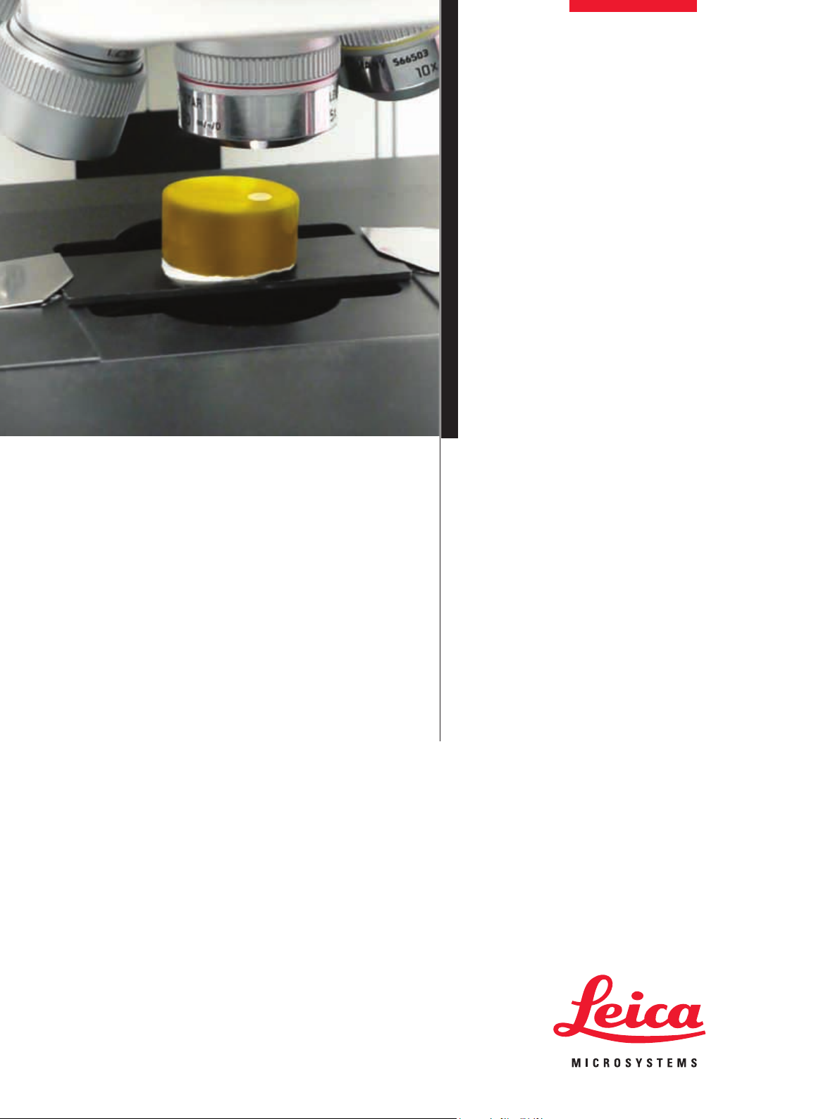
LAS Power
Mosaic
Page 2
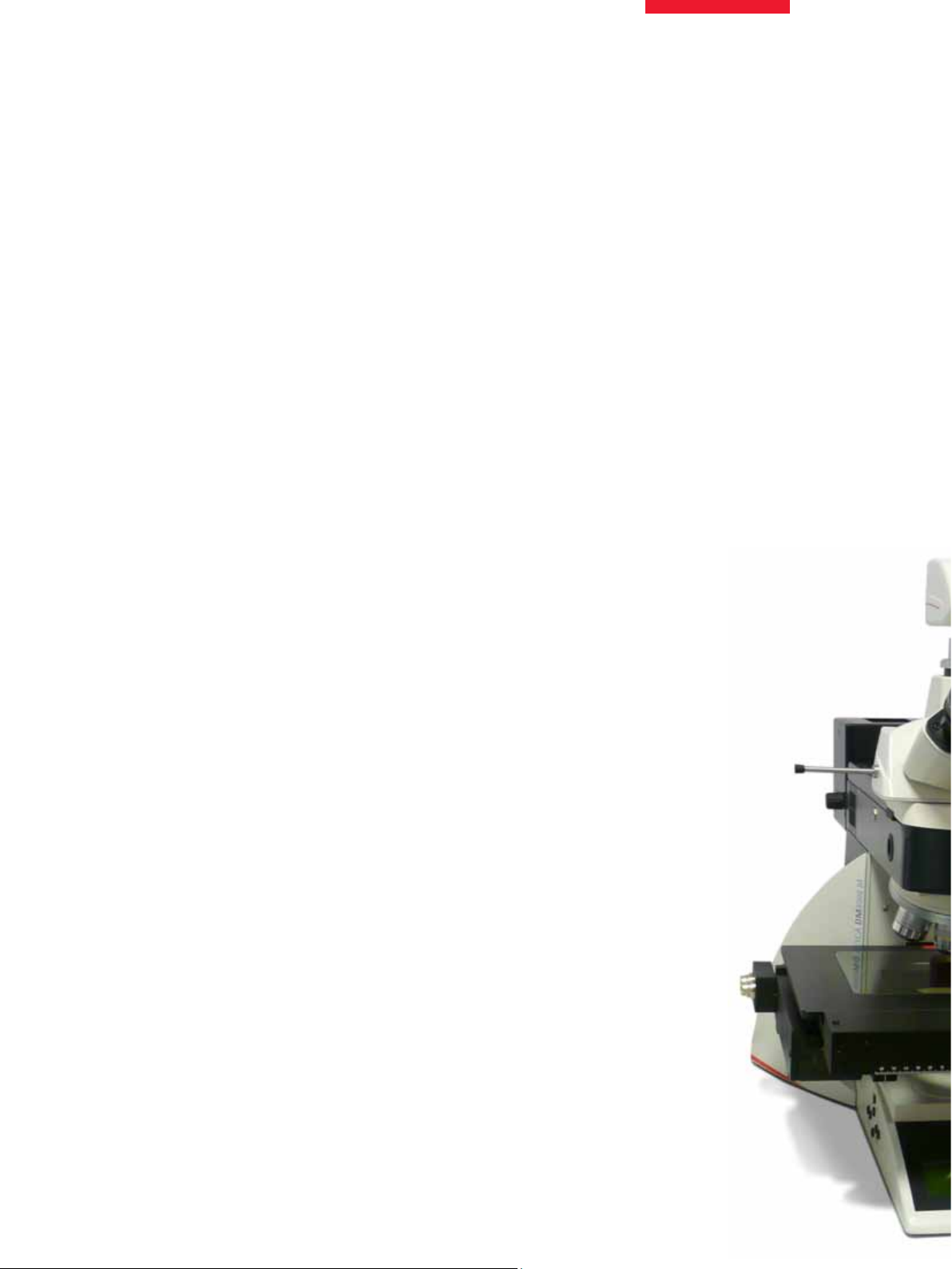
Get the Big Picture
LAS Power Mosaic is designed to bring a whole
new dimension to microscopy, providing you
with a unique understanding of the relationship
between microscopic features and the overall
structure of the specimen.
LAS Power Mosaic provides the ultimate in
high-resolution specimen visualisation. Scan
the entire specimen or select a specific region
of interest region and it’s scanned at high
speed. The captured images are immediately
combined to form a seamless mosaic image.
Once the scan is complete you can relocate
effortlessly to areas of interest with a single
mouse click and can view the live microscope
image that corresponds to a chosen location.
Additionally you can zoom and pan around the
entire digitised mosaic image using easy and
intuitive “browsing” tools.
The acquired images, including the entire
mosaic can be saved for subsequent review,
discussion with colleagues or publication.
LAS Power Mosaic is ideal for both routine
and research applications and provides
modes of scanning to suit all forms of optical
microscopy. It’s specifically developed to be
both versatile and easy to use. This powerful
LAS option provides the microscopist with a
high performance mosaic imaging solution
without compromising conventional use of the
microscope.
Key benefits include:
�
Scanning and acquisition at camera
frame rates to ensure high speed and
rapid throughput
�
Tile edge merging provides highest
possible image quality
�
No restrictions on objective or imaging
method
�
Fast and accurate relocation with easy
and intuitive review tools
�
Intelligent memory management
supports mosaic sizes limited only by
available disk space
�
Fully automated focus control and
tracking
�
User control of image size to ensure
suitability for publication and archiving.
�
One click calibration for easy camera
alignment
�
Optional advanced functions, including
powerful 3-D Z-Stack mosaic acquisition
for powerful focus visualisation
Z-Stack Mosaic
In addition to the standard scanning and review
capabilities, the Power Mosaic Plus module
further extends the capability using “Z-Stack”
acquisition to provide 3-D mosaic imaging and
visualisation, ideally suited to specimens with a
wide focal range.
Page 3
Features at a Glance
Power Mosaic Scanning
�
Uses triggered image capture for fast
continuous scan and acquire
�
Standard scan available using step and
acquire for low light applications
�
Image streaming for mosaic sizes only
limited by disk space
�
Additional scans can be easily added to
extend an initial scan
Scan Patterns
�
Rectangle, Circular, Annular, Cross (+ and x),
or Random
�
Overlap of tiles allows joins to be merged
smoothly
�
Camera rotation is automatically corrected
�
Create a scan pattern interactively or by
entering the exact details
Microscope Automation
�
An Oasis XY stage and Z focus control drive
board is used
�
A software joystick or Leica smart move can
be used for stage and focus movement
�
Fully compatible with Leica Microsystems
LAS configured microscopes handling focus,
turret, condenser and lamp controls as
available
Leica DFC Camera
�
Exposure, saturation, gain and gamma
control from LAS controls
�
Triggered acquisition from progressive scan
and DFC FX cameras for fastest scans
�
Automatic and manual white balance
�
Color or monochrome acquisition (8 or 16-bit)
�
Shading correction for smooth mosaic
results
Page 4

How it Works
When the system is initially configured a simple,
single click calibration procedure is performed
for each objective lens on the microscope. This
informs the system of the exact correlation
between a stage movement and the associated
spatial value in microns for the objective. This
calibration information enables the system
to maintain precise and accurate positional
information for each image acquired. In
particular, this also compensates for camera
rotation and stage orientation.
In addition to the spatial calibration of the
system, “shading correction” is set-up to
eliminate any uneven illumination in the acquired
images. Finally the user may, optionally, employ
a number of different automatic focussing
techniques as appropriate for the scanning
method and the nature of the specimen.
Once the scan area has been defined the system
automatically calculates the number of images
that need to be acquired to form a complete full
resolution mosaic of the chosen area. The user
selects the type of scan required and simply
starts the scan. The exact nature of the scan
performed is controlled by the user and depends
on the microscopy contrast method employed
and the format of mosaic required. Everything
else is handled automatically by the system,
using previously defined values to determine
speed, focussing method and data format.
When the scan is complete, the user may revisit
points of interest with a single mouse click or
copy all or part of the mosaic for distribution,
publication or discussion with colleagues.
Additionally, since the entire area scanned is
now available in digital form and at full resolution,
the image data is available for annotation or
further analysis within applications active in the
Leica Application Suite.
LAS Power Mosaic requires a Leica motorised
microscope, Leica Digital Camera, motorised
stepper stage, motorized focus with XY control
board, a high performance Windows PC and a
very large hard drive.
Page 5

Rock Section
PCI Board topside using extended focus
Page 6

Time and Space!
The time needed to perform a scan depends
on a number of different factors, such as the
objective lens used, the illumination method
and the area to be scanned. As a general rule,
polished specimens can be done at the highest
speeds whereas scanning of specimens with
surface roughness requiring focus adjustment
on each field or using darkfield illumination must
be performed using the standard scan method.
Some examples of the overall time needed for
different types of scan using predictive focus
are shown in the table below.
Space is also a variable. LAS Power Mosaic
saves all captured digital images to the PC
hard drive. Each individual full resolution
image captured from a Leica digital camera
will require approximately 4.5 Megabytes of
storage space. Since a typical scan may
comprise of many hundreds, or even thousands,
of individual images the storage required can
quickly become several Gigabytes. Fortunately,
modern PC technology makes this possible.
As a general rule the amount of image data that
will result from a scan will increase fourfold
for each doubling of objective magnification.
If, for example, a scan of 100 fields using a 10X
objective results in 400Mb of data then scanning
the same area at 20X magnification will require
1.6Gb of disk storage. You have the choice of
when to save scans permanently and in which
format. If a single camera frame requires about
4Mb of storage then, in the example shown, a
total of 44Mb will be used to store the Z-Stack
data. When executing Z-Stack scans on patterns
with a large number of fields you should be
aware that very large amounts of data can be
generated.
Similarly, since it is necessary in Z-Stack
operation for a number of images to be acquired
at different Z positions at the same XY location,
the total time required for the scan will increase
significantly. There are a number of factors that
may influence the scan time but as a broad
indication the time needed to perform a normal
standard scan should be multiplied by the
number of Z slices + 50% to give an estimated
time for a Z-Stack scan.
Scan Times and Data Volumes (based on DFC 300FX)
Objective C Mount Camera µm/pixel
5X 1X 1392 x 1040 1.29 12 16 97 31 0.49 406
10X 1X 1392 x 1040 0.65 24 32 388 116 1.93 1622
20X 1X 1392 x 1040 0.32 48 64 1553 461 7.18 6490
40X 1X 1392 x 1040 0.16 96 128 1842 1842 30.6 25960
15mm
Fields X
15mm
Fields Y
Total
Fields
Scan Time
Seconds
Scan Time
Minutes
BMP
Image
Size (Mb)
Page 7

Key Applications
LAS Power Mosaic is suitable for a diverse
range of applications, both research and
routine.
Due to the ability to provide appropriate
scanning methods for different microscopy
fields, the system has great versatility
and is used successfully in many different
environments and applications.
Whilst LAS Power Mosaic is the perfect
solution for the scanning of polished material
specimens and thin geological sections, it is
also well suited to recording forensic specimen
slides, inspecting electronic components,
investigating porous media and many other
microscopy applications.
As the system is designed to supplement
and enhance Leica microscopes and digital
cameras it is a very affordable means to achieve
a massive increase in imaging capability and
performance without loss of any conventional
functionality.
Mosaic Review
A separate viewer program is available to
allow Power Mosaic images to be shared
with colleagues without the need to install the
complete LAS.
Page 8

An Extra Dimension!
Many scanning applications are performed on flat
specimen sections and, with the use of either Autofocus
or Predictive Focus, it is possible to achieve a mosaic
image of acceptable quality by acquiring a single
image tile at each location within the scan pattern. The
resulting mosaic image is thus 2 dimensional (X/Y).
In other cases, the characteristics of the specimen
make acquisition of a single image inadequate for
complete visualisation because important detail is
present at different focal positions within a single
image frame. This situation occurs when inspecting
the roughness of coatings and surface preparations,
particle characterisation and when recording hairs or
textile fibres in forensic science.
The Z-Stack technique has been developed as an
optional module specifically for imaging these high
focal depth specimens and can be applied to an entire
scan pattern. Note that due to the nature of Z-Stack
acquisition it is only possible in “Standard” scanning
mode.
How does it work?
The Z-Stack technique acquires a series of images at
a pre-defined Z “spacing” at each location in the scan
pattern. You can control both the Z spacing and the total
number of images within the stack.
The resulting stack of mosaic images can be smoothly
replayed exactly as if the microscope focus knob is
being adjusted. Moreover, a single mosaic representing
the best focus image or the extended focus image can
be instantly played.
20µm
Z+4
Z+2
Z
Z-2
Z-4
Z-6
Z-8
Z-10
Z+10
Z+8
Z+6
If, for example, you require images at 2 micron intervals
over a total focal range of 20 microns the resulting
Z-Stack will comprise 11 images of which the central
image in the stack will always be the point from which
the Z-stack was initiated. This example shows 5 images
above and 5 images below the initial focal position, each
separated from its neighbour by 2 microns.
Specification subject to change
LAS Power Mosaic Module
www.leica-microsystems.com
@
Leica Microsystems (Switzerland) Ltd
Stereo and Macroscope Systems
CH 9435 Heerbrugg
Switzerland
 Loading...