

Page 1
… g o i n g o n e s t e p f u r t h e r
V2027
(4006531_1001179)
Page 2

2
Page 3

®
English Human Cell Structure
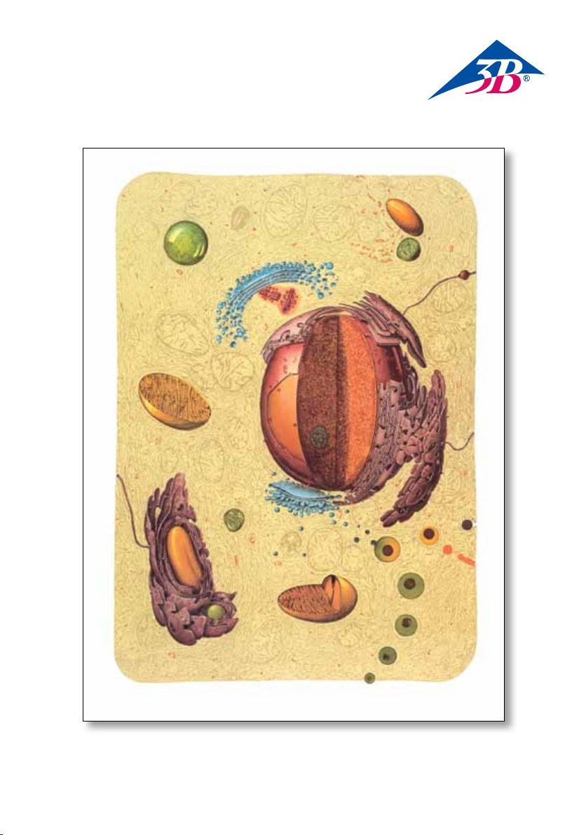
The cell is the smallest viable structural and functional unit of the multicellular organism. While the size
and shape of cells may vary widely, their underlying structural pattern, required for the performance of
vital processes, is based on characteristics which they all have in common. Eukaryont cells have a nucleus.
The cell is enclosed by a membrane (cell membrane, plasma membrane). The space between nucleus and
cell membrane is filled with cell sap (hyaloplasm) interspersed with a continuous membrane-enclosed
reticulum (endoplasmic reticulum); the hyaloplasm also contains different organelles (mitochondria, ribosomes, Golgi apparatus, lysosomes, centrioles). These organelles form the structural basis for the numerous
and manifold coordinated metabolie processes in the cell. The organelles, on the one hand, spatially
separate the individual metabolie reactions, reaction chains, enzymes, substrates and metabolins, and on
the other hand, bring them together in a purposeful way. Each organelle and its constituent parts perform
certain functions in the cell which are linked to characteristic structures (unity of morphological structure
and biochemical function).
1 Cell membrane
2 Nucleus Size: 0.5 - 500 µm
3 Nuclear membrane Thickness: about 80 Å
4 Pores of the nuclear membrane Diametre: 300 -1 000 Å
5 Nucleoles Size normally < 1 µm
6 Perinuclear spaces
7 Mitochondria Length: 0.5 - 5 µm Width: 0.1 - 1 µm
8 Outer membrane of mitochondria
9 Inner membrane of mitochondria
10 Outer matrix of mitochondria
11 Inner matrix of mitochondria
12 Granules in the inner membrane of the matrix Size: 250 - 300 Å
13 Endoplasmic reticulum (ER) (smooth-surfaced, agranulate type)
14 Ergastoplasm (rough-surfaced, granulate variant of the ER)
15 Ribosomes (monomeric ribosomes)
16 Polysomes
17 Golgi apparatus (Golgi complex)
18 Golgi lamellae
19 Golgi vesicles
20 Golgi vacuoles
21 Lysosomes
22 Phagocytosis
23 Pinocytosis
24 Heterophagosome (when destroying foreign bodies)
(Autophagosome, when destroying damaged organelles of the cell)
25 Primary lysosomes
26 Heterophagic vacuole (Autophagic vacuole, when digesting bodies from within the cell)
27 Dense body
28 Residual body
29 Excretion
30 Centrioles Length: about 150 nm Diametre: 300-350 nm
31 Bundle with three fibrils
32 Pedicular umbonate structures
33 Centrosome
Diametre: 250 - 300 Å
3
Page 4

®
DeutschDie Zelle
Die Zelle ist die kleinste, zum selbständigen Leben befähigte Struktur- und Funktionseinheit des vielzelligen Organismus. Größe und Form der Zellen können sehr verschieden sein, in dem zur Ausübung von
Lebensprozessen notwendigen Bauplan weisen sie jedoch grundsätzliche Gemeinsamkeiten auf.
Die Zellen der Eukaryonten sind durch einen Zellkern charakterisiert. Nach außen wird die Zelle von
einer Membran (Zellmembran, Plasmamembran, Plasmalemma) begrenzt. Der Raum zwischen Kern und
Zellmembran wird vom Zellsaft (Zytosol) erfüllt, der von einem kontinuierlichen Membranretikulum
(endoplasmatisches Retikulum) durchsetzt ist; außerdem sind im Zytosol verschiedene Zellorganellen
(Mitochondrien, Ribosomen, Golgi-Apparat, Lysosomen, Zentriolen) lokalisiert. Diese Zellorganellen stellen
die strukturelle Basis für die zahlreichen und vielfältigen, koordiniert ablaufenden Stoffwechselprozesse
der Zelle dar. Durch sie werden einzelne Stoffwechselreaktionen, Reaktionsketten, Enzyme, Substrate
und Stoffwechselprodukte sowohl räumlich voneinander getrennt wie andererseits auch gerichtet zusammengeführt. Jedes Zellorgan und seine Teile haben in der Zelle bestimmte Funktionen zu erfüllen, die in
charakteristischen Strukturen verankert sind (Einheit von morphologischer Struktur und biochemischer
Funktion).
1 Zellmembran
2 Zellkern Größe: 0,5-500 µm
3 Kernmembran Dicke: ca. 80 Å
4 Poren der Kernmembran Durchmesser: 300 - 1000 Å
5 Kernkörperchen Größe: meist < 1 µm
6 Perinukleäre Räume
7 Mitochondrien Länge: 0,5 - 5 µm Breite: 0,1 -1 µm
8 Äußere Membran der Mitochondrien
9 Innere Membran der Mitochondrien
10 Äußere Mitochondrienmatrix
11 Innere Mitochondrienmatrix
12 Granula im inneren Matrixraum Größe: 250 - 300 Å
13 Endoplasmatisches Retikulum (E. R.)
(glattwandige, agranuläre Form)
14 Ergastoplasma (rauhwandige, granuläre Form des E. R.)
15 Ribosomen (monomere Ribosomen)
16 Polysomen (Polyribosomen, Ergosomen)
17 Golgi-Apparat (Golgi-Feld, Golgi-Komplex)
18 Golgi-Zysternen
19 Golgi-Vesikel
20 Golgi-Vakuolen
21 Lysosomen
22 Phagozytose
23 Pinozytose
24 Heterophagosom (bei Abbau von Fremdstoffen) (= Autophagosom, bei Abbau zelleigener Stoffe)
25 Primäre Lysosomen
26 Phagolysom = Heterolysom (= sog. Verdauungsvakuole)
(= Zytolysom = Autolysom, bei Abbau zelleigener Stoffe)
27 Telolysom
28 Restkörper
29 Exkretion
30 Zentriolen Länge: ca. 150 nm Durchmesser: 300 - 350 nm
31 Bündel mit 3 Mikrotubuli
32 Gestielte knopfartige Strukturen
33 Zentrosom
Durchmesser: 250 - 300 Å
4
Page 5

®
Español
La célula es la unidad estructural y de funcionamiento más pequeña del organismo pluricelular capaz de
vida independiente. El tamaño y forma de las células puede ser muy diferente, sin embargo en la estructura necesaria para la ejecución de los procesos vitales muestran rasgos comunes fundamentales.
Las células de los eucariontes se caraterizan por tener núcleo. El exterior de la célula está delimitado por
una membrana (membrana celular, membrana plasmática, plasmalema). El espacio entre el núcleo y la
membrana celular está relleno de citoplasma, que se halla atravesado por una red continuada del retículo
endoplasmático; además de eso, en el citoplasma se hayan los diversos organelos celulares (mitocondrias,
ribosomas, aparato de Golgi, lisosomas, centriolos). Estos organelos celulares representan la base estructural de los diversos procesos que ocurren de un modo coordinado dentro del metabolismo celular. Ellos
son los que se encargan de que las reacciones metabólicas, las cadenas de reacción, encimas, sustratos y
los productos metabólicos se encuentren separados en el espacio y se reunan de manera ordenada. Cada
órgano celular y sus partes tienen una función determinada dentro de la célula, ancladas dentro de sus
respectivas estructuras características (unidad de estructura morfológica y funciones bioquímicas).
1 Membrana celular
2 Núcleo celular Tamaño: 0,5-500 µm
3 Membrana nuclear Espesor: aprox. 80 Å
4 Poros de la membrana nuclear Diámetro: 300 - 1000 Å
5 Nucleolos Tamaño: habitualmente < 1 µm
6 Espacios perinucleares
7 Mitocondrias Longitud: 0,5 - 5 µm Ancho: 0,1 -1 µm
8 Membrana externa de las mitocondrias
9 embrana interna de las mitocondrias
10 Matriz externa de las mitocondrias
11 Matriz interna de las mitocondrias
12 Gránulos en la matriz mitocondrial Tamaño: 250 - 300 Å
13 Retículo endoplasmático (R.E.) (Formada por paredes lisas libres de gránulos)
14 Ergatopalsma (Forma rugosa y de paredes ásperas del retículo endoplasmático).
15 Ribosomas (Ribosomas monómeros)
16 Polisomas (Polisomas, ergosomas)
17 Aparato de Golgi (Campo de Golgi, Complejo de Golgi)
18 Cisternas de Golgi
19 Vesículas de Golgi
20 Vacuolas de Golgi
21 Lisosoma
22 Fagocitosis
23 Pinocitosis
24 Heterofagosoma (El la digestión de cuerpos residuales)
(=Autofagosoma, en la digestión de sustancias propias de la célula)
25 Lisosomas primarios
26 Fagolisosomas = Heterolisosoma (vacuolas digestivas)
(Citosoma = Autolisosoma, en la digestión de sustancias propias de la célula)
27 Telolisosoma
28 Cuerpos residuales
29 Excreciones
30 Centriolos Longitud: aprox. 150nm Diámetro: 300 - 350 nm
31 Haces de 3 microtúbulos
32 Estructuras en forma de botón con pedúnculo
33 Centrosoma
Diámetro: 250 - 300 Å
La célula
5
Page 6

®
La cellule
La cellule est la plus petite unité structurelle et fonctionnelle apte à vivre en autonomie dans l’organisme
qui compte une myriade de cellules. Les tailles et formes des cellules peuvent être très diverses mais elles
présentent des points communs fondamentaux dans leur plan de construction nécessaire aux processus
vitaux. Les cellules des eucaryotes sont caractérisées par un noyau cellulaire. La cellule est limitée vers
l’extérieur par une membrane (membrane cellulaire, membrane plasmatique, plasmalemme). L’espace
compris entre le noyau et la membrane cellulaire est rempli de cytoplasme (cytosol) qui est traversé par
un réticulum endoplasmique ; divers organites cellulaires (mitochondries, ribosomes, appareil de Golgi,
lysosomes, centrioles) sont aussi localisés dans le cytosol. Ces organites cellulaires constituent la base structurelle des multiples processus métaboliques diversifiés de la cellule qui se déroulent en coordination. Ils
permettent de réunir des réactions métaboliques, des chaînes de réaction, des enzymes, des substrats et
des produits métaboliques isolés aussi bien séparément dans l’espace que de manière dirigée. Chacun des
organes cellulaires et ses éléments doivent remplir certaines fonctions qui s’ancrent dans des structures
caractéristiques (unité de structure morphologique et de fonction biochimique).
1 Membrane cellulaire
2 Noyau Taille : 0,5-500 µm
3 Epaisseur de la membrane du noyau : 80 Å
4 Diamètre des pores de la membrane cellulaire : 300 - 1000 Å
5 Nucleoles (sous-compartiment cellulaire du noyau) Taille : la plupart du temps < 1 µm
6 Espaces périnucléaires
7 Mitochondries Longueur : 0,5 - 5 µm Largeur : 0,1 -1 µm
8 Membrane extérieure des mitochondries
9 Membrane intérieure des mitochondries
10 Matrice extérieure des mitochondries
11 Matrice intérieure des mitochondries
12 Granules dans la membrane intérieure de la matrice Taille : 250 - 300 Å
13 Réticulum endoplasmique (R.E.) (organite présent dans les cellules eucaryotes)
14 Ergastoplasme (reticulum endoplasmique granulaire)
15 Ribosomes (ribosomes monomères)
16 Polysomes (polyribosomes, ergosomes)
17 Appareil de Golgi (le corps de Golgi)
18 Lectines marqués de Golgi
19 Vésicules de Golgi
20 Vacuoles de Golgi
21 Lysosomes
22 Phagocytose
23 Pinocytose
24 Hétérophagosome (pour décomposition de substances extérieures)
(= autophagosome pour décomposition de substances propres à la cellule)
25 Lysosomes primaires
26 Heterophagic vacuole (un organite membranaire dans lequel la digestion se
produit dans des cellules capables de phagocytose)
27 Corps dense
28 Corps résiduels
29 Excrétion
30 Centrioles Longueur : environ 150 nm Diamètre : 300 - 350 nm
31 Pack de 3 fibrilles
32 Structures pédonculées en forme de boutons
33 Centrosome
Diamètre : 250 - 300 Å
Français
6
Page 7

®
Português
A célula é a menor unidade estrutural e funcional viável dos organismos multicelulares. Enquanto o
tamanho e a forma das células podem variar muito, seu padrão estrutural de base, necessária ao desempenho dos processos vitais, é baseado em características comuns a todos as células. As células eucariontes
apresentam um núcleo celular. A célula é delimitada por uma membrana (membrana celular, membrana
plasmática). O espaço entre o núcleo e a membrana celular é preenchido pelo citoplasma (hialoplasma),
que é intercalado por um retículo membranoso contínuo (retículo endoplasmático); além disso, o hialoplasma contém diversas organelas celulares (mitocôndrias, ribossomos, aparelho de Golgi, lisossomos,
centríolos). Estas organelas celulares constituem a base estrutural para os numerosos e múltiplos processos
metabólicos coordenados da célula. As organelas, por um lado, separam espacialmente as reações metabólicas individuais, reações em cadeia, enzimas, substratos e metabólitos, e por outro lado, reúne todos
esses elementos de forma proposital. Cada órganela celular e seus componentes desempenham determinadas funções na célula, que estão conectadas a estruturas características (unidade de estrutura morfológica
e função bioquímica).
1 Membrana celular
2 Tamanho do núcleo: 0,5-500 µm
3 Membrana nuclear espessura: aprox. 80 Å
4 Poros nucleares diâmetro: 300 - 1000 Å
5 Nucléolo dimensão: geralmente < 1 µm
6 Espaços perinucleares
7 Mitocôndrias comprimento: 0,5 - 5 µm largura: 0,1 -1 µm
8 Membrana externa das mitocôndrias
9 Membrana interna das mitocôndrias
10 Matriz externa das mitocôndrias
11 Matriz interna das mitocôndrias
12 Grânulos na membrana interna da matriz; dimensão: 250 - 300 Å
13 Retículo endoplasmático (R. E.) (forma lisa não granular)
14 Ergastoplasma (forma rugosa granular do E. R.)
15 Ribossomos (ribossomos monômeros)
16 Polissomos
17 Aparelho de Golgi (Complexo de Golgi)
18 Lamelas de Golgi
19 Vesículas de Golgi
20 Vacúolos de Golgi
21 Lisossomos
22 Fagocitose
23 Pinocitose
24 Heterofagossomo (na digestão de corpos estranhos)
(Autofagossomo, na digestão de organelas danificadas da célula)
25 Lisossomos primários
26 Vacúolo heterofágico (Vacúolo autofágico, na digestão de estruturas de dentro da célula)
27 Corpo denso
28 Corpo residual
29 Excreção
30 Centríolos comprimento: aprox. 150 nm diâmetro: 300 - 350 nm
31 Conjunto com três fibrilas
32 Estruturas pediculares em forma de botão
33 Centrossomo
A Estrutura da Célula Humana
diâmetro: 250 - 300 Å
7
Page 8

1 18 17 19 20 15 16 7
21
13
30
14
21
33
13
7
A
6
4
2
7
5
3
14
22
24
7
B
23
26
25
27
21
8
7
28298917
Page 9

A
9 11 10 8
30
16
15
32
12
31
B
14
15 16
15
15
16
9
Page 10

®
La cellula
La cellula è la più piccola unità strutturale e funzionale autosufficiente dell’organismo multicellulare.
Benché le dimensioni e la forma possano variare molto, lo schema di base necessario all’esecuzione
dei processi vitali si fonda su caratteristiche comuni. Le cellule degli eucarioti sono caratterizzate da un
nucleo. All’esterno, la cellula è delimitata da una membrana (membrana cellulare, membrana plasmatica,
plasmalemma). L’area tra il nucleo e la membrana cellulare è colmata dal liquido intracellulare (citosol),
attraversato da reticolo membranoso continuo (reticolo endoplasmatico). Inoltre, all’interno del citosol
si trovano diversi organelli cellulari (mitocondri, ribosomi, apparato del Golgi, lisosomi, centrioli), i quali
costituiscono la base strutturale per i numerosi e svariati processi metabolici della cellula, che avvengono
in modo coordinato. Da un lato, gli organelli dividono a livello spaziale le singole reazioni metaboliche, le
reazioni a catena, gli enzimi, i substrati e i metaboliti, dall’altro, li riuniscono secondo uno scopo preciso.
Ogni organello e le sue parti costituenti hanno una precisa funzione da assolvere, collegata a strutture
caratteristiche (insieme di struttura morfologica e funzione biochimica).
1 Membrana cellulare
2 Nucleo Dimensioni: 0,5-500 µm
3 Membrana nucleare Spessore: ca. 80 Å
4 Pori della membrana nucleare Diametro: 300-1000 Å
5 Nucleoli Dimensioni: di norma < 1 µm
6 Spazio perinucleare
7 Mitocondri Lunghezza: 0,5-5 µm Larghezza: 0,1-1 µm
8 Membrana esterna dei mitocondri
9 Membrana interna dei mitocondri
10 Matrice mitocondriale esterna
11 Matrice mitocondriale interna
12 Granuli nella membrana interna della matrice Dimensioni: 250-300 Å
13 Reticolo endoplasmatico (RE) (tipo liscio, agranulare)
14 Ergastoplasma (tipo rugoso, granulare del RE)
15 Ribosomi (ribosomi monomerici)
16 Polisomi (poliribosomi, ergasomi)
17 Apparato del Golgi (complesso del Golgi)
18 Cisterne del Golgi
19 Vescicole del Golgi
20 Vacuoli del Golgi
21 Lisosomi
22 Fagocitosi
23 Pinocitosi
24 Eterofagosoma (quando distrugge corpi estranei,
autofagosoma quando distrugge corpi appartenenti alla cellula)
25 Lisosomi primari
26 Fagolisoma, eterolisoma (cosid. vacuolo autofagico,
citolisoma, autolisoma quando distrugge corpi appartenenti alla cellula)
27 Telolisoma
28 Corpi residui
29 Secrezione
30 Centrioli Lunghezza: ca. 150 nm Diametro: 300-350 nm
31 Fascio con 3 microtubuli
32 Strutture peduncolari umbonate
33 Centrosoma
Diametro: 250-300 Å
EnglishItaliano
10
Page 11

日本語
細胞とは多細胞生物をにおける最小の構成単位かつ機能単位です。
大きさや形状は細胞により大きく異なりますが,基礎となる構造様式,生存するうえで必要な機能,といった
基本的な特徴は全て共通しています。
細胞は膜(細胞膜,原形質膜)に包まれ,真核細胞では内部に核を持ちます。
細胞膜と核の間は細胞液(細胞質基質)で満たされ,液中には膜で包まれた網状組織(小胞体)が散在してい
ます。細胞質基質内にはその他にも多くのオルガネラ(細胞小器官:ミトコンドリア,リボソーム,ゴルジ
体,リソソーム,中心小体)が存在しています。
これらの細胞小器官が多数かつ多様な組織的代謝経路の基礎を形成しています。
細胞小器官は個別に代謝反応,反応連鎖を行い,酵素,基質,代謝産物を産生しますが,一方でそれらを目的
にそってまとめます。
細胞小器官と構成要素は,それぞれの特徴的な構造(形態的構造と生化学的機能)と結びつく特定の機能を細
胞内で示します。
1 細胞膜
2 核-大きさ:0.5-500μm
3 核膜-厚さ:約80Å
4 核膜孔-直径:300- 1000Å
5 核小体-通常<1μm
6 核周囲空間
7 ミトコンドリア-長さ:0.5-5μm,幅:0.1-1μm
8 ミトコンドリア外膜
9 ミトコンドリア内膜
10 膜間部分
11 マトリックス
12 マトリックス内の顆粒-大きさ:250 - 300Å
13 小胞体(ER)(滑面小胞体)
14 エルガストプラスム(粗面小胞体)
15 リボソーム
16 ポリソーム
17 ゴルジ体(ゴルジ複合体)
18 ゴルジ層板
19 ゴルジ小胞
20 ゴルジ液胞
21 リソソーム
22 食作用(ファゴサイトーシス)
23 飲作用(ピノサイトーシス)
24 異食胞(ヘテロファゴソーム:取り込んだ外部のものを分解)
自食胞(オートファゴソーム:細胞内の損傷した小器官を分解)
25 一次リソソーム
26 異食作用胞
自食作用胞(細胞内の器官を分解中)
27 濃密体
28 残余小体
29 分泌
30 中心小体-長さ:約150nm,直径:300 - 350nm
31 三連微小管(トリプレット微小管)
32 ダイニン
33 中心体
直径: 250 - 300 Å
ヒトの細胞の構造
11
Page 12

®
Строение клетки человека
Клетка является наименьшей функционально-структурной единицей многоклеточного организма. В
то время как размер и форма клеток может изменяться в широких пределах, лежащая в их основе
структурная модель, которая необходима для осуществления жизненных процессов, основана на общих
для всех клеток характеристиках. Эукариотические клетки имеют ядро. Клетка окружена мембраной
(клеточной мембраной, плазматической мембраной). Пространство между ядром и клеточной
мембраной заполнено клеточным соком (гиалоплазма) с вкраплениями покрытого непрерывной
мембраной ретикулума (эндоплазматический ретикулум); гиалоплазма также содержит различные
органеллы (митохондрии, рибосомы, аппарат Гольджи, лизосомы, центриоли). Эти органеллы формируют
структурную основу для многочисленных и разнообразно скоординированных метаболических
процессов в клетке. С одной стороны органеллы пространственно разделяют отдельные метаболические
реакции, цепные реакции, ферменты, субстраты и метаболиты, а с другой стороны, служат их
целенаправленному объединению. Каждая органелла и ее составные части осуществляют в клетке
определенные функции, которые связаны с характерными структурами (единство морфологической
структуры и биохимической функции).
1 Клеточная мембрана
2 Размер ядра: 0,5 – 500 мкм
3 Толщина ядерной мембраны: около 80 Å
4 Диаметр пор в ядерной мембране: 300 – 1000 Å
5 Ядрышки Обычный размер < 1 мкм
6 Перинуклеарное пространство
7 Длина митохондрии: 0,5 – 5 мкм Ширина: 0,1 – 1 мкм
8 Наружная мембрана митохондрии
9 Внутренняя мембрана митохондрии
10 Наружный матрикс митохондрии
11 Внутренний матрикс митохондрии
12 Гранулы внутренней мембраны матрикса Размер: 250 – 300 Å
13 Эндоплазматический ретикулум (ЭР) (гладкая поверхность, агранулярный тип)
14 Эргастоплазма (шероховатая поверхность, вариант гранулярного ЭР)
15 Рибосомы (мономерные рибосомы)
16 Полисомы
17 Аппарат Гольджи (комплекс Гольджи)
18 Пластинки Гольджи
19 Пузырьки Гольджи
20 Вакуоли Гольджи
21 Лизосомы
22 Фагоцитоз
23 Пиноцитоз
24 Гетерофагосома (при уничтожении инородных тел)
(аутофагосома, при повреждении клеточных органелл)
25 Первичные лизосомы
26 Гетерофагическая вакуоль (аутофагическая вакуоль,
при выведении переваренного материала за пределы клетки)
27 Плотное тело
28 Остаточное тело
29 Выведение
30 Длина центриолей: около 150 нм Диаметр: 300 – 350 нм
31 Пучок из трех фибрилл (триплет)
32 Сферические структуры на ножке (сателлиты)
33 Центросома
Диаметр: 250 – 300 Å
Русский
12
Page 13

®
英文
细胞是多细胞生物体中最小而有活力的结构和功能单元。虽然不同细胞的大小和形状变化范围大,
但是生命过程所需要的基础结构模式由它们所共有的特性来决定。真核细胞有一个核。细胞被一层
膜包绕(细胞膜、质膜)。细胞核和细胞膜之间的空间填充有细胞液(透明质),中间分布有连续
的膜封闭网(内质网)。透明质中还包含不同的细胞器(线粒体、核糖体、高尔基体、溶酶体、中
心粒)。这些细胞器构成了细胞众多协调代谢过程的结构基础。众多的细胞器,一方面在空间上有
各自的代谢反应、反应链、酶、底物和代谢产物,另一方面为了同一目的而联合在一起。每一细胞
器和它的组成部分在细胞内执行与其特征结构相关的一定的功能(形态结构和生化功能的统一)。
1 细胞膜
2 细胞核,大小:0.5-500微米
3 核膜厚度:约80埃
4 核膜孔直径:300-1000埃
5 核仁,大小通常小于1微米
6 核周间隙
7 线粒体,长度:0.5-5微米,宽度:0.1-1微米
8 线粒体外膜
9 线粒体内膜
10 线粒体外基质
11 线粒体内基质
12 基质内膜颗粒,大小:250-300埃
13 内质网(ER)(表面光滑,非颗粒型)
14 颗粒内质网(表面粗糙,内质网的颗粒变体)
15 核糖体(单体核糖体)
16 多核糖体
17 高尔基体(高尔基复合体)
18 高尔基体薄片
19 高尔基囊泡
20 高尔基体空泡
21 溶酶体
22 吞噬作用
23 胞饮作用
24 异噬体(破坏异物时)(自噬体,破坏细胞内受损的细胞器时)
25 初级溶酶体
26 异体吞噬泡(自体吞噬泡,当消化细胞内的成分时)
27 致密体
28 残体
29 排泄
30 中心粒,长:约150纳米,直径:300-350纳米
31 三根原纤维组成的束
32 弓状凸形结构
33 中心体
直径:250-300埃
人体细胞结构
13
Page 14

®14®
Page 15

15
Page 16

3B Scientific
A wo r l d w i d e g r o u p o f c o m p a n i e s
V2027 (4006531_1001179)-01/13-25002131
3B Sci entific GmbH
Rudorffweg 8 • 21031 Hamburg • Germany
Tel.: + 49-40-73966-0 • Fax: + 49-40-73966-100
www.3bscientific.com • 3b@3bscientific.com
© Copyright 2002 / 2013 for instruction manual and design of product:
3B Scientific GmbH, Germany
 Loading...