

3B Scientific Femoral Fracture and Hip Osteoarthritis User Manual [en, es, de, fr]
A88

English Femoral Fracture and Hip Osteoarthritis
Femoral fracture is a bone injury to the upper leg. These fractures most commonly occur in the area of
the femoral neck or in the bony prominences located below (greater and lesser trochanter). Femoral
neck fracture is the most common bone fracture in elderly persons as the bone structure is less dense
for age-related reasons (osteoporosis), and the bone may consequently be unstable. Even a minor fall
may cause a bone fracture.
Coxarthrosis or hip osteoarthritis is a chronically progressive, painful disease of the hip joint caused
by wear and tear on the cartilage and bone tissue in older persons resulting e.g. from inflammations
or metabolic disorders. Hip osteoarthritis leads to the formation of bony outgrowths at the edges of the
socket of hip and the femoral head as well as deformation of the femoral head. The joint cartilage is
progressively destroyed, perhaps also the ligament of head of femur (ligamentum capitis). Movement
of the hip joint is increasingly restricted.
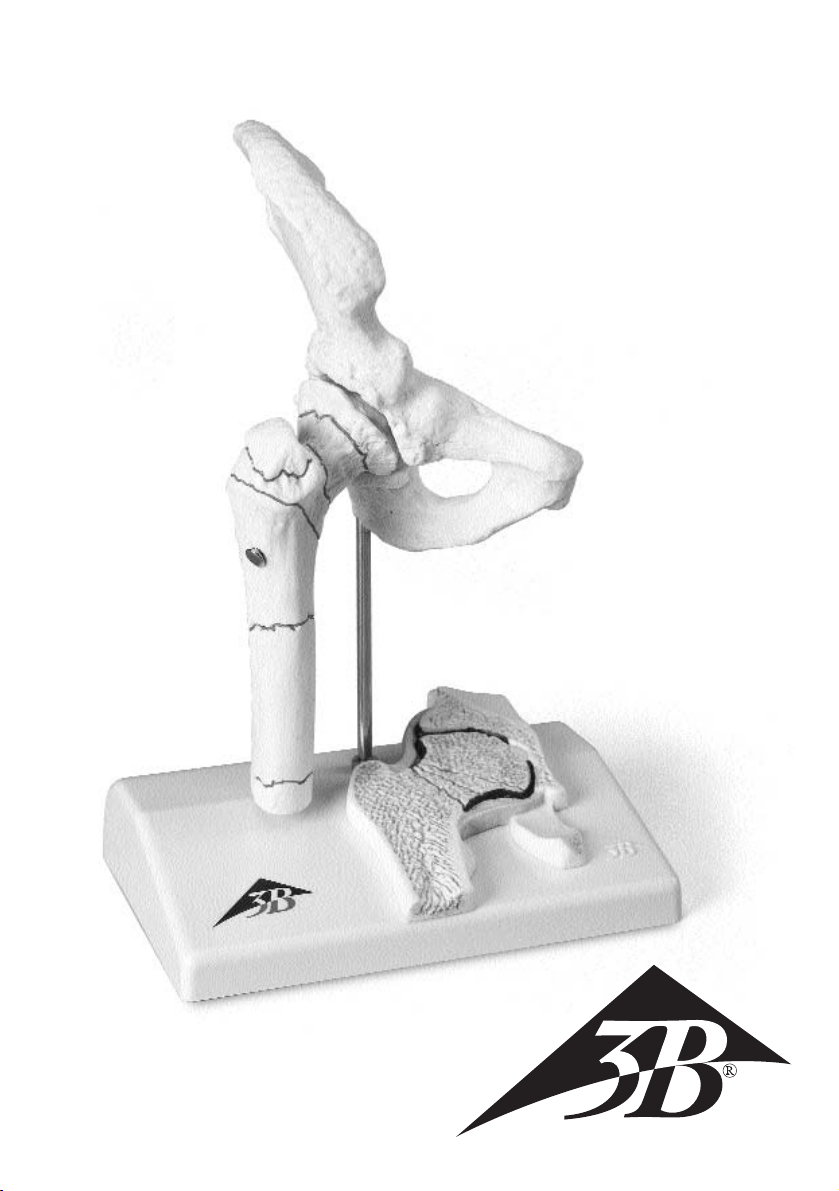
The model shows the right hip joint of an elderly person, reduced to a half of his natural size; the relief
representation on the base shows a frontal section through the femoral neck.
1. Medial femoral neck fracture
Depending on the type of fall (while leg is drawn up or stretched out) the femoral neck tears at
various angles in the area of the joint capsule (intraarticular), with or without compression of the
joint head. These angles are the basis of Pauwel’s classification: the steeper the angle the higher
the risk that the bone fragments shift against each other in opposite directions.
Pauwel’s classification:
1a: Type I: Angle of up to 30° to the horizontal (stable under pressure load).
1b: Type II: Angle of up to 70° to the horizontal (increasing effect of shearing forces).
1c: Type III: Angle of over 70° to the horizontal (bone healing is prevented).
2. Lateral femoral neck fracture
Typical lateral extracapsular injury in older persons, rare.
3. Fracture through the trochanteric region (”pertrochanteric femoral fracture”)
Typical injury in older persons.
4. Fracture below the trochanters (”subtrochanteric femoral fracture”)
Also occurs in younger patients as a result of a high impact of force (turning or
bending) below the lesser trochanter, always unstable.
5. Fracture in the area of the femoral shaft (”femoral shaft fracture”)
Caused by an impact of force on the femur.
6. Fracture in the area of the femoral head (”femoral head fracture”)
Caused by direct or indirect impact of force, usually in case of dislocation of the
femoral head.
7. Fracture of the greater trochanter (”greater trochanter fracture”)
Caused by direct impact of force.
8. Fracture or avulsion of the lesser trochanter (”avulsion fracture of the lesser
trochanter”) Common sports injury in younger persons.
9. Degenerative hip osteoarthritis (”coxarthrosis”)
Causes formation of bony outgrowths at the edges of the socket of hip and the femoral head as well as
deformation of the femoral head. The joint cartilage (painted blue on the model) is destroyed, in some
cases also the ligament of head of femur (ligamentum capitis).
9a: Remainders of joint cartilage (blue)
9b: Spurlike bony outgrowths
9c: Femoral head deformation
9d: Ligament of head of femur
Español Fractura de fémur y desgaste de la articulación
de la cadera
La fractura del cuello del fémur es una lesión ósea traumática del fémur. Este se rompe especialmente a
nivel del cuello del fémur o a la altura de las tuberosidades óseas que se encuentran situadas por debajo
(trocánter mayor y trocánter menor). La fractura del cuello de fémur es la fractura ósea más frecuente en
las personas mayores puesto que la estructura ósea se altera con la edad (osteoporosis) y, por tanto, los
huesos son menos estables. Como consecuencia, un ligero resbalón puede causar una fractura ósea.
El desgaste de la articulación de la cadera (coxartrosis) es una enfermedad crónica, progresiva y dolorosa
de la articulación de la cadera, determinada por el desgaste del cartílago y del tejido óseo propio de la
edad, o como consecuencia de trastornos inflamatorios o metabólicos. El desgaste óseo da lugar a la
formación de osteofitos en la cavidad cotiloidea y en la cabeza del fémur, así como a la deformación
de la cabeza del fémur. El cartílago articular se altera progresivamente y, eventualmente, también el
ligamento de la cabeza del fémur (ligamentun capitis). La movilidad de la articulación de la cadera se
limita cada vez más.
El modelo muestra la articulación de la cadera derecha de una persona mayor, reducida la mitad de su
tamano natural; la figura en rieleve sobre el soporte de sección frontal a través del cuello del fémur.
1. Fractura medial del cuello del fémur
Según el tipo de resbalón (sobre la pierna encogida o estirada), el cuello del fémur se rompe dentro
de la cápsula (intraarticular) en diferentes ángulos, con o sin reducción de la cabeza del fémur. Estos
ángulos sirven de base para la clasificación de Pauwels; cuanto más pronunciado es el ángulo, mayor
es el peligro de desplazamiento de las partes óseas entre sí.
Clasificación de Pauwels:
1a: Grado I: ángulo hasta 30º sobre la horizontal (bajo carga de presión estable)
1b: Grado II: hasta 70º sobre la horizontal (actúan fuerzas de torsión crecientes).
1c: Grado III: más de 70º sobre la horizontal (la curación ósea es limitada)
2. Fractura lateral del cuello del fémur
Típica lesión lateral por fuera de la cápsula articular en la edad madura, rara.
3. Fractura a través del trocánter (fractura pertrocantérea del fémur)
Típica lesión de la edad avanzada.
4. Fractura por debajo del trocánter (fractura subtrocantérea del fémur)
También la sufren pacientes jóvenes por acciones violentas (rotación o flexión), por debajo del
trocánter menor, siempre inestable.
5. Fractura a nivel de la diáfisis del fémur
Por una acción de fuerza sobre el fémur.
6. Fractura a nivel de la cabeza del fémur
Por una acción de fuerza directa o indirecta, habitualmente por luxación de la cabeza del fémur.
7. Fractura del trocánter mayor
Por acción de fuerza directa.
8. Fractura o arrancamiento del trocánter menor
A menudo es una lesión deportiva en edad joven.
9. Desgaste progresivo de la articulación de la cadera (coxartrosis)
Conduce a la formación de osteofitos en la cavidad cotiloidea y en la cabeza del fémur,y a
deformaciones de la cabeza del fémur. El cartílago articular (de azul en el modelo) se altera y,
eventualmente, también el ligamento de la cabeza del fémur (ligamentum capitis).
9a: Restos de cartílago articular (azul)
9b: Formaciones óseas del tipo de los osteofitos
9c: Deformación de la cabeza del fémur
9d: Ligamento de la cabeza del fémur
Der Oberschenkelbruch (Femurfraktur) ist eine knöcherne Verletzung des Oberschenkels. Dieser bricht am
häufigsten im Bereich des Oberschenkelhalses oder an den darunter liegenden Knochenvorwölbungen
(Trochanter major und -minor). Der Oberschenkelhalsbruch ist der häufigste Knochenbruch bei älteren
Menschen, da die Knochenstruktur altersbedingt aufgelockert ist (Osteoporose) und der Knochen dadurch
instabil sein kann. Bereits ein leichter Sturz kann zu einem Knochenbruch führen.
Der Hüftgelenkverschleiß (Koxarthrose oder Hüftgelenkarthrose) ist eine chronisch fortschreitende,
schmerzhafte Erkrankung des Hüftgelenks, bedingt durch den Verschleiß von Knorpel- und Knochengewebe
im Alter z. B. infolge von Entzündungen- oder Stoffwechselstörungen. Der Hüftgelenkverschleiß führt zu
Bildung von Randzacken an der Hüftpfanne und am Hüftkopf sowie Hüftkopfverformung. Der
Gelenkknorpel wird fortschreitend zerstört, evtl. auch das Schenkelkopfband (Ligamentum capitis). Das
Hüftgelenk ist in seiner Beweglichkeit zunehmend eingeschränkt. Das Modell zeigt das rechte Hüftgelenk
eines alten Menschen in halber natürlicher Größe, die Reliefabbildung auf dem Sockel einen Frontalschnitt
durch den Schenkelhals.
1. Mittlerer Schenkelhalsbruch („Mediale Schenkelhalsfraktur“)Je nach Art des Sturzes (auf
das angezogene oder abgespreizte Bein) reißt der Schenkelhals im Bereich der Gelenkkapsel
(intraartikulär) in verschiedenen Winkeln, mit oder ohne Einstauchung des Gelenkkopfes. Diese Winkel
liegen der Einteilung nach „Pauwels“ zugrunde: je steiler der Winkel,desto größer ist die Gefahr einer
Verschiebung der Knochenstücke gegeneinander.
Pauwels Klassifikation:
1a: Grad I: Winkel von bis zu 30° zur Horizontalen (unter Druckbelastung stabil).
1b: Grad II: Winkel von bis zu 70° zur Horizontalen (Es wirken zunehmend Scherkräfte).
1c: Grad III: Winkel ab 70° zur Horizontalen (Knöcherne Ausheilung verhindert).
2. Seitlicher Schenkelhalsbruch („Laterale Schenkelhalsfraktur“)
Typische Verletzung seitlich außerhalb der Gelenkkapsel im hohen Alter, selten.
3. Bruch durch den Bereich der Rollhügel („Pertrochantäre Femurfraktur“)
Typische Verletzung im hohen Alter.
4. Bruch unterhalb der Rollhügel („Subtrochantäre Femurfraktur“)
Auch jüngere Patienten bei hoher Gewalteinwirkung (Drehung oder Biegung),
unterhalb des kleinen Rollhügels (Trochanter minor), immer instabil.
5. Bruch im Bereich der Röhre des Oberschenkelknochens („Femurschaftfraktur")
Durch Gewalteinwirkung auf den Oberschenkelknochen.
6. Bruch im Bereich des Oberschenkelkopfes („Femurkopffraktur“)
Durch direkte oder indirekte Gewalteinwirkung, in der Regel bei Ausrenkung des Hüftkopfes.
7. Bruch des großen Rollhügels („Trochanter major Fraktur“)
Durch direkte Gewalteinwirkung.
8. Bruch oder Abriss des kleinen Rollhügels („Abrissfraktur des Trochanter minor“)
Oft Sportverletzung im jugendlichen Alter.
9. Fortschreitender Hüftgelenkverschleiß („Koxarthrose“)
Führt zu Bildung von Randzacken an der Hüftpfanne und am Hüftkopf sowie Hüftkopfverformung.
Der (am Modell blau bemalte) Gelenkknorpel wird zerstört, evtl. auch das Schenkelkopfband
(Ligamentum capitis).
9a: Reste von Gelenkknorpel (blau)
9b: Randzackenartige Knochenanbauten
9c: Hüftkopfverformung
9d: Schenkelkopfband
Oberschenkelbruch und Hüftgelenkverschleiß Deutsch
 Loading...