

Page 1
3B SCIENTIFIC
Bedienungsanleitung
04/09 Hh
®
PHYSICS
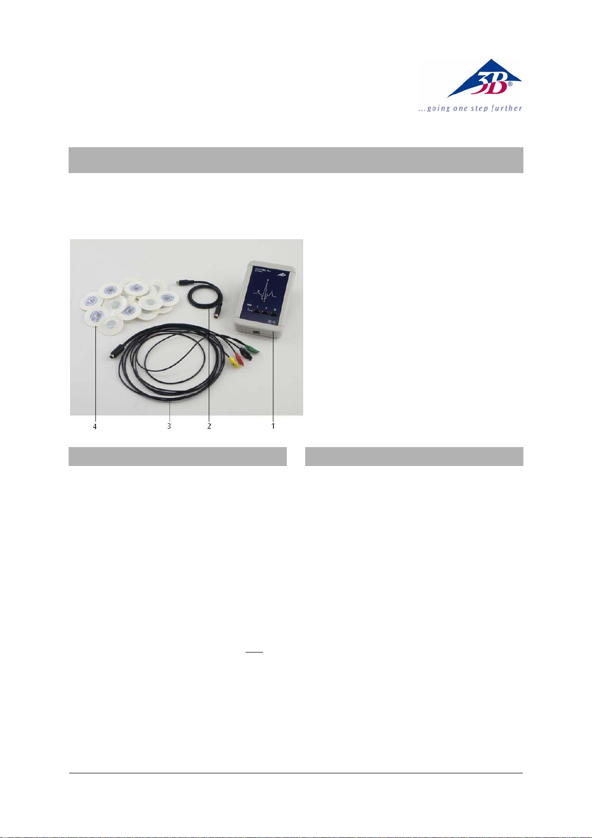
EKG / EMG-Box U11396
1 EKG / EMG-Box
2 miniDIN-Anschlusskabel
3 Extremitätenkabel
4 EKG-Klebeelektroden
1. Sicherheitshinweise
Die EKG / EMG-Box ist ausschließlich für Unterrichtszwecke bestimmt! Die hiermit ermittelten Messwerte
und -kurven dürfen niemals zur Beurteilung des
Gesundheitszustandes einer Person verwendet werden!
• Die EKG / EMG-Box nicht zu diagnostischen Zwe-
cken einsetzen!
• Die EKG / EMG-Box nicht als Kontrollgerät für
therapeutische Maßnahmen einsetzen!
• Die EKG / EMG-Box unter keinen Umständen
öffnen oder manipulieren!
• Die EKG / EMG-Box nicht in der Nähe von Herz-
schrittmachern oder anderen elektrischen Anregungs- und Reizgeräten betreiben!
• Die EKG / EMG-Box nur an jeweils eine Ver-
suchsperson anschließen!
Die EKG / EMG-Box ist nach den derzeit gültigen
Sicherheitsbestimmungen „Schutzklasse II, Klassifikation BF (body float)“ gebaut!
• Die Gerätekonfiguration aus EKG / EMG-Box und
3B NETlog
aktuellen CE-Bestimmungen entspricht!
TM
nur an einem PC betreiben, der den
2. Beschreibung
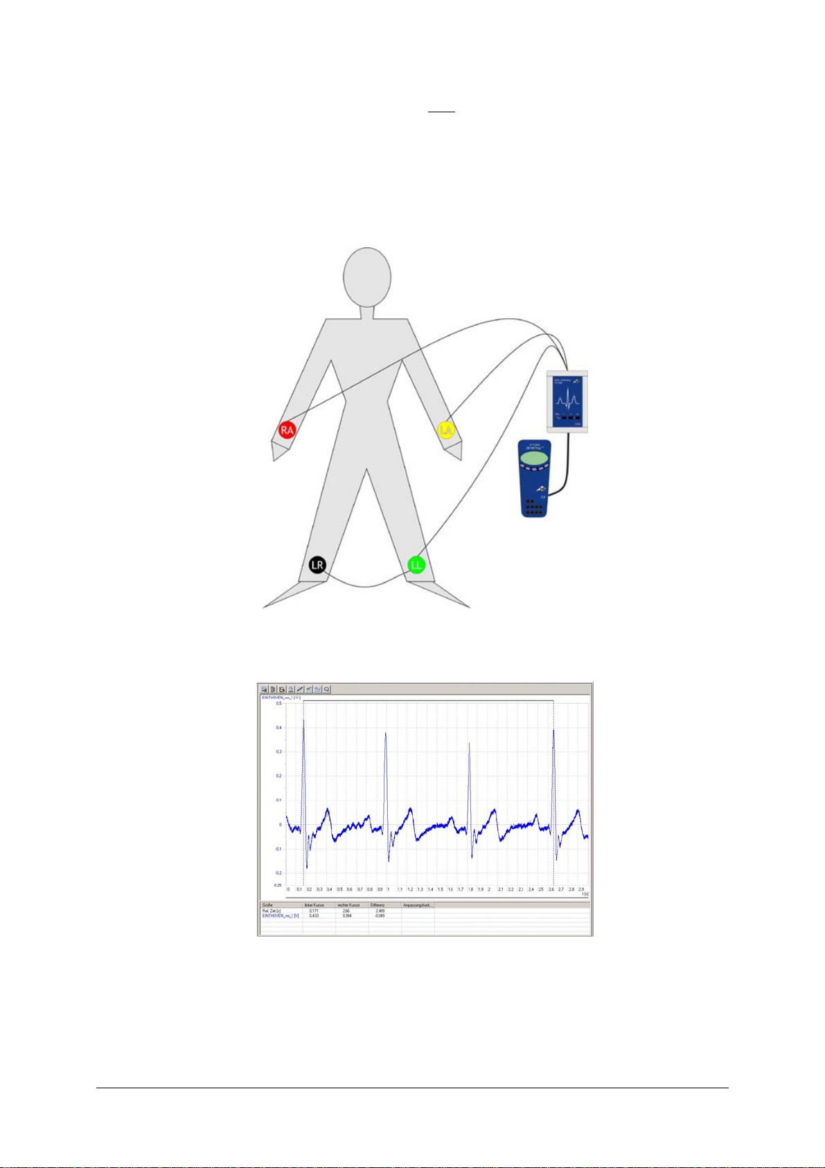
Sensorbox zur Messung eines Elektrokardiogramms
(EKG) an der Skelettmuskulatur in Form der drei
Standardableitungen I, II, III nach EINTHOVEN:
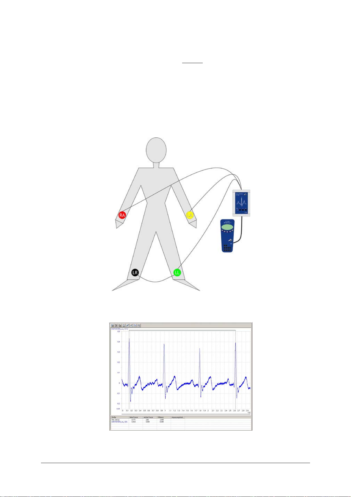
Ableitung I: Vom rechten Arm zum linken Arm;
Ableitung II: Vom rechten Arm zum linken Bein;
Ableitung III: Vom linken Arm zum linken Bein.
Auswahl der Ableitung per Tastendruck und Anzeige
durch Leuchtdioden.
Messung der durch die Herzkontraktion erzeugten
Potenzialänderungen an der Hautoberfläche.
Messung der Aktionspotenziale von Muskeln (Elektromyogramm (EMG)) im ruhenden und erregten
Zustand.
Automatische Sensorboxerkennung durch das 3B
TM
NETlog
.
1
Page 2
3. Lieferumfang
1 EKG / EMG-Box
1 Extremitätenkabel, 4-fach (RA, LA, LL, RL), mit
Druckknopfkontakten, Länge 1,50 m
2 Packungen (60 Stück) EKG-Klebeelektroden F55,
Ag/AgCl, vorgeliert
1 miniDIN-Anschlusskabel 8-pin, 1 m lang
1 Bedienungsanleitung für U11396
4. Technische Daten
Eingangswiderstand: > 10 MΩ
Ausgangsspannung: max. ± 1 V
Sperrfrequenz: 50 - 60 Hz
5. Bedienung
Hinweis: Beim Anlegen der Extremitätenkabel ("Patientenkabel") darauf achten, dass sich die Leitungen
möglichst nicht überkreuzen und dass keine stromführenden Leitungen in direkter Nähe sind.
Heftige körperliche Bewegungen der Versuchsperson
können Artefakte (Störungen) in den Kurvenaufzeichnungen verursachen. Die Person sollte sich in
einer entspannten und ruhigen Rückenlage befinden.
• Zur Aufzeichnung des EKGs je eine Klebeelektro-
de (insgesamt 4 Stück) an der Innenseite des linken und rechten Unterarms sowie an der Innenseite des rechten und linken Unterschenkels ankleben.
• Die Extremitätenkabel mit der korrekten Far-
benzuordnung an den EKG-Klebeelektroden an-
knüpfen:
ROT an den rechten Unterarm (RA), GELB an den
linken Unterarm (LA), GRÜN an den linken
Unterschenkel (LL), SCHWARZ an den rechten
Unterschenkel, siehe hierzu die Fig. 1.
• Die EKG / EMG-Box mit dem miniDIN-
Anschlusskabel wahlweise an einen der beiden
Analogeingänge U
A
oder U
in
B
des 3B NETlogTM an-
in
stecken.
• Das 3B NETlog
TM
einschalten und die automatische Erkennung ("Probe Detect") der Box abwarten.
• Die EKG / EMG-Box in der Nähe der Versuchsper-
son platzieren.
• Per Tastendruck auf der Box die gewünschte
Standardableitung I, II oder III auswählen.
• Für die Messung des EMG (Elektromyogramms)
vier Klebeelektroden auf dem Arm der Versuchsperson gemäß Fig. 3 anbringen:
ROT an die untere Innenseite des linken Unterarms, GELB an die obere Innenseite des linken
Unterarm, GRÜN und SCHWARZ an die obere
Aussenseite des linken Unterarms.
• Mit einem ca. 2 s andauernden Druck auf die
Taste "I" die EMG-Funktion auswählen.
6. Anwendungen
Messung des Ruhe-EKG auf den drei Standardableitungen nach EINTHOVEN.
Studium der P, Q, R, S, T und U-Wellenformen.
Messung des EKG im Anschluss an eine leichte kör-
perliche Aktivität.
Einfluss verschiedener Körperhaltungen auf die EKG-
Messkurven.
Auswirkungen äußerlicher Einflüsse (Erregung,
Schreck) auf das EKG.
Ermittelung der Pulsfrequenz aus den Messkurven.
Messung eines EMG (Elektromyogramm) von Muskel-
kontraktionen; Erfassung der elektrischen Potenziale
von "Spontanaktivitäten" entspannter Muskelbereiche.
7. Versuchsbeispiele
1. Messung des EKG (Elektrokardiogramms) einer
Versuchsperson
Benötigte Geräte:
1 3B NETlog
1 3B NETlab
TM
U11300
TM
U11310
1 EKG / EMG-Box U11396
• Vorbereitung des Versuchs gemäß Fig. 1.
• 3B NETlab
TM
-Anwendung (Template) zum EKG-
Experiment mit der EKG / EMG-Box öffnen.
• Das Template starten und die Messkurve des
vorgewählten EKG (hier: Ruhe-EKG auf der Standardableitung I nach EINTHOVEN) aufzeichnen
(Fig. 2).
• Die EKG-Kurve anhand der Informationen in den
Grundlagen im Template bewerten.
• Die Pulsfrequenz der Versuchsperson bestim-
men.
Hinweis:
Sollte das aktuell aufgezeichnete EKG nicht
vollständig mit der Beispielkurve übereinstimmen,
so besteht kein Anlass zur Sorge! Bei jeder Versuchsperson werden Veränderungen feststellbar sein;
auch gesunde Herzen zeigen typische Verläufe! Die
präzise Auswertung eines EKG erfordert eine große
medizinische Erfahrung. Diese EKG / EMG-Box ist
kein Hilfsmittel für eine entsprechende Diagnose!
2
Page 3
2. Messung des EMG (Elektromyogramms) einer
Versuchsperson
Benötigte Geräte:
1 3B NETlog
1 3B NETlab
TM
U11300
TM
U11310
1 EKG / EMG-Box U11396
• Vorbereitung des Versuchs gemäß Fig. 3.
• 3B NETlab
TM
-Anwendung (Template) zum EMG-
Experiment mit der EKG / EMG-Box öffnen.
• Das Template starten und die Messkurve des
Elektromyogramms aufzeichnen (Fig. 4).
• Die EMG-Kurve anhand der Informationen in
den Grundlagen im Template bewerten.
Hinweis:
In der medizinischen "Elektrodiagnostik"
liefert das EMG Aussagen über Erkrankungen der
Nerven- und Muskelzellen (Neuro- und Myopathien).
In der "Biomechanik" untersucht man die Zusammenhänge zwischen den Frequenzen und Amplituden der elektrischen Signale und der Kraft eines
Muskels, um z.B. die Bewegungen eines Sportlers zu
optimieren.
Fig. 1 Messung des EKG an einer Versuchsperson
Fig. 2 Bildschirmdarstellung der Messung des Ruhe-EKG auf der Standardableitung I nach EINTHOVEN in 3B NETlab
(U11310)
TM
3
Page 4

Fig. 4 EMG von Muskelkontraktionen des linken Unterarms; Ruhe- und Aktivphasen im Wechsel
Fig. 3 Aufzeichnung des Elektromyogramms am linken Unterarm einer Versuchsperson
3B Scientific GmbH • Rudorffweg 8 • 21031 Hamburg • Deutschland • www.3bscientific.com
Technische Änderungen vorbehalten
© Copyright 2009 3B Scientific GmbH
Page 5
3B SCIENTIFIC
Instruction sheet
04/09 Hh
®
PHYSICS
ECG/EMG Box U11396
1 ECG/EMG box
2 miniDIN connecting cable
3 Limb attachment leads
4 ECG attachment electrodes
1. Safety instructions
The ECG/EMG box is only suitable for educational
purposes. The measurement data and curves
obtained with it must never be used for assessing the
state of health of a person.
• Do not use the ECG/EMG box for diagnostic
purposes.
• Do not use the ECG/EMG box for monitoring the
effects of therapeutic treatments.
• The ECG/EMG box must not be opened or
interfered with under any circumstances.
• Do not operate the ECG/EMG box near heart
pacemakers or other electrical stimulation or
therapeutic devices.
• Only connect the ECG/EMG box to one test
person at any time.
The ECG/EMG box is constructed in accordance with
the current safety requirements for “Protection Class
II, Classification BF (body float)”!
• An instrument configuration consisting of the
ECG/EMG box and the 3B NETlog
be operated with a PC that conforms to the
current CE regulations!
TM
unit may only
2. Description
Sensor box for the measurement of an
electrocardiogram (ECG) on the skeletal musculature
using the three standard Einthoven limb leads I, II
and III:
Lead I: from right arm to left arm;
Lead II: from right arm to left leg;
Lead III: from left arm to left leg.
The three leads can be selected by push-button and
the choice is indicated by an LED.
The equipment works by measuring the changes in
skin surface potential caused by contraction of the
heart.
The action potentials of muscles in both relaxed and
energised states can also be measured
(electromyogram, EMG).
The sensor box is detected automatically by the 3B
TM
NETlog
unit.
1
Page 6

3. Equipment supplied
6. Applications
1 ECG/EMG box
1 Limb attachment leads, 4-position (right arm, left
arm, left leg, right leg), with push-button contacts,
length 1.50 m
2 Packs (60 in each) of ECG attachment electrodes
F55, Ag/AgCl, with gel pre-applied
1 miniDIN connecting cable, 8-pin, 1 m long
1 Instruction sheet for U11396
4. Technical data
Input resistance: > 10 MΩ
Output voltage: max. ± 1 V
Blocked frequency: 50 - 60 Hz
5. Operation
Important: When attaching the limb leads (“patient
leads”), try to avoid crossing them over, and ensure
that there are no current-carrying conductors
nearby.
Vigorous movements by the person being tested can
cause artefacts (disturbances) in the recorded curves.
The person should remain lying back in a quiet and
relaxed state.
• To prepare for recording the ECG, attach one
electrode each (4 altogether) to the inner
surfaces of both forearms and on the inner
surfaces of both calves.
• Connect the limb leads to the ECG electrodes,
with the correct colour identifications:
RED for the right forearm (right arm), YELLOW
for the left forearm (left arm), GREEN for the left calf
(left leg), and BLACK for the right calf (see Fig. 1).
• Using the miniDIN connecting cable, connect the
ECG/EMG box to one of the two analog inputs U
B
or U
of the 3B NETlogTM unit, whichever is
in
A
in
preferred.
• Switch on the 3B NETlog
TM
unit and wait for the
box to be detected automatically ("Probe
Detect").
• Place the ECG/EMG box near the test person.
• By means of the push-button on the box, choose
the required standard lead I, II or III.
• For recording the EMG (electromyogram), attach
four electrodes to the arm of the test person as
shown in Fig. 3:
RED to the inner surface of the lower part of the
left forearm, YELLOW to the inner surface of the
upper part of the left forearm, GREEN and
BLACK to the outer surface of the left forearm.
• Select the EMG mode by pressing the button "I"
for about 2 seconds.
Recording an ECG in a relaxed state with the three
standard EINTHOVEN leads.
Studying the P, Q, R, S, T and U waveforms.
Recording an ECG after light physical activity.
Investigating the effects of different body postures
on the ECG curves.
Investigating the effects of external influences
(excitement, fright) on an ECG.
Determining pulse rate from the ECG curves.
Recording an EMG (electromyogram) produced by
muscle contractions; measuring the electric
potentials for involuntary movements of relaxed
muscular regions.
7. Sample experiments
1. Recording the ECG (electrocardiogram) of a
person
Equipment required:
1 3B NETlog
1 3B NETlab
TM
unit U11300
TM
software installation U11310
1 ECG/EMG box U11396
• Prepare the experiment as shown in Fig. 1.
• In 3B NETlab
TM
, open the application program
(template) for the ECG experiment using the
ECG/EMG box.
• Start the template and record the curve for the
chosen ECG variant (in this case, relaxed-state
ECG with Einthoven standard lead I, see Fig. 2).
• Evaluate the ECG curve in accordance with the
information about basic principles in the
template.
• Determine the pulse rate of the person being
tested.
Important
: If the ECG just recorded does not
completely resemble the example provided, this is
not a cause for concern. Every person can show
differences from the norm; even healthy hearts show
such variations. The precise interpretation of an ECG
requires much medical experience. This ECG/EMG
box is not an instrument for diagnoses of that kind.
2. Recording the EMG (electromyogram) of a
person
Equipment required:
1 3B NETlog
1 3B NETlab
TM
unit U11300
TM
software installation U11310
1 ECG/EMG box U11396
• Set up the experiment as shown in Fig. 3.
2
Page 7
• In 3B NETlab
TM
, open the application program
(template) for the EMG experiment using the
ECG/EMG box.
• Start the template and record the
electromyogram curve (Fig. 4).
• Evaluate the EMG curve in accordance with the
information about basic principles in the
template.
Note
: In the practice of medical “electrodiagnostics”,
an EMG yields information about diseases of the
nerve and muscle cells (neuropathy and myopathy).
In “biomechanics” the relationships between the
frequencies and amplitudes of the electrical signals
and the performance of muscles are investigated, for
example in order to optimise the movements of an
athlete or sportsperson.
Fig. 1 Recording the ECG of a person
Fig. 2 A relaxed-state ECG recorded using Einthoven standard lead I and displayed on the screen in 3B NETlab
TM
(U11310)
3
Page 8
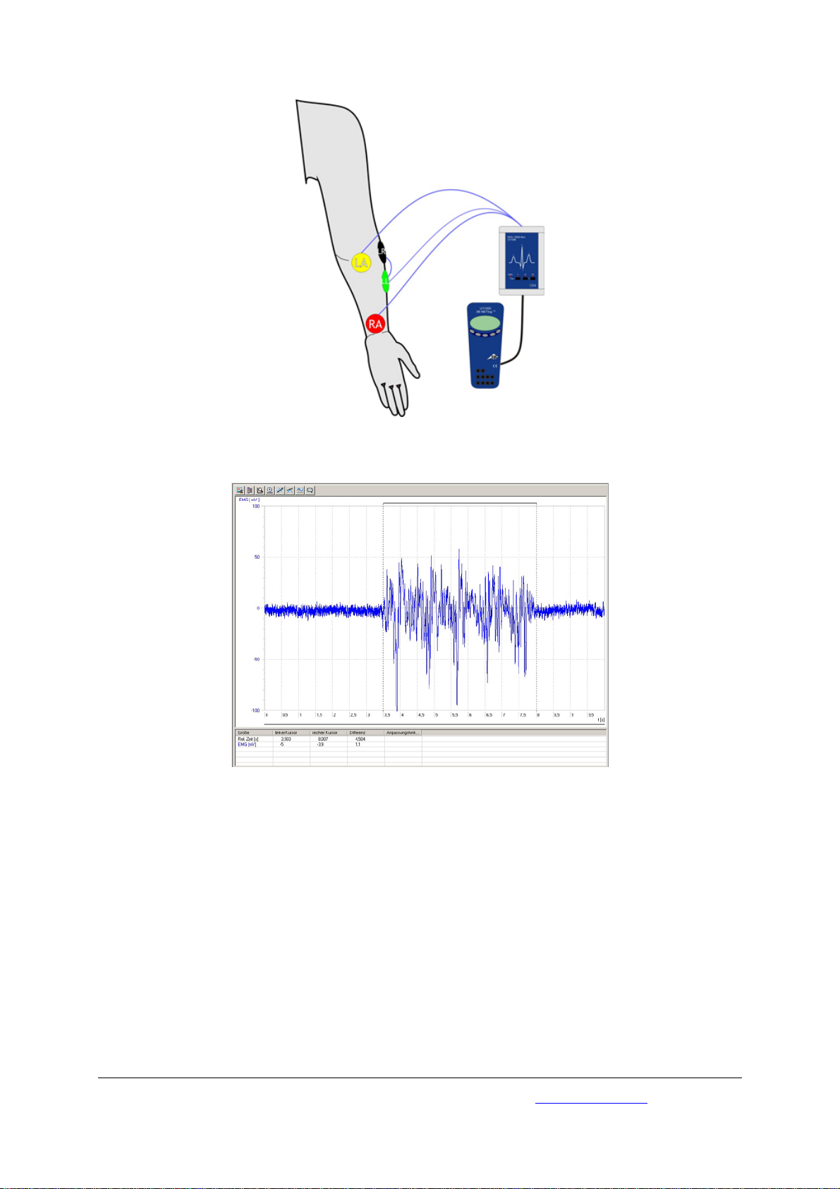
Fig. 3 Recording an electromyogram of the left forearm of a person
Fig. 4 EMG of muscular contractions of the left forearm; effect of switching between relaxed and active phases
3B Scientific GmbH • Rudorffweg 8 • 21031 Hamburg • Germany • www.3bscientific.com
Subject to technical amendments
© Copyright 2009 3B Scientific GmbH
Page 9

3B SCIENTIFIC
Instructions d’utilisation
04/09 Hh
®
PHYSICS
Boîtier ECG / EMG U11396
1 Boîtier ECG / EMG
2 Câble de raccordement miniDIN
3 Câbles d'extrémité
4 Électrodes autocollantes pour ECG
1. Consignes de sécurité
Le boîtier ECG / EMG est exclusivement destiné à
l'enseignement ! Les valeurs et courbes de mesure ainsi
déterminées ne peuvent en aucun cas être utilisées
pour l'évaluation de l'état de santé d'une personne !
• N'utilisez jamais le boîtier ECG / EMG dans des
buts diagnostiques !
• N'utilisez jamais le boîtier ECG / EMG en tant que
dispositif servant au contrôle de traitements
thérapeutiques !
• N'ouvrez en aucun cas le boîtier ECG / EMG, ne le
manipulez pas non plus !
• N'utilisez jamais le boîtier ECG / EMG à
proximité de stimulateurs cardiaques ou
d'autres appareils de stimulation ou d'excitation
électrique !
• Ne raccordez toujours le boîtier ECG / EMG qu'à
un seul
sujet d'expérience !
La construction du boîtier ECG / EMG se conforme aux
directives relatives à la sécurité actuellement en vigueur
« Classe de protection II, type Body Floating (BF) » !
• Veillez à n'utiliser la configuration de
périphériques, composée du boîtier ECG / EMG et
du 3B NETlog
aux directives CE actuellement en vigueur !
TM
que sur un ordinateur conforme
2. Description
Boîtier capteurs permettant de réaliser la mesure
d'un électrocardiogramme (ECG) sur les muscles
squelettiques, sous forme des trois dérivations
bipolaires des extrémités I, II, III d'EINTHOVEN :
Dérivation bipolaire I : entre bras droit et bras
gauche ;
Dérivation bipolaire II : entre bras droit et jambe
gauche ;
Dérivation bipolaire III : entre bras gauche et jambe
gauche.
Sélection de la dérivation souhaitée par simple
pression de touche et affichage par diodes
luminescentes.
Mesure des modifications de potentiels à la surface
de la peau, provenant de contractions cardiaques.
Mesure des potentiels d'action musculaire
(électromyogramme (EMG)) à l'état de repos et
d'excitation.
Reconnaissance automatique du boîtier capteurs via
le 3B NETlog
TM
.
1
Page 10

3. Étendue de la livraison
1 Boîtier ECG / EMG
1 Câbles d'extrémité quadruples (RA, LA, LL, RL),
équipés de contacts bouton poussoir, d'une
longueur de 1,50 m
2 Emballages (60 unités) d'électrodes autocollantes
F55 pour ECG, Ag/AgCl, prégélifiées
1 Câble de raccordement miniDIN, 8 broches, d'une
longueur de 1 m
1 Instructions d’utilisation pour U11396
4. Caractéristiques techniques
Résistance d'entrée : > 10 MΩ
Tension de sortie : au maximum
± 1 V
Fréquence limite : entre 50 et 60 Hz
5. Manipulation
Remarque : en plaçant les câbles d'extrémité (« câble
patient »), veillez dans la mesure du possible à ce
que les câbles ne se croisent pas et qu'aucun cordon
conducteur ne se trouve à proximité directe.
Des mouvements physiques brusques du sujet
d'expérience peuvent résulter dans des artefacts
(élément faussant les résultats) sur les courbes
enregistrées. Le sujet sera allongé en décubitus
dorsal, dans une position tranquille et détendue.
• Pour enregistrer l'ECG, fixez une électrode
autocollante (4 en tout) respectivement sur la
partie intérieure des avant-bras droit et gauche
ainsi que sur la partie intérieure des jambes
inférieures droite et gauche.
• Reliez les câbles d'extrémité aux électrodes
autocollantes pour ECG en veillant à respecter
les couleurs correctes :
ROUGE sur l'avant-bras droit (RA), JAUNE sur
l'avant-bras gauche (LA), VERT sur la jambe
inférieure gauche (LL), NOIR sur la jambe
inférieure droite (veuillez comparer à
l'illustration 1).
• Connectez le boîtier ECG / EMG à l'une des deux
entrées analogiques U
A
ou U
in
B
du 3B NETlogTM
in
en utilisant le câble de raccordement miniDIN.
• Mettez le 3B NETlog
TM
en marche, puis attendez la
reconnaissance automatique du boîtier (« Probe
Detect »).
• Placez le boîtier ECG / EMG à proximité du sujet
d'expérience.
• Sélectionnez la dérivation bipolaire souhaitée (I,
II ou III) par une pression de touche sur le
boîtier.
• Pour la mesure de l'EMG (électromyogramme),
placez quatre électrodes autocollantes sur le
bras du sujet d'expérience, conformément à
l'illustration 3 :
ROUGE à l'intérieur de la partie inférieure de
l'avant-bras gauche, JAUNE à l'intérieur de la
partie supérieure de l'avant-bras gauche, VERT
et NOIR à l'extérieur de la partie supérieure de
l'avant-bras gauche.
• Sélectionnez la fonction EMG en exerçant une
pression constante d'environ deux secondes sur
la touche « I ».
6. Applications
Mesure de l'ECG de repos sous forme des trois
dérivations bipolaires des extrémités d'EINTHOVEN.
Examen des formes d'onde P, Q, R, S, T et U.
Mesure de l'ECG après une activité physique légère.
Influence de diverses positions du corps sur les
courbes de mesure de l'ECG.
Effets d'influences externes (excitation, frayeur) sur
l'ECG.
Détermination de la fréquence de pouls à partir des
courbes de mesure.
Mesure d'un EMG (électromyogramme) de
contractions musculaires ; saisie de potentiels
électriques des « activités spontanées » de zones
musculaires détendues.
7. Exemples d’expérience
1. Mesure de l'ECG (électrocardiogramme) d'un
sujet d'expérience
Dispositifs nécessaires :
1 3B NETlog
1 3B NETlab
TM
U11300
TM
U11310
1 boîtier ECG / EMG U11396
• Préparation de l'essai conformément à
l'illustration 1.
• Ouvrez l'application 3B NETlab
TM
(modèle) pour
procéder à l'essai expérimental ECG avec le
boîtier ECG / EMG.
• Démarrez le modèle, puis enregistrez la courbe
de mesure de l'ECG présélectionné (dans ce cas :
ECG de repos pour la dérivation bipolaire
d'EINTHOVEN I) (Fig. 2).
• Évaluez la courbe ECG en vous appuyant sur les
informations des notions de base du modèle.
• Déterminez la fréquence de pouls du sujet
d'expérience.
Remarque :
si l'ECG actuellement enregistré ne
correspondait pas exactement à la courbe
d'exemple, nous n'avez aucune raison de vous
inquiéter ! Vous pourrez constater des modifications
chez tous les sujets d'expérience ; des cœurs en
bonne santé présentent eux aussi des courbes
typiques ! L'interprétation précise d'un ECG exige et
présuppose une grande expérience médicale. Ce
2
Page 11

boîtier ECG / EMG ne représente pas un appareil
auxiliaire permettant un diagnostic approprié !
2. Mesure de l'EMG (électromyogramme) d'un
sujet d'expérience
Dispositifs nécessaires :
1 3B NETlog
1 3B NETlab
TM
U11300
TM
U11310
1 boîtier ECG / EMG U11396
• Préparation de l'essai conformément à
l'illustration 3.
• Ouvrez l'application 3B NETlab
TM
(modèle) pour
procéder à l'essai expérimental EMG avec le
boîtier ECG / EMG.
• Démarrez le modèle, puis enregistrez la courbe
de mesure de l'électromyogramme (Fig. 4).
• Évaluez la courbe EMG en vous appuyant sur les
informations des notions de base du modèle.
Remarque :
en « électrodiagnostic » médical, l'EMG
permet de faire des pronostics sur des troubles des
cellules nerveuses et musculaires (neuropathies et
myopathies). La « biomécanique » examine les
corrélations existant entre d'une part les fréquences
et les amplitudes des signaux électriques et d'autre
part la force d'un muscle — ce qui permet par
exemple d'optimiser les mouvements d'un sportif.
Fig. 1 Mesure de l'ECG d'un sujet d'expérience
Fig. 2 Visualisation à l'écran de la mesure de l'ECG de repos pour la dérivation bipolaire d'EINTHOVEN I avec 3B NETlab
(U11310)
TM
3
Page 12

Fig. 3 Enregistrement de l'électromyogramme sur l'avant-bras gauche d'un sujet d'expérience
Fig. 4 EMG pour des contractions musculaires de l'avant-bras gauche ; alternance de phases d'activité et de phases de repos
3B Scientific GmbH • Rudorffweg 8 • 21031 Hamburg • Allemagne • www.3bscientific.com
Sous réserve de modifications techniques
© Copyright 2009 3B Scientific GmbH
Page 13

3B SCIENTIFIC
Istruzioni per l'uso
04/09 Hh
®
PHYSICS
Scatola ECG / EMG U11396
1 Scatola ECG / EMG
2 Cavo di collegamento miniDIN
3 Cavo estremità
4 Elettrodi adesivi ECG
1. Norme di sicurezza
La scatola ECG / EMG è destinata unicamente a scopi
didattici! I valori e le curve di misurazione con essa
determinati non devono mai essere utilizzati per la
valutazione dello stato di salute di una persona!
•
Non utilizzare la scatola ECG / EMG a scopi
diagnostici!
• Non utilizzare la scatola ECG / EMG come unità
di controllo né come strumento terapeutico!
• Non è consentito aprire né manipolare la scatola
ECG / EMG!
• Non azionare la scatola ECG / EMG in prossimità
di pacemaker o altri apparecchi elettrici di
eccitazione e stimolazione!
• Collegare la scatola ECG / EMG di volta in volta
soltanto a una persona da testare!
La scatola ECG /EMG è realizzata conformemente alle
disposizioni di sicurezza attualmente vigenti “Classe
di protezione II, classificazione BF (body float)”!
• Azionare la configurazione dell'apparecchio di
scatola ECG / EMG e 3B NETlog
PC conforme alle attuali disposizioni CE!
TM
soltanto su un
2. Descrizione
Scatola del sensore per la misurazione di un
elettrocardiogramma (ECG) in corrispondenza della
muscolatura scheletrica sotto forma delle tre
derivazioni standard I, II, III secondo EINTHOVEN:
Derivazione I: Dal braccio destro al braccio sinistro;
Derivazione II: Dal braccio destro alla gamba
sinistra;
Derivazione III: Dal braccio sinistro alla gamba
sinistra.
Selezione della derivazione mediante pressione di
pulsante e visualizzazione mediante diodi luminosi.
Misurazione delle variazioni di potenziale prodotte
dalla contrazione del cuore, in corrispondenza della
superficie della pelle.
Misurazione del potenziale di azione di muscoli
(elettromiogramma (EMG)) allo stato a riposo e
eccitato.
Riconoscimento automatico del sensore mediante 3B
NETlog
TM
.
1
Page 14

3. Fornitura
1 Scatola ECG / EMG
1 Cavo estremità, quadruplo (RA, LA, LL, RL) con
contatti a pulsante, lunghezza 1,50 m
2 Confezioni (60 pezzi) di elettrodi adesivi ECG F55,
Ag/AgCl, pregelificati
1 Cavo di collegamento miniDIN a 8 pin, lungo 1 m
1 Istruzioni per l’uso per U11396
4. Dati tecnici
Resistenza d'ingresso: > 10 MΩ
Tensione di uscita: max. ± 1 V
Frequenza di blocco 50 - 60 Hz
5. Utilizzo
Nota: Nell'applicazione dei cavi per le estremità
("cavi paziente") badare al fatto che i conduttori non
si incrocino e che conduttori percorsi da corrente
non si trovino in vicinanza diretta .
Movimenti fisici bruschi della persona da testare
possono dare luogo ad alterazioni (disturbi) nel
tracciamento delle curve. La persona dovrebbe
trovarsi una posizione supina rilassata e ferma.
• Per il tracciamento dell'ECG attaccare ogni
elettrodo adesivo (complessivamente 4 pezzi) sul
lato interno dell'avambraccio sinistro e destro e
sul lato interno della gamba destra e sinistra.
• Collegare i cavi per le estremità agli elettrodi
adesivi ECG con la corretta associazione dei
colori:
ROSSO sull'avambraccio destro (RA), GIALLO
all'avambraccio sinistro (LA), VERDE sulla gamba
sinistra (LL), NERO sulla gamba destra, vedere al
riguardo la fig. 1.
• Connettere la scatola ECG / EMG con il cavo di
collegamento miniDIN a scelta a uno dei due
ingressi analogici U
• Accendere 3B NETlog
in
A
B
o U
del 3B NETlogTM.
in
TM
e attendere il
riconoscimento automatico ("Probe Detect")
della scatola.
• Posizionare la scatola ECG / EMG vicino alla
persona da testare.
• Selezionare la derivazione standard I, II o III
desiderata premendo il tasto sulla scatola.
• Per la misurazione dell'EMG (elettromiogramma)
applicare quattro elettrodi adesivi sul braccio
della persona da testare secondo la fig. 3:
ROSSO sul lato interno inferiore
dell'avambraccio sinistro, GIALLO sul lato
interno superiore dell'avambraccio sinistro,
VERDE e NERO sul lato esterno superiore
dell'avambraccio sinistro.
• Premendo per circa 2 s il tasto "I", selezionare la
funzione EMG.
6. Applicazioni
Misurazione dell'ECG a riposo sulle tre derivazioni
standard secondo EINTHOVEN.
Studio delle forme d'onda P, Q, R, S, T e U.
Misurazione dell'ECG in relazione a una leggera
attività fisica.
Influenza di posture diverse sulle curve di misura
ECG.
Ripercussioni di influssi esterni (agitazione,
spavento) sull'ECG.
Determinazione della frequenza del polso dalle
curve di misura.
Misurazione di un EMG (elettromiogramma) di
contrazioni muscolati; rilevamento dei potenziali
elettrici di "attività spontanee" di aree muscolari
rilassate.
7. Esempi di esperimenti
1. Misurazione dell'ECG (elettrocardiogramma)
della persona da testare)
Apparecchi necessari:
1 3B NETlog
TM
U11300
1 3B NETlabTM U11310
1 Scatola ECG / EMG U11396
• Preparazione dell’esperimento secondo la fig. 1.
• Aprire l'applicazione 3B NETlab
TM
(Template) per
l'esperimento ECG con la scatola ECG / EMG.
• Avviare il template e tracciare la curva di misura
dell'ECG preselezionato (qui: ECG a riposo sulla
derivazione standard I in base a EINTHOVEN)
(fig. 2)
• Valutare la curva ECG in base alla informazioni
dei principi contenuti nel template.
• Determinare la frequenza del polso della
persona da testare.
Nota:
Se l'ECG attualmente tracciato non dovesse
coincidere completamente con la curva
esemplificativa, non c'è ragione di preoccuparsi. Per
ogni persona da testare si possono individuare delle
variazioni; anche cuori sani mostrano andamenti
tipici! La valutazione precisa di un ECG richiede una
grande esperienza in campo medico. Questa scatola
ECG / EMG non è un mezzo ausiliario per una
diagnosi adeguata!
2
Page 15

2. Misurazione dell'EMG (elettromiogramma)
della persona da testare)
Apparecchi necessari:
1 3B NETlog
TM
U11300
1 3B NETlabTM U11310
1 Scatola ECG / EMG U11396
• Preparazione dell’esperimento secondo la fig. 3.
• Aprire l'applicazione 3B NETlab
TM
(Template) per
l'esperimento EMG con la scatola ECG / EMG.
• Avviare il template e tracciare la curva di misura
dell'elettromiogramma (fig. 4).
• Valutare la curva EMG in base alle informazioni
dei principi contenuti nel template.
Nota:
Nel settore dell'"elettrodiagnostica" medica,
l'EMG fornisce informazioni riguardo a patologie
delle cellule nervose e muscolari (neuropatie e
miopatie). Nella "biomeccanica" si esaminano le
relazioni tra le frequenze e le ampiezze dei segnali
elettrici e della forza di un muscolo, ad esempio per
ottimizzare i movimenti di uno sportivo.
Fig. 1 Misurazione dell'ECG (elettrocardiogramma) della persona da testare
Fig. 2 Rappresentazione della schermata della misurazione dell'ECG a riposo sulla derivazione standard I secondo
EINTHOVEN in 3B NETlab
TM
(U11310)
3
Page 16

Fig. 3 Tracciamento dell'elettromiogramma sull'avambraccio sinistro di una persona da testare
Fig. 4 EMG delle contrazioni muscolati del braccio sinistro; fasi di riposo e di attività in alternanza
3B Scientific GmbH • Rudorffweg 8 • 21031 Amburgo • Germania • www.3bscientific.com
Con riserva di modifiche tecniche
© Copyright 2009 3B Scientific GmbH
Page 17

3B SCIENTIFIC
Instrucciones de uso
04/09 Hh
®
PHYSICS
Caja de ECG / EMG U11396
1 Caja de sensor de EKG / EMG
2 Cable de conexión miniDIN
3 Cables de extremidades
4 Electrodos adhesivos
1. Advertencias de seguridad
¡La caja de ECG / EMG se ha diseñado única y
exclusivamente para propósitos didácticos. Los
valores de medida y las curvas determinadas con ella
no se deben nunca utilizar para evaluar el estado de
salud de una persona!
• ¡No se debe utilizar la caja de ECG / EMG para
propósitos de diagnosis!
• ¡No se debe aplicar la caja de ECG / EMG como
aparato de control para medidas terapéuticas!
• ¡En ningún momento se debe abrir o manipular
la caja de ECG / EMG!
• ¡No se debe trabajar con la caja de ECG / EMG en
la cercanía de marcadores paso y de ningún
aparato eléctrico de excitación o de
estimulación!
• ¡La caja de ECG / EMG se conecta cada vez en
una sola persona de prueba al mismo tiempo!
¡La caja de ECG / EMG ha sido construida según las
prescripciones de seguridad “Clase de protección II,
Clasificación BF (body float)” vigentes!
• La configuración compuesta de caja de ECG / EMG y
3B NETlog
corresponda a las determinaciones de CE vigentes!
TM
se pone a trabajar sólo en un PC que
2. Descripción
Caja de sensor para la medición de un
electrocardiograma (ECG) en la musculatura
esquelética en la forma de las tres derivaciones
estándares I, II, III según EINTHOVEN:
Derivación I: Del brazo derecho al brazo izquierdo;
Derivación II: Del brazo derecho a la pierna
izquierda;
Derivación III: Del brazo izquierdo a la pierna
izquierda.
Selección de la derivación por botones e indicación
por medio de diodos luminosos.
Medición de la variación de potencial producida en
superficie de la piel debida a la contracción cardíaca.
Medición de los potenciales de acción musculares
(Electromiograma EMG) en los estados de reposo y de
excitación.
Reconocimiento automático de la caja de sensor por
medio de 3B NETlog
TM
.
1
Page 18

3. Volumen de suministro
1 Caja de sensor de EKG / EMG
1 Cables de extremidades, 4 unidades, (RA, LA, LL,
RL), con contacto por botón, longitud 1,50 m
2 Paquetes (60 unidades) de electrodos adhesivos
F55, Ag/AgCl, gelatinados
1 Cable de conexión miniDIN de 8 pines, 1 m de
largo
1 Instrucciones de uso para U11396
4. Datos técnicos
Resistencia de entrada: > 10 MΩ
Tensión de salida: max. ± 1 V
Frecuencia de bloqueo: 50 - 60 Hz
5. Manejo
Observación: Al conectar los cables de extremidades
(“Cables de paciente“) téngase en cuenta lo más
posible que los conductores no se crucen y que no se
encuentren en directa cercanía de conductores que
lleven corriente.
Movimientos bruscos de la persona de prueba
pueden originar señales parásitas de interferencia en
el registro de las curvas. La persona se debe
encontrar acostada sobre la espalda tranquila y
relajada.
• Para el registro del ECG se pegan electrodos
adhesivos (4 unidades en total) en la parte
interna de los antebrazos derecho e izquierdo
así como en los lados internos de las piernas
izquierda y derecha.
• Los cables de extremidades con las asignaciones
correctas se conexionan con los electrodos de
ECG adhesivos:
ROJO en el antebrazo derecho (RA), AMARILLO
en el antebrazo izquierdo (LA), VERDE en la pierna
izquierda (LL), NEGRO en la pierna derecha, para ello
observe la Fig. 1.
• Con el cable de conexión miniDIN se enchufa la
caja ECG / EMG en una de las dos entradas
analógicas U
• Se conecta el 3B NETlog
A
B
o U
del 3B NETlogTM .
in
in
TM
y se espera el
reconocimiento automático de la caja ("Probe
Detect").
• Se coloca la caja de ECG / EMG en la cercanía de
la persona de prueba.
• Pulsando una tecla en la caja se selecciona la
derivación estándar deseada I, II, o III.
• Para la medición del EMG (Electromiograma) se
fijan cuatro electrodos adhesivos en el brazo de
la persona de prueba de acuerdo con la Fig. 3:
ROJO en el lado interno inferior del antebrazo
izquierdo, AMARILLO en el lado interno superior
del antebrazo izquierdo, VERDE y NEGRO en la
parte superior externa del antebrazo izquierdo.
• Haciendo una presión constante durante aprox.
2 min. sobre la tecla ”I” se selecciona la función
EMG.
6. Aplicaciones
Medición del ECG en reposo en las tres derivaciones
estándares según EINTHOVEN.
Estudio de las formas de onda P, Q, R, S, T y U.
Medición del ECG a continuación de una actividad
corporal leve.
Influencia de diferentes posiciones corporales sobre
las curvas de medida del ECG.
Efectos de influencias externas (excitación, susto)
sobre el ECG.
Determinación de la frecuencia del pulso a partir de
las curvas de medida.
Medición de un EMG (electromiograma) de
contracciones de músculos; Captación de potenciales
eléctricos de “actividades espontáneas” de sectores
musculares en reposo.
7. Experimentos ejemplares
1. Medición del ECG (electrocardiograma) de una
persona de prueba
Aparatos requeridos:
1 3B NETlog
1 3B NETlab
TM
U11300
TM
U11310
1 Caja de ECG / EMG U11396
• Preparación del experimento de acuerdo con la
Fig. 1
• Se abre la aplicación de 3B NETlab
TM
(Templete)
para el experimento de ECG con la caja de ECG /
EMG.
• Se inicia el templete y se registra la curva de
medida del ECG previsto (aquí: El ECG en reposo
en la derivación estándar I según EINTHOVEN,
Fig. 2).
• Se evalúa la curva de ECG en base a las
informaciones en los fundamentos del templete.
• Se determina la frecuencia del pulso de la
persona de prueba.
Observación:
Si el ECG registrado actualmente no
concuerda con la curva ejemplar, no debe ser motivo
de preocupación. En cada persona de prueba se
podrán observar cambios; también corazones sanos
muestran cursos atípicos. La evaluación precisa de
un ECG requiere una gran experiencia médica. Esta
caja de ECG / EMG no es un medio auxiliar para una
diagnosis correspondiente!
2
Page 19

2. Medición del EMG (electromiograma) de una
persona de prueba
Aparatos requeridos:
1 3B NETlog
1 3B NETlab
TM
U11300
TM
U11310
1 Caja de ECG / EMG U11396
• Preparación del experimento de acuerdo con la
Fig. 3.
• Se abre la aplicación de 3B NETlab
TM
(Templete)
para el experimento de EMG con la caja de ECG /
EMG.
• Se inicia el templete y se registra la curva de
medida del electromiograma (Fig. 4).
• Se evalúa la curva de EMG en base a las
informaciones en los fundamentos del templete.
Observación:
En la “Electrodiagnosis“ médica el EMG
entrega informaciones sobre enfermedades de las
células nerviosas o musculares (neuropatía o
miopatía). En la “Biomecánica“ se estudia la relación
entre las frecuencias y amplitudes de las señales
eléctricas y la fuerza de un músculo, para, por
ejemplo, optimizar los movimientos de un atleta.
Fig. 1 Medición del ECG en una persona de prueba
Fig. 2 Reprentación en pantalla del ECG en reposo sobre la derivación estándar I según EINTHOVEN en el 3B NETlab
(U11310)
TM
3
Page 20

Fig. 4 EMG de contracciones de músculos del antebrazo izquierdo; fases de reposo y activas alternadas
Fig. 3 Registro del electromiograma del antebrazo izquierdo de una persona de prueba
3B Scientific GmbH • Rudorffweg 8 • 21031 Hamburgo • Alemania • www.3bscientific.com
Nos reservamos el derecho a cambios técnicos
© Copyright 2009 3B Scientific GmbH
Page 21

3B SCIENTIFIC
Instruções de operação
04/09 Hh
®
PHYSICS
Caixa ECG / EMG U11396
1 Caixa de ECG / EMG
2 Cabo de conexão miniDIN
3 Cabo de extremidades
4 Eletrodos adesivos para ECG
1. Indicações de segurança
A caixa ECG / EMG é indicada exclusivamente para
finalidades de ensino! Os valores de medição e
gráficos obtidos por este meio jamais devem ser
usados para a avaliação do estado de saúde de uma
pessoa!
• Não empregar a caixa ECG / EMG para
finalidades diagnósticas!
• Não empregar a caixa ECG / EMG como aparelho
de controle para medidas terapêuticas!
• Em nenhum caso abrir ou manipular a caixa
ECG / EMG!
• Não operar a caixa ECG / EMG na proximidade
de marca-passos cardíacos ou outros incitadores
e estimuladores elétricos!
• Ligar a caixa ECG / EMG somente a uma pessoa
de teste por vez!
A caixa de ECG / EMG construída segundo a determinação de segurança válida da época “Classe de proteção
II, Classificação BF (body float = corpo flutuante)“!
• Operar a configuração dos dispositivos da caixa
de ECG / EMG e do 3B NETlog
que esteja conformado às atuais determinações
da certificação CE!
TM
somente num CP,
2. Descrição
Caixa sensorial para a medição de um
eletrocardiograma (ECG) na musculatura esquelética
em forma das três derivações I, II, III segundo
EINTHOVEN:
Derivação I: Do braço direito ao braço esquerdo;
Derivação II: Do braço direito à perna esquerda;
Derivação III: Do braço esquerdo à perna esquerda.
A seleção da derivação é feita por aperto de tecla e a
indicação por diodos luminosos.
A medição das variações potenciais produzidas pela
contração do coração na superfície da pele.
A medição dos potenciais de ação dos músculos
(Eletromiograma – [EMG]) em estado de descanso e
excitado.
Reconhecimento automático da caixa de sensores
pelo 3B NETlog
TM
.
1
Page 22

3. Fornecimento
1 Caixa de ECG / EMG
1 Cabo de extremidades, quádruplo (RA, LA, LL, RL),
com contatos de botões de pressão, Comprimento
1,50 m.
2 Embalagens (60 Unidades) de eletrodos adesivos
F55, Ag/AgCl para ECG, pré-fornecidos.
1 Cabo de conexão miniDIN de 8-pinos,
comprimento de 1 m
1 Instruções de operação para U11396
4. Dados técnicos
Resistência de entrada: > 10 MΩ
Tensão de saída: máx. ± 1 V
Freqüência máxima: 50 - 60 Hz
5. Operação
Indicação: Ao prender os cabos de extremidades
("Cabos do paciente“) prestar atenção, para que, no
possível, os fios não se entrecruzam e que não exista
nenhuma fiação condutora de corrente elétrica nas
proximidades imediatas.
Movimentos bruscos da pessoa cobaia podem
provocar artefatos ("Interferências“) nas gravações
das curvas. A pessoa deveria colocar-se numa
posição deitada, relaxada e quieta.
• Para a gravação do ECG aderir um por vez, os
eletrodos de aderência (Total de 4 unidades) no
lado interno da parte inferior do braço esquerdo
e direito, assim como nos lados interiores direito
e esquerdo das pernas.
• Conectar os cabos de extremidades aos eletrodos
de adesão ECG na ordem correta da designação
das cores:
VERMELHO no braço inferior direito (RA),
AMARELO no braço inferior esquerdo (LA), VERDE na
perna esquerda (LL), NEGRO na perna direita (RL),
para isto ver a Fig. 1.
• Seletivamente ligar a caixa de ECG / EMG com o
cabo de conexão miniDIN a uma das duas
entradas análogas U
• Ligar o 3B NETlog
A
B
ou U
in
TM
e aguardar o reconhecimento
do 3B NETlogTM.
in
automático ("Probe Detect" = detectar a sonda)
da caixa.
Colocar a caixa de ECG / EMG na proximidade da
pessoa cobaia.
• Com um aperto de tecla selecionar a derivação
padrão I, II ou III desejada.
• Para a medição do EMG (Eletromiograma) aderir
os quatro eletrodos de aderência no braço da
pessoa cobaia, segundo Fig. 3: VERMELHO no
lado inferior interior do braço inferior esquerdo,
AMARELO no lado superior interior do braço
inferior esquerdo, VERDE e NEGRO no lado
superior exterior do braço inferior esquerdo.
• Com um aperto de aproximadamente 2 seg. de
duração sobre a tecla "I", selecionar a função
EMG.
6. Aplicações
Medição do ECG em descanso nas três derivações
padrões segundo EINTHOVEN.
Estudo das formas de ondas de P, Q, R, S, T e U.
Medição do ECG em conexão a uma ligeira atividade
física.
Influência das posturas corporais diferentes nas
curvas de medição do ECG.
Efeitos de influências externas (Agitação, susto) sobre
o ECG.
Averiguação da freqüência do pulso por meio das
curvas de medição.
Medição de um EMG (Eletromiograma) de contrações
musculares; compreensão dos potenciais elétricos
das “atividades espontâneas” nas áreas de músculos
relaxados.
7. Exemplos de experiências
1. Medição do ECG (Eletrocardiograma) de uma
pessoa cobaia
Aparelhos necessários:
1 3B NETlog
1 3B NETlab
TM
U11300
TM
U11310
1 Caixa de ECG / EMG U11396
• Preparação da experiência segundo a Fig. 1.
• Abrir a utilidade (Template) do 3B NETlab
TM
para
a experiência ECG com a caixa de ECG / EMG.
• Iniciar a utilidade e desenhar a curva de
medição pré-selecionada do ECG (Aqui: ECG em
descanso da derivação I segundo EINTHOVEN)
(Fig. 2).
• Avaliar a curva do ECG considerando as
informações de base na utilidade.
• Determinar a freqüência do pulso da pessoa
cobaia.
Indicação:
Se o EKG desenhado não coincidir
completamente com a curva do exemplo, não existe
nenhum motivo para a preocupação! Em cada
cobaia poderão ser verificadas variações; também
corações sadios mostram evoluções típicas! A
avaliação precisa de um ECG exige uma ampla
experiência médica. Esta caixa de ECG / EMG não é
um recurso de ajuda para uma diagnose
correspondente!
2
Page 23

2. Medição do EMG (Eletromiograma) de uma
pessoa cobaia
Aparelhos necessários:
1 3B NETlog
1 3B NETlab
TM
U11300
TM
U11310
1 Caixa de ECG / EMG U11396
• Preparação da experiência segundo a Fig. 3.
• Abrir a utilidade (Template) do 3B NETlab
TM
para
a experiência EMG com a caixa de ECG / EMG.
• Iniciar a utilidade e desenhar a curva de
medição do eletromiograma (Fig. 4).
• Avaliar a curva do EMG considerando as
informações de base na utilidade.
Indicação:
No “eletro-diagnose” medicinal, o EMC
fornece confirmações de doenças das células de
nervos e músculos (Neuro- e miopatias). Na
“biomecânica” se estudam as relações entre as
freqüências e amplitudes dos sinais elétricos e a
força de um músculo, para, por exemplo,
aperfeiçoar a movimentação de um esportista.
Medição do ECG numa pessoa cobaia
Fig. 1
Fig. 2 Representação na tela da medição do ECG em descanso sobre a derivação padrão I segundo EINTHOVEN no
3B NETlab
TM
(U11310)
3
Page 24

Fig. 3 Desenho do eletromiograma no braço inferior esquerdo de uma pessoa cobaia
Fig. 4 EMG de contrações musculares do braço inferior esquerdo, fases alternativas em descanso e atividade
3B Scientific GmbH • Rudorffweg 8 • 21031 Hamburg • Alemanha • www.3bscientific.com
Modificações técnicas reservadas
© Copyright 2009 3B Scientific GmbH
 Loading...