

Page 1
3B SCIENTIFIC
Instruction sheet
04/09 Hh
®
PHYSICS
ECG/EMG Box U11396
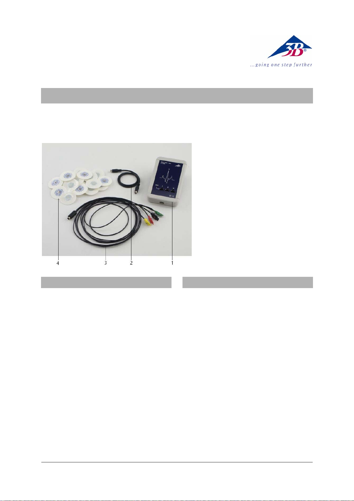
1 ECG/EMG box
2 miniDIN connecting cable
3 Limb attachment leads
4 ECG attachment electrodes
1. Safety instructions
The ECG/EMG box is only suitable for educational
purposes. The measurement data and curves
obtained with it must never be used for assessing the
state of health of a person.
• Do not use the ECG/EMG box for diagnostic
purposes.
• Do not use the ECG/EMG box for monitoring the
effects of therapeutic treatments.
• The ECG/EMG box must not be opened or
interfered with under any circumstances.
• Do not operate the ECG/EMG box near heart
pacemakers or other electrical stimulation or
therapeutic devices.
• Only connect the ECG/EMG box to one test
person at any time.
The ECG/EMG box is constructed in accordance with
the current safety requirements for “Protection Class
II, Classification BF (body float)”!
• An instrument configuration consisting of the
ECG/EMG box and the 3B NETlog
be operated with a PC that conforms to the
current CE regulations!
TM
unit may only
2. Description
Sensor box for the measurement of an
electrocardiogram (ECG) on the skeletal musculature
using the three standard Einthoven limb leads I, II
and III:
Lead I: from right arm to left arm;
Lead II: from right arm to left leg;
Lead III: from left arm to left leg.
The three leads can be selected by push-button and
the choice is indicated by an LED.
The equipment works by measuring the changes in
skin surface potential caused by contraction of the
heart.
The action potentials of muscles in both relaxed and
energised states can also be measured
(electromyogram, EMG).
The sensor box is detected automatically by the 3B
TM
NETlog
unit.
1
Page 2

3. Equipment supplied
6. Applications
1 ECG/EMG box
1 Limb attachment leads, 4-position (right arm, left
arm, left leg, right leg), with push-button contacts,
length 1.50 m
2 Packs (60 in each) of ECG attachment electrodes
F55, Ag/AgCl, with gel pre-applied
1 miniDIN connecting cable, 8-pin, 1 m long
1 Instruction sheet for U11396
4. Technical data
Input resistance: > 10 MΩ
Output voltage: max. ± 1 V
Blocked frequency: 50 - 60 Hz
5. Operation
Important: When attaching the limb leads (“patient
leads”), try to avoid crossing them over, and ensure
that there are no current-carrying conductors
nearby.
Vigorous movements by the person being tested can
cause artefacts (disturbances) in the recorded curves.
The person should remain lying back in a quiet and
relaxed state.
• To prepare for recording the ECG, attach one
electrode each (4 altogether) to the inner
surfaces of both forearms and on the inner
surfaces of both calves.
• Connect the limb leads to the ECG electrodes,
with the correct colour identifications:
RED for the right forearm (right arm), YELLOW
for the left forearm (left arm), GREEN for the left calf
(left leg), and BLACK for the right calf (see Fig. 1).
• Using the miniDIN connecting cable, connect the
ECG/EMG box to one of the two analog inputs U
B
or U
of the 3B NETlogTM unit, whichever is
in
A
in
preferred.
• Switch on the 3B NETlog
TM
unit and wait for the
box to be detected automatically ("Probe
Detect").
• Place the ECG/EMG box near the test person.
• By means of the push-button on the box, choose
the required standard lead I, II or III.
• For recording the EMG (electromyogram), attach
four electrodes to the arm of the test person as
shown in Fig. 3:
RED to the inner surface of the lower part of the
left forearm, YELLOW to the inner surface of the
upper part of the left forearm, GREEN and
BLACK to the outer surface of the left forearm.
• Select the EMG mode by pressing the button "I"
for about 2 seconds.
Recording an ECG in a relaxed state with the three
standard EINTHOVEN leads.
Studying the P, Q, R, S, T and U waveforms.
Recording an ECG after light physical activity.
Investigating the effects of different body postures
on the ECG curves.
Investigating the effects of external influences
(excitement, fright) on an ECG.
Determining pulse rate from the ECG curves.
Recording an EMG (electromyogram) produced by
muscle contractions; measuring the electric
potentials for involuntary movements of relaxed
muscular regions.
7. Sample experiments
1. Recording the ECG (electrocardiogram) of a
person
Equipment required:
1 3B NETlog
1 3B NETlab
TM
unit U11300
TM
software installation U11310
1 ECG/EMG box U11396
• Prepare the experiment as shown in Fig. 1.
• In 3B NETlab
TM
, open the application program
(template) for the ECG experiment using the
ECG/EMG box.
• Start the template and record the curve for the
chosen ECG variant (in this case, relaxed-state
ECG with Einthoven standard lead I, see Fig. 2).
• Evaluate the ECG curve in accordance with the
information about basic principles in the
template.
• Determine the pulse rate of the person being
tested.
Important
: If the ECG just recorded does not
completely resemble the example provided, this is
not a cause for concern. Every person can show
differences from the norm; even healthy hearts show
such variations. The precise interpretation of an ECG
requires much medical experience. This ECG/EMG
box is not an instrument for diagnoses of that kind.
2. Recording the EMG (electromyogram) of a
person
Equipment required:
1 3B NETlog
1 3B NETlab
TM
unit U11300
TM
software installation U11310
1 ECG/EMG box U11396
• Set up the experiment as shown in Fig. 3.
2
Page 3

• In 3B NETlab
TM
, open the application program
(template) for the EMG experiment using the
ECG/EMG box.
• Start the template and record the
electromyogram curve (Fig. 4).
• Evaluate the EMG curve in accordance with the
information about basic principles in the
template.
Note
: In the practice of medical “electrodiagnostics”,
an EMG yields information about diseases of the
nerve and muscle cells (neuropathy and myopathy).
In “biomechanics” the relationships between the
frequencies and amplitudes of the electrical signals
and the performance of muscles are investigated, for
example in order to optimise the movements of an
athlete or sportsperson.
Fig. 1 Recording the ECG of a person
Fig. 2 A relaxed-state ECG recorded using Einthoven standard lead I and displayed on the screen in 3B NETlab
TM
(U11310)
3
Page 4

Fig. 3 Recording an electromyogram of the left forearm of a person
Fig. 4 EMG of muscular contractions of the left forearm; effect of switching between relaxed and active phases
3B Scientific GmbH • Rudorffweg 8 • 21031 Hamburg • Germany • www.3bscientific.com
Subject to technical amendments
© Copyright 2009 3B Scientific GmbH
 Loading...