

3B Scientific Diseases of the Esophagus User Manual [en, es, de, fr, it]
K18

®
English Illnesses of the Oesophagus
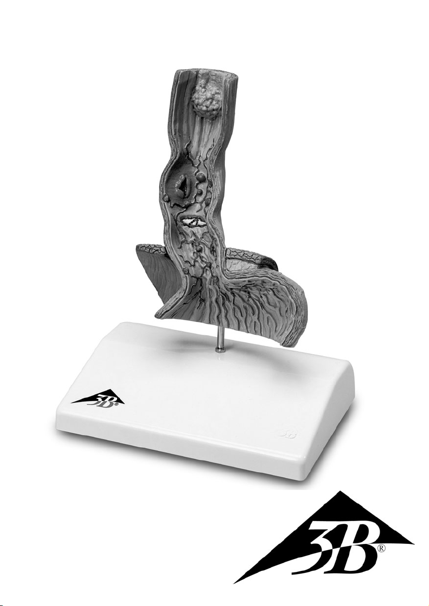
The life-size model is a frontal section of the lower part of the oesophagus from normal to columnar oesophageal epithelium. The diaphragm is recognisable in area of the oesophagus' point of passage.
Furthermore, the upper part (cardia and fundus) of the stomach is shown. The back of the oesophagus,
diaphragm and stomach is anatomically replicated with unstriated muscles.
The following illnesses are shown:
1. Reflux oesophagitis: infection of the oesophagus caused by stomach acid reflux
2. Ulcus: ulcer of the oesophageal epithelium caused by chronic reflux
3. Barrett's Ulcer: changes of the mucous lining after chronic reflux and ulcers
4. Oesophageal carcinoma: malignant cancer of the oesophagus
5. Oesophageal varices: varicose veins in the oesophagus
6. Hiatal hernia: occurs when the stomach pushes through the hiatus oesophagus into
the thoracic cavity

®
Español Enfermedades del esófago
El modelo representa de tamaño natural un corte frontal de la parte inferior del esófago con la transición
entre la mucosa esofágica normal y la cilíndrica (epitelio). Se puede percibir el diafragma en la zona
del orificio para el paso del esófago. Además, se representa la parte superior (cardias y fondo) del
estómago. Las caras posteriores del esófago, del diafragma y del estómago han sido modelados anatómicamente con musculatura lisa.
Se representan las siguientes enfermedades:
1. Esofagitis por reflujo: inflamación del esófago por reflujo de ácido gástrico
2. Úlcera: úlcera de la mucosa por reflujo crónico
3. Úlcera de Barrett: alteración de la mucosa tras reflujo crónico y úlceras
4. Carcinoma esofágico: cáncer de esófago maligno
5. Varices esofágicas: varices del esófago
6. Hernia hiatal: protrusión del estómago al tórax a través del hiato esofágico
®
Erkrankungen der Speiseröhre Deutsch
Das Modell zeigt in natürlicher Größe einen Frontalschnitt durch den unteren Anteil der Speiseröhre
(Ösophagus) mit dem Wechsel von normaler auf zylindrische Ösophagusschleimhaut (Epithel). Das
Zwerchfell ist im Bereich der Durchtrittsstelle für die Speiseröhre errkennbar. Weiterhin ist der obere
Anteil (Kardia und Fundus) des Magens dargestellt. Die Rückseite der Speiseröhre, des Zwerchsfells und
des Magens sind anatomisch mit glatter Muskulatur modelliert.
Folgende Erkrankungen sind dargestellt:
1. Refluxösophagitis: Entzündung der Speiseröhre bei Rückfluss von Magensäure (Reflux)
2. Ulkus: Schleimhautgeschwür bei chronischem Reflux
3. Barrett Ulkus: Veränderung der Schleimhaut nach chronischem Reflux und Geschwüren
4. Ösophaguskarzinom: bösartiger Speiseröhrenkrebs
5. Ösophagusvarizen: Speiseröhrenkrampfadern
6. Hiatushernie: Vorwölbung des Magens in den Brustraum durch den Hiatus oesophageus
 Loading...