

Welch Allyn CP50 User Manual

CP 50™ and CP 50 Plus™ 12-lead resting electrocardiograph
Quick reference guide
Powering the electrocardiograph
1
The electrocardiograph runs on AC or battery power. Plug the electrocardiograph into AC
power as often as possible so that the built-in charger can keep the battery charged.
Regardless of the battery condition, you can use the electrocardiograph whenever it is
plugged in.
WARNING When you use AC power, always plug the electrocardiograph
into a hospital-grade outlet to avoid the risk of shock.
WARNING If the integrity of the building’s safety ground is in doubt,
operate this device on battery power to avoid the risk of shock.
To power up or power down
Press .

2 CP 50™ and CP 50 Plus™ 12-lead resting electrocardiograph
Attaching the leads to the patient
Proper lead attachment is important for a successful ECG. The most common ECG
problems are caused by poor electrode contact and loose leads. Follow your local
procedures for attaching the leads to the patient. Here are some common guidelines.
To attach the leads to the patient
1. Prepare the patient.
• Describe the procedure. Explain the importance of holding still during the test.
(Movement can create artifact.)
• Verify that the patient is comfortable, warm, and relaxed. (Shivering can create
artifact.)
• Put the patient in a reclining position with the head slightly higher than the heart
and legs (the semi-Fowler’s position).
2. Select the electrode locations. (See the “Electrode locations” chart.)
• Look for flat areas.
• Avoid fatty areas, bony areas, and major muscles.
3. Prepare the electrode locations.
• Shave or clip the hair.
• Thoroughly clean the skin, and lightly rub it dry. You may use soap and water,
isopropyl alcohol, or skin preparation pads.
4. Attach the lead wires to the electrodes.
5. Apply the electrodes to the patient.
Electrode examples, left to right: arm clamp (reusable), Welsh cup (reusable), tab
electrode (disposable), monitoring electrode (disposable).
• For reusable electrodes: Use electrode paste, gel, or cream to cover an area
the size of each electrode but no larger. Secure the arm and leg clamps. Apply
the Welsh cups (suction electrodes) to the chest.

CP 50™ and CP 50 Plus™ 12-lead resting electrocardiograph 3
• For disposable tab electrodes: Place the electrode tab between the “jaws” of
the connector. Keep the tab flat. Verify that the metal portion of the connector
makes contact with the skin side of the electrode tab.
• For all disposable electrodes: Lightly tug on the connector to ensure that the
lead is securely attached. If the electrode comes off, replace it with a new
electrode. If the connector comes off, reconnect it.
4 CP 50™ and CP 50 Plus™ 12-lead resting electrocardiograph
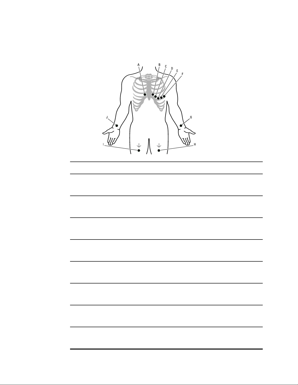
Electrode locations
AHA IEC Location
A
B
C
D
E
F
V1
(red)
V2
(yellow)
V3
(green)
V4
(blue)
V5
(orange)
V6
(purple
C1
(red)
C2
(yellow)
C3
(green)
C4
(brown)
C5
(black)
C6
(purple)
Fourth intercostal space at the right sternal border.
Fourth intercostal space at the left sternal border.
Midway between V2 and V4.
Fifth intercostal space to the left of the midclavicular line.
Anterior axillary line at the same horizontal level as V4.
Mid-axillary line at the same horizontal level as V4 and V5.
G
H
LA
(black)
LL
(red)
L
(yellow)
R
(green)
Just above the left wrist on the inside of the arm.
Just above the left ankle.

CP 50™ and CP 50 Plus™ 12-lead resting electrocardiograph 5
AHA IEC Location
I
J
RL
(green)
RA
(white)
N
(black)
R
(red)
Just above the right ankle.
Just above the right wrist on the inside of the arm.
6 CP 50™ and CP 50 Plus™ 12-lead resting electrocardiograph
About the test types
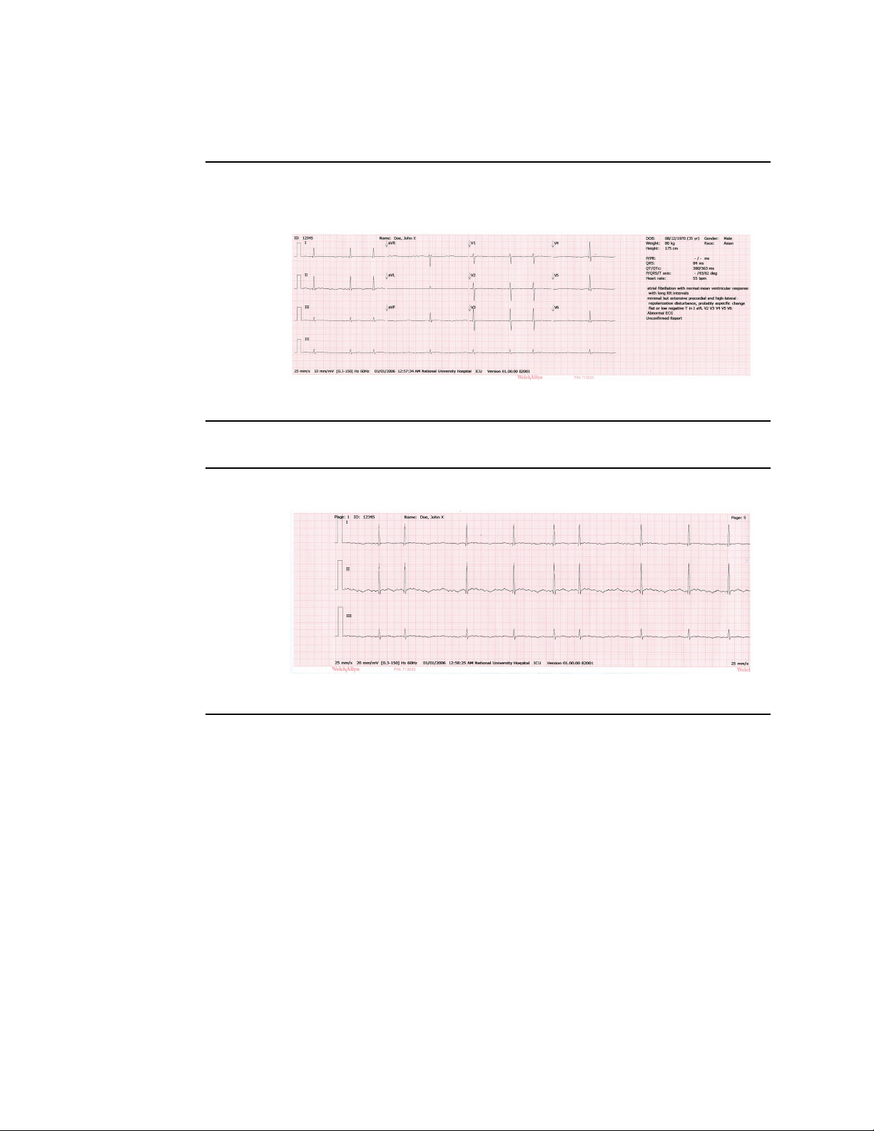
Auto ECG
Stat ECG
Rhythm ECG
A report typically showing a 10-second acquisition of 12 leads of ECG information combined with
patient data, measurements, and optional interpretation. Auto ECGs can be saved to the
electrocardiograph’s test directory or to a USB mass-storage device. In CP 50 Plus models, auto ECGs
can also be sent to a Welch Allyn CardioPerfect workstation.
Auto ECG report example
An auto ECG that starts instantly without waiting for you to enter patient data or adjust the
waveforms.
A continuous, real-time printout of rhythm strips with a user-defined lead configuration. Rhythm ECGs
are printouts only. They cannot be saved.
Rhythm ECG report example
 Loading...