

Olympus Discover FV10i System Features And Benefits
Laser Scanning Confocal Microscope
Discover FV10i
F V10i
®
I Discovered a new way to look at high-end imaging.
I needed a system that would give me the same high-end imaging as a confocal,
but would fit on my benchtop. With the FV10i, I got all that and more—superior
image quality, a compact solution, and an easy-to-use instrument—with no darkroom
required. Now I can focus on the science, instead of the system.
Isn’t it time you saw high-end imaging in a new light?
Scan this code or visit
www.olympusamericaFV10i.com
to learn more and to
schedule a demo.
®
SYSTEM FEATURES AND BENEFITS
F V10i
No experience is required with the FV10i, even for sophisticated confocal imaging.
The navigation function leads a first-time user to operate the FV10i with ease.
FV10i-LIV
Simplied Built-in Incubator
The system has a simplied built-in incubator, allowing easy
time-lapse imaging of live cells without losing valuable time
in setting up equipment. The environment in the culture
chamber is maintained at temperature - 37 degrees Celsius,
humidity of - 90%, and CO
Dedicated Culture Pod
The system is provided with a dedicated culture pod for
dia. 35mm cover glass bottom dishes. Recirculation of the
culture media and addition of reagents during time-lapse
is possible. In addition, the culture pod system can be
autoclaved for sterilization.
Stable Time-Lapse Imaging
Not only the incubator but also the surrounding air space
is maintained at 37 degrees Celsius. Long-term time-lapse
imaging is possible while maintaining cell activity.
Water is Automatically Supplied to the
Water-Immersion Objective
The newly developed automatic water dispensing system
enables the FV10i to supply water to the top of the waterimmersion objective. You can continue long-term time-lapse
imaging without worrying about insufcient immersion
media. Water is supplied automatically when the objective is
moved into the observation position.
Detection of Cover Glass Thickness and Automatic
Adjustment of the Correction Collar
The system is equipped with the capability to detect the
thickness of the cover glass, allowing it to adjust the
correction collar automatically, when using the waterimmersion objective. This assures imaging is performed
each time with optimal conditions.
2 concentration of - 5%.
FV10i-LIV/FV10i-DOC
Equipped with Four Wavelength Diode Lasers
for Imaging up to Five Channels
The system is equipped with four (405/473/559/635nm)
lasers. Multi-stained specimens can be imaged with up to
four uorescence dyes. Transmitted light phase images can
also be acquired. Maintenance-free and power-saving diode
lasers with longer operating lives are employed in all the laser
units, and operate with low noise levels.
Detector Utilizes a Newly Developed Spectrum Method
The detecting mechanism has two uorescence channels,
and one phase contrast channel. The uorescent channels
use a newly developed spectrum method comprising grating,
beam splitter, and slit. In addition, they are equipped with
the variable barrier lter function where the most suitable
wavelength width is set automatically in accordance with the
characteristics of the uorescence dye.
Objectives of 10
The system is equipped with 10x and 60x phase objectives
specically designed for the FV10i to maintain high resolution.
By changing lens and scanning zoom, magnication can be
changed continually from 10x to 600x. The most suitable
imaging area can be set depending on size of the specimen.
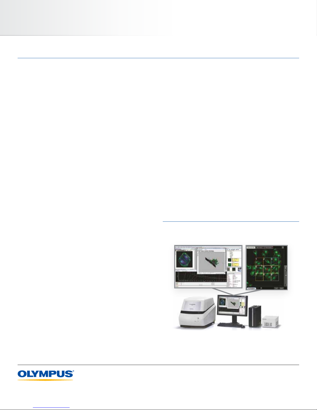
Software dedicated for exclusive use for FluoView is provided
to easily perform various editing / analysis operations.
x and 60x are Mounted on the System
The System Supports Multi-Area Time-Lapse
The system is equipped with a motorized stage, and repeat
imaging is possible through multi-area time-lapse. Ten point
locations can be assigned within a single dish (well). For
example, in the case of a dia. 35mm glass bottom dish,
three dishes can be mounted, allowing a maximum of up to
30 locations to be captured.
olympus america inc.
3500 Corporate Parkway, Center Valley, PA 18034-0610, U.S.A.
olympus canada inc.
25 Leek Crescent, Richmond Hill, Ontario L4B 4B3
www.olympusamerica.com
• ©2012 Olympus America Inc. All rights reserved.
• Olympus and FV are registered trademarks of Olympus Corporation, Olympus America Inc., and/or
their afliates, in the U.S. and/or other countries.
• Images on the PC monitors are simulated.
• Specications and appearances are subject to change without any notice or obligation on the part of
the manufacturer.
olympus latin america inc.
5301 Blue Lagoon Drive, Suite 290 Miami, FL 33126, U.S.A.
Printed in USA. FV10IFLYER12.06
 Loading...