

Page 1

Spectacular Imaging!
Leica TCS SPE: High Resolution Spectral Confocal
Affordable Excellence for Your Daily Research
Page 2
1
2
•
Spectacular Imaging
•
Easy to Achieve
•
A Reliable System
•
Affordable Excellence
2
Page 3
Confocal microscopy applications have in-
3
4
5
3
creased greatly in the last decade, and high
quality fluorescent images have been an important key to new discoveries. In addition, clinical,
pharmaceutical and biotechnological research
shows a growing demand for high-resolution 3D
images. Confocal systems offer the advantage
of the best image quality, but most instruments
currently offered still require intensive training
before they can be usefully operated. They also
need specific room conditions not found in
every facility.
Spectacular
Imaging
Affordable Excellence
for your Daily Research
To make confocal technology accessible to a
wide range of users in their daily research, we
have developed the Leica TCS SPE, a high resolution spectral confocal, easy to use, extremely
compact and robust – yet still affordable.
Without compromising our innovative concepts
and high quality standards, the Leica TCS SPE
offers outstanding spectral detection technology developed and patented by Leica Microsystems. New optical concepts for true colocalization plus a new Leica software platform
common to our complete range of confocal
and widefield products, simplify operation and
reduce training time. You’ll find it all in the
Leica TCS SPE.
The highly integrated Leica TCS SPE has all
you need for your daily research. It is optimized
for applications such as live cell imaging and
morphological studies in small research groups
and multi user environments.
Page 4
6
7 8
Page 5
The advantages of true confocal imaging
Spectacular Imaging
•
3D confocal imaging
•
Variable motorized pinhole to
match objectives
•
488, 532 and 635 nm excitation
•
405 nm excitation for
nuclear stainings
•
Low-noise solid state lasers
•
Highly efficient prism for
emission spectrometer
•
High dynamic photomultiplier
with photon booster technology
“Brilliant images, great
technology and excellent
cost performance ratio –
Leica’s TCS SPE
is the ideal tool for our
laboratory.”
Jean-Luc Vonesch
Head of Imaging Center at the IGBMC (INSERM,
CNRS, ULP), Strasbourg-Illkirch, France
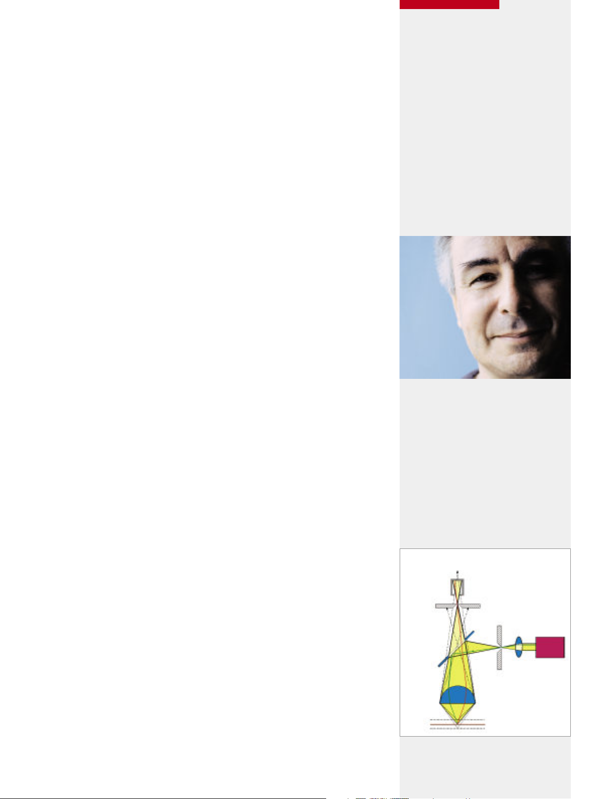
The Confocal Principle
Detector
Confocal pinhole
Laser
Beam
Splitter
Objective
Focal plane
out
in
out
Scanning the specimen in thin optical layers and detecting the
fluorescence signal point by point results in images free from
the stray light of adjacent elements. The result – brilliant images
at very high resolution. The information from each signal in
each optical section is reconstructed by intelligent software
into excellent 3D images, resolving the smallest detail of the
specimen’s structure.
Crystal clear 3D confocal images are our standard
Our confocal systems are famous for crystal clear high-resolution images with minimum noise, due to high performance
components unrivalled in the confocal field. We have integrated
this highly innovative and unsurpassed technology into the new
Leica TCS SPE. To ensure high quality imaging, optimal excitation
is provided by up to four low noise solid-state lasers with 488,
532 and 635 nm excitation lines for common dyes. The broad
range of applications is extended by the 405 nm option for
nuclear staining.
Ultra-high sensitivity with maximum spectral efficiency is ensured
by our spectrophotometer based on a prism, which spreads the
light into its spectrum. Maximum signal strength and optimal
resolution is provided by a special high dynamics photomultiplier with photon booster technology, usually found in systems
of a higher price range. The continuously adjustable pinhole
diameter automatically adapts to all objectives. Channel multiplexing by sequential scanning prevents any cross-talk of dyes
and results in excellent dye separation. Optimally integrated with
our high performance research microscopes and optics, we
fully harness our own technology to deliver the best images.
5
Page 6

6
Spectral detection – exclusive to this class of system
A freely tunable spectral range, maximizing signal independent
of the fixed barriers of standard filters, is the ultimate goal for
many researchers. The Leica TCS SPE uses the prism spectral
detection system developed and patented by Leica Microsystems – well known for its unrivalled detection efficiency.
It provides full flexibility in adjusting the wavelength to your
specimen as you can continuously tune bandwidths from 430 nm
to 750 nm. With the Leica spectral detection system you are
completely independent of fixed filter settings.
Perfect characterization of your dyes
Characterize your dyes directly within the specimen and gain
new information via a lambda scan. Profit from the freedom of
using various dyes – today and tomorrow.
Better protection against cell damage will provide more reliable
results. Confocal systems from Leica Microsystems are designed
to ensure longevity of your specimens – whether they are fixed
samples or living cells. The Acousto Optical Tunable Filter
(AOTF) minimizes exposure to light by individual tuning of the
laser power.
•
Freely tunable spectral detection
(430 -750 nm)
•
Fully flexible adjustment to specimen
•
AOTF tuning for minimizing
cell damage
•
Motorized beam splitter
•
Perfect colocalization with
new ACS optics
The Spectral Principle
Light beam
Prism
Variable spectral
detector
Collimation optics
Detector
1.0
0.9
0.8
0.7
0.6
0.5
0.4
0.3
0.2
0.1
0
460
480
500
520
540
560
580
600
620
640
660
680
700
720
740
1.0
0.9
0.8
0.7
0.6
0.5
0.4
0.3
0.2
0.1
0
460
480
500
520
540
560
580
600
620
640
Sequential Scan
-Scan
y
Wavelength [nm]
Intensity [arbitrary units]
Wavelength [nm]
Intensity [arbitrary units]
Page 7

Perfect colocalization
Leica ACS-Technology: Ideal for confocal
0.5
0.4
0.3
0.2
0.1
0
400 500 600 700 800 900
Wavelength [nm]
The whole spectral range from 400 nm to near IR is covered by ACS
standard
red
blue
ACS
useful for
confocal
Focal plane shift
“Top technology made
available to non-expert
users. TCS SPE has the
advantages of a latest
generation confocal,
and its simplicity makes
it really easy to use.”
Dr. Maria C. Montoya
Confocal Microscopy and Cytometry Unit
Biotechnology Program
Spanish National Cancer Center (CNIO)
Madrid, Spain
uncorrected corrected
405 nm causes focal plane
shifts
ACS corrects focal plane
shifts
Benefit from clear and true confocal signals over the full spectral
range by perfect super positioning of all laser foci at only one
point in the focal plane. For example, a nucleus stained with
DAPI is projected in its natural position in the center of the cell.
For perfect colocalization in the specimen, the Leica TCS SPE
confocal system applies the brand new Advanced Correction
System (ACS) technology by Leica Microsystems. This truly
innovative optical design of the light paths, from excitation to
detection, guarantees ideal transmission from 400 nm to 830 nm.
With ACS technology, additional correction optics and beam
splitters in the light path are redundant. Maximum transmission
is achieved within the entire light band from 405 nm to infrared.
Profit from ultra-bright pin sharp images with ACS.
7
Page 8

Minimal training effort
Easy to Achieve
True Confocal is not Complicated
Easy confocal imaging: 6 steps to 3D
1 Start the system
2 Insert and focus specimen
3 Select instrument settings
4 Define z-range and acquire
5 Calculate 3D image
6 Save and close
Wavelength selection and laser attenuation
•
Reliable, ergonomic software LAS AF
•
Software upgradable to
•
deconvolution, Motion Spy,
Dye Finder and more
Preinstalled system settings
•
Easy data transfer
•
Personal USB for storage of
•
individual instrument settings
Multiple export functions
•
Analysis of specimen with up to
•
8 color stainings
The Leica TCS SPE is an instrument designed to make your work
as uncomplicated as possible. It requires minimal training effort
and first results can be achieved immediately. Its reliable and
ergonomic software leads you straight through your experiment
without losing precious time.
Easy to use
The standardized, self-explanatory user interface of the Leica
Application Suite Advanced Fluorescence (LAS AF) software
enables you to start your work autonomously with the first click of
a button. Newcomers to confocal will appreciate the uncomplicated software with its workflow-oriented screens. They guide
you through your experiment from the selection of the objective up
to the reconstruction of the first 3D image. Full application flexibility is offered by extending the software capabilities with additional modules, opening up your project possibilities to applications such as deconvolution, time lapse and spectral unmixing.
Easy interfacing
Preinstalled and optimized system settings for defined dyes ensure fast and excellent results right from the start. More experienced users profit from the full flexibility of the automated system
for individual tuning of different experiments. With the wavelength selector, you can easily adjust the detection range as
both excitation laser line and emission spectra of the dye are
displayed in the same window. Just align the detection range
sliders and you are ready to start spectral imaging. For future
applications the full software suite of LAS AF is open to you.
8
Page 9

Your data – easy to transfer, easy to share
Z-range definition
9
After your work session you can ensure repeatable results by
saving your individual settings on a USB stick. Continue working
any time at from exactly the same point – even if other users
have used the system in the meantime for other applications with
different settings.
Exchange of data is easy with the multiple export functions of
the Leica TCS SPE. Aside from the USB stick you can use the
LAN connectivity interface for local server connection. Benefit
from fast transfer of your experimental results to PCs or Apple
Macintosh computers or send data direct to the printer. With
such easy data sharing, you and your colleagues will harness
true teamwork for accelerated results.
Designed for target applications in research laboratories ...
The Leica TCS SPE is the ideal instrument for morphologic investigations as well as for live cell studies into cell division or cell
growth. Analyze up to eight colors of multi stained samples or
track GFP development in time lapse experiments. Generate
overlays of transmitted light images with fluorescent information
to investigate the developmental biology of your specimen.
... and pharma-biotechnology
The Leica TCS SPE is an extremely stable and reliable partner
for routine applications in biotechnology. Screen your cells at
defined intervals to discover new effects of active agents on
target cells or test the impact of new compounds on tissues
while discovering new drugs. Gain a closer insight into cells
involved in fermentation processes, routinely monitor culture
processes or simply document growth experiments in microwell
plates. The Leica TCS SPE provides the perfect opportunity to
optimize many aspects of bio-production.
9
Page 10

Page 11

11
A reliable system is a must for routine laboratory work. The
robust and durable hardware with long-life components and
easy to use software, make the Leica TCS SPE an indispensable
workhorse that totally fulfils these exacting demands.
Reliability and robustness in your every day work
Long-life solid-state lasers are combined with additional optical
elements installed on the robust interior optical bench. Instead
of filter wheels, we have integrated the AOTF and kept the
number of moving parts to a minimum. This shortened light
path with a single fiber coupling requires minimum maintenance. The workflow oriented and self-explanatory LAS AF software enables smooth and fast operation. The system requires
little maintenance and minimises the workload of system administrators. Conflicts with other software or from Internet downloads are avoided as the system runs exclusively with the Leica
operating software, keeping the system virus free and secure.
Highly integrated system
The Leica TCS SPE is a highly integrated system – extremely
compact and robust with a supply unit no larger than a standard
PC. Equipped with solid-state lasers, the system needs no extra
cooling. With its small footprint and standard room requirements,
the Leica TCS SPE fits into any laboratory.
A Reliable System
Concentrate on your Work – Not on Your System
•
Long lifetime components
•
Minimal maintenance
•
Smooth and fast operation
•
Highly integrated system
•
Small footprint
•
Only standard room requirements
Transmitted light detector
“High precision, robust
technique and easy to use
software is what we always
looked for. Leica’s new
confocal will become our
workhorse for routine
research.”
Dr. Markus Dürrenberger
Microscopy Center (ZMB)
University Basel
Basel, Switzerland
Page 12

12
From high resolution fluorescence imaging to 3D reconstruction
and time lapse, the new Leica TCS SPE provides all the features
you need – at a highly attractive price.
Enter the world of high resolution 3D fluorescence imaging
Profit from Leica’s years of experience and enter the world of
top confocal microscopy. Achieve excellent results and detailed
information in your specimen and reach new goals with your
research. The Leica TCS SPE platform opens new horizons for
scientific research and offers an affordable start to high quality
3D fluorescence imaging. The system can grow with your changing requirements, thanks to a range of easy to install upgrade
kits. Regardless of whether your chosen option is a transmitted
light detector or a new software module, single-supplier compatibility is assured.
The flexible system is easy to operate – forget about time consuming instructions and complex tutorials. Our new software
platform is self-explanatory and guides you through the entire
workflow from image acquisition to high-resolution 3D reconstruction. Predefined and optimized Leica settings guarantee
high-end results every time.
Spectacular images can be printed immediately for discussion
and larger data sets can be stored on DVD or server. Should
you have new applications or further questions, our application
specialists will be happy to share their wealth of experience
with you.
Affordable Excellence
•
High resolution
•
True confocal imaging
•
Freely tunable spectral detection
•
Ready for new dyes
•
Broad laser excitation range
•
Stable solid state lasers
•
Fully automated
•
100% tunable (AOTF, pinhole)
•
Perfect colocalization throughout
the spectrum with ACS
•
No special room requirements
•
Flexible to upgrade by defined kits
•
Modular software and hardware
10
Page 13

11
Leica TCS SPE sets new standards in your imaging center
Leica Microsystems stands for excellent quality and ingenuity.
Whether top-range or entry-level, we never compromise on the
quality of our confocal microscopes.
Increase your capacity: the new Leica TCS SPE offers high-end
imaging at an affordable price. After a short installation time,
the system is ready for action. Easy to use software minimizes
training effort, allowing scientists to work with the confocal
straight away. As the Leica LAS AF software is the only software
installed, internet downloads or other software installations are
not possible, minimising maintenance and administration. This
also facilitates large user groups. With no compromise in image
quality, the SPE will also relieve the workload of your high-end
imaging systems.
LAN connectivity and external devices such as USB or DVD
burners ensure full compatibility with your environment and easy
data exchange. To achieve reproducible results, scientists are
able to store individual instrument settings together with their
results on their own USB stick. For highest utilization of the
system, post processing of images can be performed on a separate workstation thanks to easy data export functions.
Specially selected long-life components, such as solid-state
lasers and robust technologies, make the TCS SPE stable and
reliable and reduce the cost of ownership. Service contracts
provide maximum system uptime. The high-resolution spectral
confocal system Leica TCS SPE will become the ideal workhorse for your daily research.
The Leica TCS SPE is your reliable partner in research, providing
you with spectacular results – easily and at an affordable price.
Leica image quality
•
Minimal administration
•
Little training required
•
Predefined instrument settings
•
Single software platform
•
Easy data transfer
•
Individual settings on USB stick
•
Reliable system
•
Highly efficient, maximum capacity
•
Service contracts
•
13
Page 14

Leica Design by Christophe Apothéloz
Page 15

Acknowledgements:
We gratefully acknowledge the following scientists
for providing images:
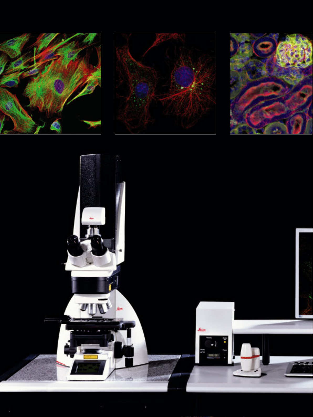
1 Mouse fibroblasts
Green: F-Actin, FITC; Red: Tubulin, Cy5; Blue: Nuclei, DAPI
Courtesy of Dr. Günter Giese, Max Planck Institute for Medical
Research, Heidelberg, Germany
2, 8 COS 7 cells
Green: uncharacterized protein, GFP; Red: α-Tubulin, Cy3;
Blue: Nuclei, DAPI
Courtesy of Prof. Wei Bian, Cell Research Center, Institute of
Biochemistry and Cell Biology, SIBS, CAS, Shanghai, China
3 Mouse kidney section
Green: glomeruli and convoluted tubules, Alexa 488 WGA;
Red: F-Actin (prevalent in glomeruli and brush border);
Blue: Nuclei, DAPI
Leica Microsystems CMS GmbH, Mannheim, Germany
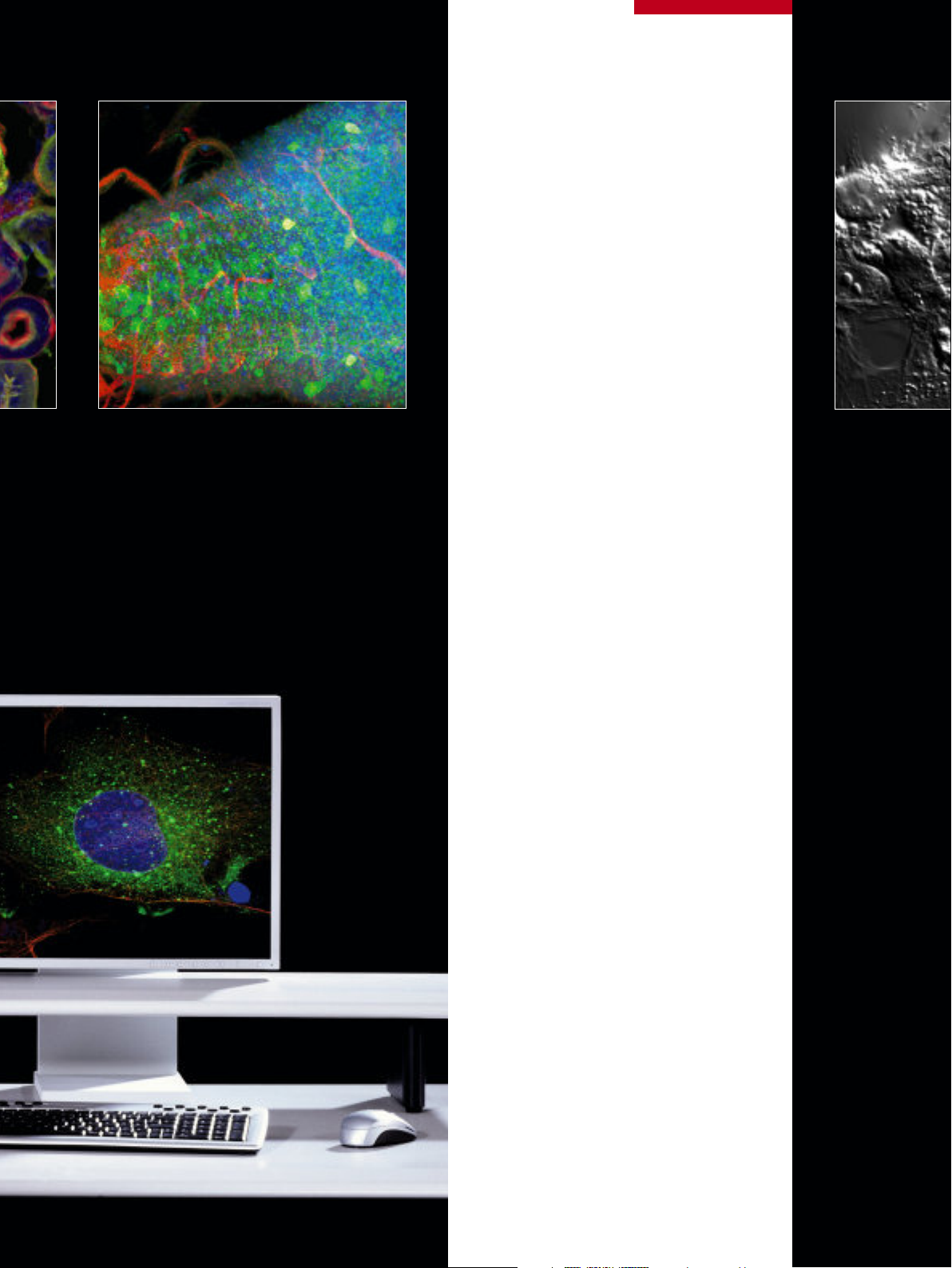
4 Drosophila melanogaster, larval stadium
Green: Feb211 positive neurons and their axons, Alexa 488;
Red: fibrous part of the cns (i.e all axons), Cy3; Blue: Nuclei, DAPI
Courtesy of Dr. Christoph Melcher, Research Institute Karlsruhe,
Institute for Toxicology and Genetics, Eggenstein-Leopoldshafen,
Germany
5 Mouse fibroblasts
DIC
Courtesy of Dr. Günter Giese, Max Planck Institute for Medical
Research, Heidelberg, Germany
SPE
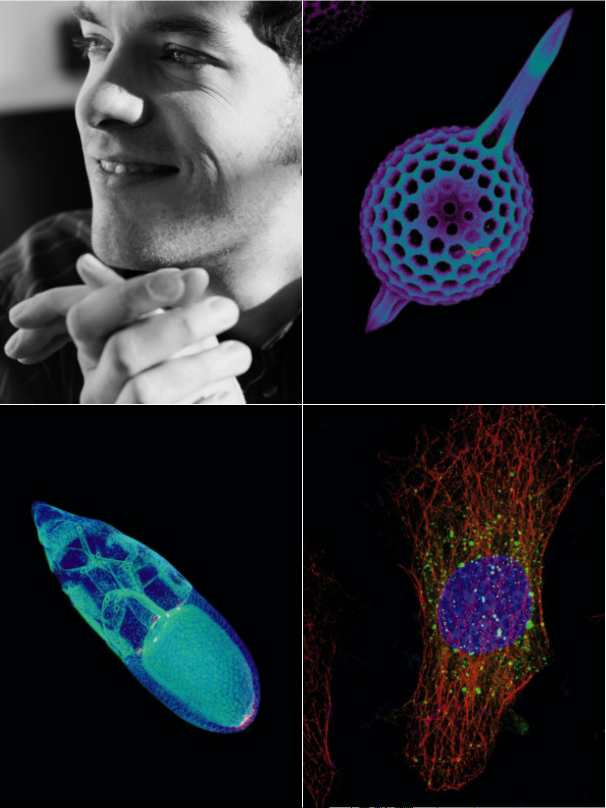
6 Radiolaria
Silica-skeleton, reflection mode
Leica Microsystems CMS GmbH, Mannheim, Germany
7 Drosophila melanogaster, egg chamber
Red: Nuclei, Cy5; Blue: Cytoplasmatic and Nuclear GFP, GFP;
Cyan: Actin, Phalloidin-Rhodamin
Dr. Juliette Mathieu, Rørth Lab, European Molecular Biology
Laboratory; EMBL, Heidelberg, Germany
9 Phaseolus vulgaris, native plant stipe
Autofluorescence with 488 nm, 532 nm, 635 nm excitation and
transmitted light; overlay image
Courtesy of Dr. Markus Dürrenberger, Microscopy Center (ZMB),
University Basel, Switzerland
10 Drosophila melanogaster, egg chamber
Green: Actin, Phalloidin-Rhodamin; Red: Cytoplasmatic and
Nuclear GFP, GFP; Blue: Nuclei, Cy5
Dr. Juliette Mathieu, Rørth, European Molecular Biology Laboratory;
EMBL, Heidelberg, Germany
11 Mouse fibroblasts
Green: F-Actin, FITC; Red: Vimentin, Cy3, Blue: Nuclei, DAPI
Courtesy of Dr. Günter Giese, Max Planck Institute for Medical
Research, Heidelberg, Germany
www.leica-microsystems.com/TCS_
15
Page 16

Leica Microsystems –
www.leica-microsystems.com/Confocal_Microscopes
the brand for outstanding products
Leica Microsystems’ mission is to be the world’s first-choice provider of innovative
solutions to our customers’ needs for vision, measurement and analysis of microstructures.
Leica, the leading brand for microscopes and scientific instruments, developed
from five brand names, all with a long tradition: Wild, Leitz, Reichert, Jung and Cambridge Instruments. Yet Leica symbolizes innovation as well as tradition.
Leica Microsystems – an international company
with a strong network of customer services
Australia: Gladesville Tel. +61 2 9879 9700 Fax +61 2 9817 8358
Austria: Vienna Tel. +43 1 486 80 50 0 Fax +43 1 486 80 50 30
Canada: Richmond Hill/Ontario Tel. +1 905 762 2000 Fax +1 905 762 8937
Denmark: Herlev Tel. +45 4454 0101 Fax +45 4454 0111
France: Rueil-Malmaison Tel. +33 1 47 32 85 85 Fax +33 1 47 32 85 86
Germany: Bensheim Tel. +49 6251 136 0 Fax +49 6251 136 155
Italy: Milan Tel. +39 0257 486.1 Fax +39 0257 40 3475
Japan: Tokyo Tel. + 81 3 5421 2800 Fax +81 3 5421 2896
Korea: Seoul Tel. +82 2 514 65 43 Fax +82 2 514 65 48
Netherlands: Rijswijk Tel. +31 70 4132 100 Fax +31 70 4132 109
People’s Rep. of China: Hong Kong Tel. +852 2564 6699 Fax +852 2564 4163
Portugal: Lisbon Tel. +351 21 388 9112 Fax +351 21 385 4668
Singapore Tel. +65 6779 7823 Fax +65 6773 0628
Spain: Barcelona Tel. +34 93 494 95 30 Fax +34 93 494 95 32
Sweden: Sollentuna Tel. +46 8 625 45 45 Fax +46 8 625 45 10
Switzerland: Glattbrugg Tel. +41 1 809 34 34 Fax +41 1 809 34 44
United Kingdom: Milton Keynes Tel. +44 1908 246 246 Fax +44 1908 609 992
USA: Bannockburn/lllinois Tel. +1 847 405 0123 Fax +1 847 405 0164
and representatives of Leica Microsystems
in more than 100 countries.
The companies of the Leica Microsystems Group operate internationally
in three business segments, where we
rank with the market leaders.
Microscopy Systems
•
Our expertise in microscopy is the basis for all our solutions for visualization,
measurement and analysis of microstructures in life sciences and industry.
With confocal laser technology and image analysis systems, we provide
three-dimensional viewing facilities
and offer new solutions for cytogenetics, pathology and materials sciences.
Specimen Preparation
•
We provide comprehensive systems
and services for clinical histo- and cytopathology applications, biomedical
research and industrial quality assurance. Our product range includes instruments, systems and consumables
for tissue infiltration and embedding,
microtomes and cryostats as well as
automated stainers and coverslippers.
Medical Equipment
•
Innovative technologies in our surgical
microscopes offer new therapeutic approaches in microsurgery.
Fax +49 (0)621-7028 1028 LEICA and the Leica Logo are registered trademarks of Leica IR GmbH.
•
Tel. +49 (0)621-7028 0
•
Germany 2006
•
68165 Mannheim
•
Am Friedensplatz 3
•
Printed on chlorine-free bleached paper. II/06/SK
•
Leica Microsystems CMS GmbH
©
Order nos.: English 1593031008
Copyright
 Loading...