

Leica
TCS SP5 MP
Broadband Confocal and
Multiphoton Microscope
The Solution for Deep Imaging
2
Deep Imaging
Since the advent of confocal microscopy, immense progresses
have been made in cellular biology, neurosciences, medical
research. Today, it is a major challenge to penetrate deeper
into samples for improved studies of cells, organs or tissues. An
effi cient method to achieve a deep penetration into samples
is two-photon and multiphoton excitation with laser scanning
microscopes which are equipped with pulsed infrared lasers.
Thanks to reduced absorption and scattering of the excitation
light, two-photon and multiphoton confocal microscopes reach a
penetration depth of about 400 µm.
In the case of two-photon excitation, the dye is excited by the
simultaneous absorption of two photons. Due to the non-linearity
nature of two-photon absorption, the excitation is limited to the
focal volume and the photobleaching outside the focal plane is
reduced. Only inside the confocal volume the photon density is
suffi ciently high to allow two photon absorption by the fl uorophore.
Multiphoton excitation performance improves with pulsed laser
excitation in the NIR spectra. Longer excitation wavelengths are
scattered less in biological tissue allowing a deeper penetration
in very thick specimen. Emission/Fluorescence signal is not degraded either by scattering from within the sample.
Advantages of multiphoton exitation:
• Greater penetration depth due to lower scattering
• Intrinsical optical sectioning properties – no need for
a detection pinhole
• Bleaching restricted to focal plane – no volume bleaching
• Reduced phototoxicity due to spatial confi nement, which is
ideal for living cells.
• Uncaging, photoactivation or photobleaching in a diffraction-
limited volume
The Leica TCS SP5 MP covers a wide range of imaging applications (multiphoton and one photon) by combining two technologies in one system: a conventional scanner for maximum resolution and a resonant scanner for high time resolution.
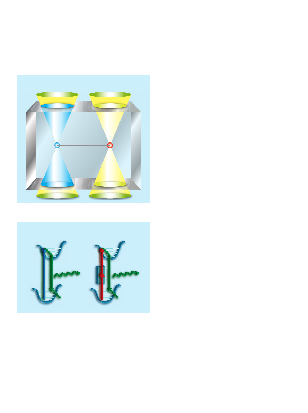
In 2-photon excitation fl uorescence emission occurs only on the focal plane.
Energy diagram of fl uorescence with 1-photon and 2-photon excitations
1-photon excitation
1-photon excitation
excitation
fl uorescence
emission
fl uorescence
emission
2-photon excitation
2-photon excitation
focal plane
Applications
The invention of multiphoton microscopy in the 1990’s raised
a tremendous interest and has become a widespread imaging
method in the biological sciences since then. Meanwhile there
is plethora of applications and publications involving multiphoton
microscopy.
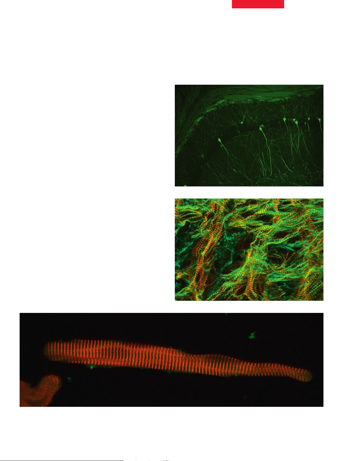
It is now established as the method of choice for non-invasive
deep-penetration fl uorescence microscopy of thick highly scattering samples and has been used for a diversity of specimen,
e.g. lymphatic organs, kidney, heart, skin and brain (slices as well
as intact organs).
Various research fi elds, e.g. immunology (lymphocyte tracking,
embryology, cancer research and particularly neuroscience (e.g.
for the study of calcium dynamics and neuronal plasticity) take
the advantage of the deep in vivo imaging with multiphoton.
Top: hyppocampal region in mouse brain slice.
Courtesy of Dr. Michael E. Calhun, Hertie Institute, Tübingen, Germany.
Middle: mouse embryo, detail of the heart.
Courtesy of Dr. Elisabeth Ehler, King’s College, London, UK.
Bottom: adult rat cardiomyocytes
3
 Loading...