

Page 1

Leica TCS LSI
The World‘s First Super Zoom Confocal!
From Gene to Cell - from Cell to Embryo
Page 2

1
The World‘s First Super Zoom Confocal
•
2
In Vivo
•
Freedom for New Reserach
•
Easy to Achieve
•
- Large Scale Imaging
2
Page 3

An increasing number of scientists extend their
focus of bio-research from single cell studies to
entire organisms, analyzing the complex interaction within whole animals. Thus, modern
developmental biology is an emerging field of
research, studying the dynamics of cell growth,
differentiation processes and the development
of organs
As living organisms grow in three dimensions
these studies require imaging systems which provide - beside high resolution – a large workspace
and field of view. Leica Microsystems introduces
a pioneering imaging system for developmental
3
4
biology which provides all these features in one:
the new Leica TCS LSI.
in vivo
.
Leica TCS LSI
The world‘s first super zoom
confocal!
Leica TCS LSI is the first super zoom 3D-confocal,
offering high resolution plus large field of view for
in vivo
(LSI) platform provides generous workspace and
adapts perfectly to the experiment needs of
native specimen analysis.
True confocal technology is used to provide crystal clear images of highest spectral resolution,
revealing finest details of the model organism, no
matter if drosophila fly, mouse, plant or zebra fish.
An automated optical zoom system allows for
seamless magnification change on demand,
easy switching from overview to details and
free 3D navigation through the specimen.
imaging. The new Large Scale Imaging
Gain new insights with
TCS LSI and study processes of life – from egg to
fly, from embryo to adult.
in vivo
imaging by Leica
3
Page 4

5
4
Page 5

Benefit from freedom for new applications offered by Leica TCS
LSI, feel free to zoom in and out and travel time resolved in all dimensions
in vivo
through your specimen.
Leica TCS LSI
New perspectives of life: From cell to animal
A system tailored to your needs
With the Leica TCS LSI, study the process of life from embryo to
adult. Due to the functional and workflow oriented design, even
sample preparation and orientation can be directly performed on
the imaging system.
Take advantage of the combination of high-resolution confocal
technology and large field macro zoom imaging. A generous
workspace provides extended freedom for
Zoom in and out, from overview to area of interest at highest resolution and obtain fascinating results.
in vivo
experiments.
Features
Zoom factor up to 16x
•
Field of view up to 16 mm
•
Generous workspace
•
Free sample access by wing door
•
Easy sample manipulation
•
Largest motorized z-control
•
Large xy-travel range for optimal
•
positioning
Precise z-control by galvo stage
•
From cell to embryo
Leica TCS LSI enables you to visualize cell growth and the fascinating differentiation of cells into organs in real life from cell to
embryo – in one system!
Identify new pathways from gene to cell, from cell to animal; or examine the influence of genomic defects on the whole animal.
Study time resolved 4D processes at highest resolution easily as
Advanced Time Lapse software is provided for in vivo studies.
Analyze protein interactions or test the influence of drugs
in bio-medical research.
Visualize the development of life: the Leica TCS LSI Large Scale
Imaging System provides highest image quality, easy operation
and maximum flexibility.
in vivo
Largest workspace...
...for finest insights
6
5
Page 6
Leica TCS LSI – The Large Scale Ima
High Resolution in 2D and 3D
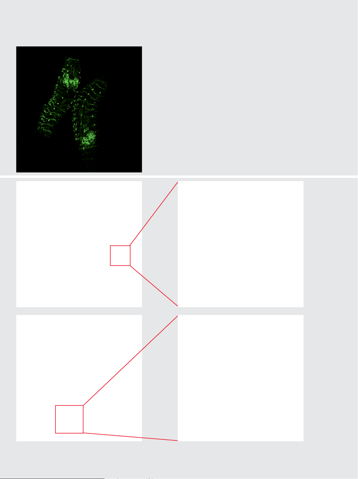
From micro to macro ...
Get the big picture of large specimen in 2D or 3D. Use the
Leica LAS AF 3D-visualisation package for reconstruction of
Drosophila larvae. (Left)
... zoom in seamlessly into the finest details!
The motorized optical zoom offers the advantage of flexible
magnification to identify the finest details of your model
organism. Backbone details of zebra fish or eye of the
7
drosophila larvae, both in highest resolution. (Bottom)
8
10
6
9
11
Page 7

4D-Advanced Time Tapse
From larvae to fly ...
Tracking the development of life over time offers exciting
insights for embryology and morphogenesis – all in the same
specimen! (Right)
... from egg to fish, achieve excellent results in 4D!
To obtain exciting views of the development of organs with
LAS AF Live Data Mode software and see the backbone formation during the growth of a zebra fish. (Bottom)
T= 0 h
T= 10 h
T= 7 h
T= 15 h
12
7
Page 8

Dynamic studies
2D, 3D and 4D analysis
•
Advanced Time Lapse
•
Spectral analysis
•
Photo activation with 405 nm
•
Micromanipulation
•
in vivo
More freedom than ever: For various specimen sizes and research
questions, micro- and macro objectives can be used. High resolution
and largest field of view enable a wide range of new applications.
For excellent new research opportunities the Leica TCS LSI offers
variable magnification with a field of view up to 16 mm plus large
and precise z-positioning – all in one system.
In Vivo –
Large Scale Imaging
High resolution from micro to macro
A comfortable start
Instead of engineering the living object through holes into incubators, start to place your specimen easy and securely through
wide-open wing doors of the laser safety chamber. Gain overview
first by digital camera for easy orientation. Fine-focus precisely
either with the tuning wheel of Universal Microscope Control or
by LAS AF software.
Study the changes of life in 3D
With Leica TCS LSI, you navigate through your model organism
from large to small and back, identifying functions and obtaining
insights never seen before. From the entire animal up to the finest
detail the motorized zoom offers the advantage to select any area
of interest – without changing the objective! Fine-tune the z-position through the highly precise SuperZ Galvo stage and orientate
the sample perfectly in xy-direction with a high precision motorized stage.
Ultra dynamic z-control
The flexibility of the Leica TCS LSI is unique in confocal microscopy. The SuperZ Galvo stage offers backlash free sensitive
vertical positioning with a maximum travel range of 1500 µm. The
fine focus integrated in the motorized zoom system allows further
to extend the focus range by 10 mm. Finally, ultimate z-position control is achieved by the motor focus itself, travelling up to 150 mm.
Seamless zooming in and out
8
13
Page 9

Free xy-sample positioning
Leica TCS LSI systems provide a maximum travel range, independent of the type of stage. Both, manual and automated stages offer a wide range for ideal sample positioning.
Increased cell-viability and highest resolution
Obtain brilliant fluorescent images over long time. Ultimate image
quality is provided by true confocal point scanning technology.
The Leica TCS LSI offers a variety of automated tools to adjust excitation and emission perfectly to your individual sample conditions. Maximize signal efficiency with the freely tunable spectral
detector, the highly dynamic photomultiplier and minimize laser
excitation power with the 100%-tuneable AOTF-attenuation. This
maximizes the lifetime of your specimen.
Developmental
Biology
Plant Research
Growth of
Animal
Structure of
the Leaf
Studies of the
Anatomy
I S L S C T a c i e L
Crop Design
Research areas
Developmental biology
•
Embryology studies
•
Morphogenesis
•
Embryo genetics
•
Plant science
•
Genetics
•
Proteomics
•
Neurology
•
Applications
Cancer research
•
Agriculture investigations
•
Pharmaco screening
•
Seed development
•
Cell development
•
Heart diseases
•
Brain development
•
Genetics
Neurology
Examples of Leica TCS LSI applications
Inuence of
new Drugs
Structure of
the Brain
Lead Finding
Research
Neuro-
Research in
degenerative
Diseases of the
Diseases
Elderly
Field of view depending on objective and zoom adjustment
14
9
Page 10

New Dimensions
Generous workspace
•
Free sample access by wing doors
•
Easy sample manipulation
•
Largest motorized z-control
•
Hardware for most flexible use
•
Large xy-travel range for optimal
•
positioning
Precise z-control by galvo stage
•
Motorized and manual xy-stages
•
Accessories for environmental
•
control of temperature, CO2,
humidity
15
10
Page 11

Making life visible in 4D
Even more than the static view, it is fascinating to observe the development of whole organisms over time. The Leica TCS LSI with LAS
AF Live Data Mode Software offers perfect automation for cell development studies, from egg to embryo. Individual experiments can
be easily combined to a fully automated workflow. The door is open
for continuous studies from cell to adult.
Advanced Time Lapse
High resolution from cell to embryo
Perfect climate included
Optimal growth conditions for living animals can be provided by a
wide range of accessories. The laser safety cabinet is converted into a climate chamber just by adopting a heating unit for precise temperature control. Stage adapters for CO
fer optimal sample conditions. Even during the experiment, remote
controlled manipulators enable active specimen handling within the
native environment.
-gas and humidification of-
2
All accessories are offered in modular and approved kits for easy
system upgrade on demand, ready for todays and future experiments.
Following the changes of life
Studying movements of cancer cells in bio-medical research, investigating translocations after photo-activation, studying the growth of
bio films on large implants: The creativity for future experiments will
come from you, the extended freedom for new research applications is provided by Leica TCS LSI.
16
Zebra fish development video
11
Page 12

Confocal technology
Highest resolution
•
True confocal point scanner
•
Spectral detector
•
Solid state laser
•
405, 488, 532, 635 nm excitation
•
AOTF controlled
•
Fully automated
•
Easy to use
•
Crystal clear 3D images – The benefits of true spectral confocal
imaging
Leica TCS LSI is equipped with a true spectral confocal scanner that
provides ultimate crisp resolution. Scanning the specimen in thin optical layers and detecting the fluorescence signal point-by-point results in images free from the stray light of adjacent elements. The result – brilliant images at very high resolution.
The World’s First Super
Zoom Confocal
Technique for new perspectives
Beam
Splitter
The Confocal Principle
Detector
Confocal pinhole
Laser
Objective
Focal plane
The Spectral Principle
Information from each signal in all optical sections is reconstructed
by intelligent software into excellent 3D images, resolving the
smallest detail of the specimen's structure. To ensure high quality
imaging, optimal excitation is provided by up to four solid-state
lasers with 488, 532 and 635 nm lines for common dyes. The broad
range of applications is extended by the 405 nm laser option for
nuclear staining.
Maximum signal efficiency by spectral detection
Maximize the signal independent from barriers posed by fixed filters and tune freely the emission band from 430 nm to 750 nm.
Cross-talk of different dyes in one sample is prevented when tuning the spectral detector precisely in 10 nm steps to the respective
dye. For unrivalled detection efficiency, the Leica TCS LSI uses a
Light beam
Prism
Variable
Spectral
Detector
Collimation
Optics
Detector
12
Page 13

prism spectral detector. Minimize bleaching and cell damage by optimizing the detection range and reduce the excitation power via
Acousto-Optical Tuneable Filter (AOTF).
Highly innovative
Zoom freely in and out, switching between overview and smallest
detail without changing the objectives. The Leica Z16 APO A 16:1 super zoom offers the largest magnification range from 0,57x to 9,2x,
whereas the Z6 APO A ranges from 0,57x to 3.6x – at excellent optical performance. The Z-zoom systems are fully apochromatic and
allow to adjust magnification continuously and parallax-free.
Benefit from the motorized versions Z16 APO A and Z6 APO A: Fully
software controlled, the magnification can be completely altered
without touching the imaging system. To achieve precise focusing
for sharpest images over a wide range of 10 mm, all motorized zooms
are equipped additionally with a motorized fine focus lens.
Macro and Micro: New dimensions for largest specimen
Benefit from the high quality macro objectives of Leica Microsystems. A novelty in confocal imaging: the tremendous working distance (WD) of 97 mm and a field of view (FOV) of 16 mm provided by
the 1x apochromatic macro objective. Additionally, classic microobjectives can be adapted to use the Leica TCS LSI as a classical
confocal. With high resolution, high numerical aperture lens systems, finest details at maximum resolution become visible.
New zoom optics
Change magnification by fingertip
•
Continuously variable magnification
•
by 16x
Motorized
•
No objective change required
•
Apochromatic, parallax-free optics
•
Macro and micro objectives
•
1x, 2x, 5x, 10x, 40x, 63x
•
Adjusting of magnification by motorized
zoom
Intensity (arbitrary units)
Wavelenght (nm)
Sequential Scan
λ-Scan
Intensity (arbitrary units)
Wavelenght (nm)
13
Page 14

Highest Flexibility
Dynamic confocal imaging
•
Spectral band tuning
•
0 – 100% AOTF laser control
•
Variable pinhole
•
Variable magnification
•
Motorized z-zooms
•
Full objective band from 1x to 63x
•
FOV max: 16 mm
•
WD max: 97 mm
•
Z-range max: 150 mm
•
Observation and handling of large specimen becomes facile: Overcome the limits of fixed magnifications, small fields of view and low
working distances.
Freedom for New Research
Enter new dimensions for flexible applications
The new Leica TCS LSI offers unrivaled flexibility for high resolution
imaging. Enjoy the freedom and navigate through your model organism in all three dimensions.
Stage inserts for various applications
MicroLenses
A system flexible for all experiment needs
Observe any specimen straightforward: The large workspace allows for studying even an entire mouse. Select the objective which
corresponds best to your experiment. Profit from the fundamentally
new design of the laser safety chamber and modify your sample actively by adding a drug to the test population, as even manipulators
fit in easily. Through the wide-open wing doors, specimen insertion
is facile and handling is comfortable.
Open the door to new applications
Leica TCS LSI is the true first high resolution imaging system offering
this extraordinary flexibility. Instead of switching between different
instruments, you can do your research and design new experiments
with one system. From micro to macro, Leica TCS LSI opens up future for ultimate new research topics.
14
Page 15

Easy to use software and workflow-oriented hardware minimizes
the training effort and allows scientists to work with the Leica TCS
LSI confocal straight away.
Easy to Achieve
Efficient operation for faster success
Easy interfacing: requires less training and enables faster working
From magnification up to z-position, remote control the instrument
easily. Minimum training is required even for newcomers. The
standardized intuitive user interface of Leica LAS AF enables you
to start your work autonomously from the first button click.
Ergonomic software with workflow-oriented screens guides you
through your experiment from the first confocal image up to the full
3D reconstruction of your specimen. The effect: Highly efficient
imaging and best results on the first shot.
Minimal training effort
•
Large workspace
•
Variable magnification
•
Workflow orientated
•
Fully automated system
•
Comfortable operation
•
Easy sample access and
•
manipulation
Compact design
•
Additional functionality like deconvolution or spectral unmixing ensure application flexibility for the future.
Easy sample handling
15
Page 16

Efficient Operation for
Faster Success
Workflow orientated hardware design
The time for preparation, pre-selection and orientation of the specimen reduces enormously as macro and high-resolution confocal
are combined in one in vivo system.
Saving costs and lab space
The Leica TCS LSI combines both, a macro and a confocal imaging
system. The provision of various imaging tools becomes obsolete.
Less stress for the specimen
By avoiding the transportation between different imaging tools,
stress to living specimen is reduced, the survival rate increased.
Easy confocal imaging:
6 Steps to 3D
1. Start the system
2. Insert specimen, align focus
3. Select instrument settings
4. Define z-range and acquire
5. Calculate 3D image
6. Save and close
Comfortable handling
Profit from workflow orientated design of the Leica TCS LSI: specimen exchange can be performed fast and secure through wide
open wing doors.
Minimal pre-processing
As all sizes fit easily into one system
test organisms by avoiding histopathological tissue processing.
Efficiency for research
Enjoy the efficient operation of the fully automated system: Change
magnification quickly on demand and avoid the possible risks during
objective changes.
Fast results
Save the time for post processing algorithms as the Leica TCS LSI
provides excellent true confocal raw data immediately from your
experiment.
in vivo
, reduce the number of
16
Page 17

Less specimen needed
With Leica TCS LSI, more experiments now can be performed with
a single specimen to ensure highest reproducibility and optimal
results.
Time Saving
Operate the Leica TCS LSI where your research equipment is,
shorten the lane and place the instrument directly in your lab environment.
No need for special room conditions
The dark acrylic glass protects the experiment against light and
as the system works at room temperature, additional costs for
special room conditions are not required.
A robust system
Stable technology and long lifetime components make the Leica
TCS LSI robust and minimize maintenance. Predefined upgrade
kits offer extensions for future research.
Ergonomic software
Common Leica LAS AF platform
•
Ergonomic and standardized user
•
interface
Predefined settings
•
Automated long term observation
•
with Advanced Time Lapse
Easy data transfer
•
Analyse specimen with up to eight
•
colors
Efficient operation and excellent results
Brilliant images in short time with less test organisms ensure fast
success.
Prepared for the future
Achieve excellent results from micro to macro today and investigate ultimate new research topics in the future with the new
Leica TCS LSI.
Leica TSC LSI was developed in cooperation with Jean Luc Vonesch and
Didier Hentsch of the Institut de
Génétique et de Biologie Moléculaire
et Cellulaire (IGBMC), Illkirch, France.
17
17
Page 18

1
1 Confocal scanhead
2
8
5
3
1
7
15
Supply unit
3
Laser safety chamber
4
Wing doors
5
Z-zoom, motorized
6
SuperZ Galvo stage
7
Motor focus drive
8
DFC camera option
9
Anti-vibration table, passive
10
Monitors
11
Mouse
12
Keyboard
13
UMC control
14
XY-stage control
15
Heat pipe adapter
16
EL6000 fluorescence
llumination
17
Computer table
1
8
10
57
4
3
6
9
4
16
13
14
12
11
17
2
Leica Design by Christophe Apothéloz
18
Page 19

Acknowledgements:
1. Mouse transgenic embryo
E10.5 mouse transgenic embryo: EpaxialMyf5 eGFP; immuno-stained for GFP-Alexa 488; the embryonic
muscle fibers and the heart are stained with Desmin-Cy3. From top to bottom around 3.5 mm. Courtesy
of: Aurélie Jory and Shahragim Tajbakhsh, Cellules Souches et Développement, Institut Pasteur, Paris,
France and Imaging centre of IGBMC, IGBMC, Illkirch, France.
2. Drosophila melanogaster, head of larvae
Head with nerve system and photoreceptors, Red: Cy3, Green: Alexa 488.
3, 13. Lilly of the valley, rhizome
Convallaria sp.: Rhizome with concentric vascular bundles. Red: cell wall, Green: chloroplasts.
4. Mouse cerebellum
Mus musculus. Cerebellum of mouse P21. Green: Alexa 488, antibodies against GFAP, glial marker,
Blue: DAPI. Courtesy of: Giovanni Marchetti, Team E. Georges-Labouesse, Imaging centre of IGBMC,
IGBMC, Illkirch, France.
5. Mouse transgenic embryo
Mus musculus. E10.5 mouse transgenic embryo: EpaxialMyf5 eGFP; immuno-stained for GFP-Alexa
488; embryonic muscle fibers and heart stained with Desmin-Cy3. Size from top to bottom: 3.5 mm. Top:
RGB-channels. Courtesy of: Aurélie Jory, Cellules Souches et Développement, Institut Pasteur, Paris,
France.
6. Chicken, spinal cord
Gallus gallus. Dorsal view of a whole chicken embryo at 10-12 somite-stage, 6h post-electroporation
using two DNA constructs: CMV-GFP and neural specific enhancer β−Galactosidase reporter gene.
Courtesy of: Isabelle Le Roux and Shahragim Tajbakhsh, Cellules Souches et Développement, Institut
Pasteur, Paris, France, Imaging centre of IGBMC, Illkirch, France.
7.
Drosophila melanogaster
Drosophila melanogaster
Ventral view, larvae L1.
8, 9. Mouse transgenic embryo, interlimb somites
Five interlimb somites of an E10.5 mouse transgenic embryo: EpaxialMyf5 eGFP; immuno-stained for
GFP-Alexa 488; embryonic muscle fibers stained with Desmin-Cy3, the nuclei are revealed with
Hoechst Size from top to bottom: 3.5 mm left, 800 µm right. Courtesy of: Aurélie Jory and Shahragim
Tajbakhsh, Cellules Souches et Développement, Institut Pasteur, Paris, France and Imaging centre of
IGBMC, IGBMC, Illkirch, France.
10.
Drosophila melanogaster
Larvae, Head with nerve system, Red: Cy3, Green: Alexa 488, Eye: Photoreceptors with nerve system.
Red: Cy3.
12. Zebra fish, novocord development
Danio Rerio. Red: Rhodamine-dextran, Green: GFP, labelling of the notochord. Courtesy of Sophie DalPra, Team B&C Thisse, Imaging centre of IGBMC, IGBMC, Illkirch, France
14. Mouse cerebellum
Mus musculus. Cerebellum of mouse P21. Violet: Alexa488, antibodies against GFAP, glial marker,
Blue: DAPI. Courtesy of: Giovanni Marchetti, Team E. Georges-Labouesse, Imaging centre of IGBMC,
IGBMC, Illkirch, France
15. Zebra fish
Danio Rerio. Red: Cy3, z-disk staining. Blue: DAPI, nucleoli.
, central nerve system
. Green: GFP. Transgenic fluorescent protein fluorescence in larval CNS.
, head and eye.
www.leica-microsystems.com/tcs_lsi
16. Zebra fish development
Danio Rerio. Embryo, T=0h, 6h, 11h, 17h. Bodipy TR. Courtesy of: Jabier Gallego Llamas, Team P. Dolle,
Imaging centre of IGBMC, IGBMC, Illkirch, France.
17.
Drosophila melanogaster
Larvae Blue: DAPI, nucleoli, Green: Alexa 488, CNS.
, nerve system development.
19
Page 20

Leica Microsystems –
the brand for outstanding products
Leica Microsystems operates internationally in four divisions, where we rank with the market leaders.
Life Science Research Division
•
Leica Microsystems’ Life Science Research Division supports the imaging needs of the scientific community with
advanced innovation and technical expertise for the
visualization, measurement and analysis of microstructures. Our strong focus on understanding scientific applications puts Leica Microsystems’ customers at the leading edge of science.
Industry Division
•
The Leica Microsystems Industry Division’s focus is to
support customers’ pursuit of the highest quality end
result by providing the best and most innovative imaging
systems for their needs to see, measure and analyze the
microstructures in routine and research industrial applications, in materials science and quality control, in
forensic science investigations, and educational applications.
Biosystems Division
•
The Biosystems Division of Leica Microsystems brings
histopathology labs and researchers the highest-quality,
most comprehensive product range. From patient to
pathologist, the range includes the ideal product for each
histology step and high-productivity workflow solutions
for the entire lab. With complete histology systems featuring innovative automation and Novocastra™ reagents,
the Biosystems Division creates better patient care
through rapid turnaround, diagnostic confidence and
close customer collaboration.
Surgical Division
•
The Leica Microsystems Surgical Division’s focus is to
partner with and support micro-surgeons and their care
of patients with the highest-quality, most innovative surgical microscope technology today and into the future.
Leica Microsystems’ mission is to be the world’s first-choice provider of innovative solutions to our
customers’ needs for vision, measurement and analysis of micro-structures.
Leica, the leading brand for microscopes and scientific instruments, developed from five brand
names, all with a long tradition: Wild, Leitz, Reichert, Jung and Cambridge Instruments. Yet Leica
symbolizes innovation as well as tradition.
Leica Microsystems – an international company
with a strong network of customer services
Australia: North Ryde Tel. +61 2 8870 3500 Fax +61 2 9878 1055
Austria: Vienna Tel. +43 1 486 80 50 0 Fax +43 1 486 80 50 30
Belgium: Groot Bijgaarden Tel. +32 2 790 98 50 Fax +32 2 790 98 68
Canada: Richmond Hill/Ontario Tel. +1 905 762 2000 Fax +1 905 762 8937
Denmark: Herlev Tel. +45 4454 0101 Fax +45 4454 0111
France: Rueil-Malmaison Tel. +33 1 47 32 85 85 Fax +33 1 47 32 85 86
Germany: Wetzlar Tel. +49 64 41 29 40 00 Fax +49 64 41 29 41 55
Italy: Milan Tel. +39 0257 4861 Fax +39 0257 40 3475
Japan: Tokyo Tel. + 81 3 5421 2800 Fax +81 3 5421 2896
Korea: Seoul Tel. +82 2 514 65 43 Fax +82 2 514 65 48
Netherlands: Rijswijk Tel. +31 70 4132 100 Fax +31 70 4132 109
People’s Rep. of China: Hong Kong Tel. +852 2564 6699 Fax +852 2564 4163
Portugal: Lisbon Tel. +351 21 388 9112 Fax +351 21 385 4668
Singapore Tel. +65 6779 7823 Fax +65 6773 0628
Spain: Barcelona Tel. +34 93 494 95 30 Fax +34 93 494 95 32
Sweden: Kista Tel. +46 8 625 45 45 Fax +46 8 625 45 10
Switzerland: Heerbrugg Tel. +41 71 726 34 34 Fax +41 71 726 34 44
United Kingdom: Milton Keynes Tel. +44 1908 246 246 Fax +44 1908 609 992
USA: Bannockburn/lllinois Tel. +1 847 405 0123 Fax +1 847 405 0164
and representatives of Leica Microsystems
in more than 100 countries.
LEICA and the Leica Logo are registered trademarks of Leica IR GmbH.
•
X/07/AX/K.H.
•
Order nos. of the editions in: English 1593071008
www.leica-microsystems.com
 Loading...