

Page 1
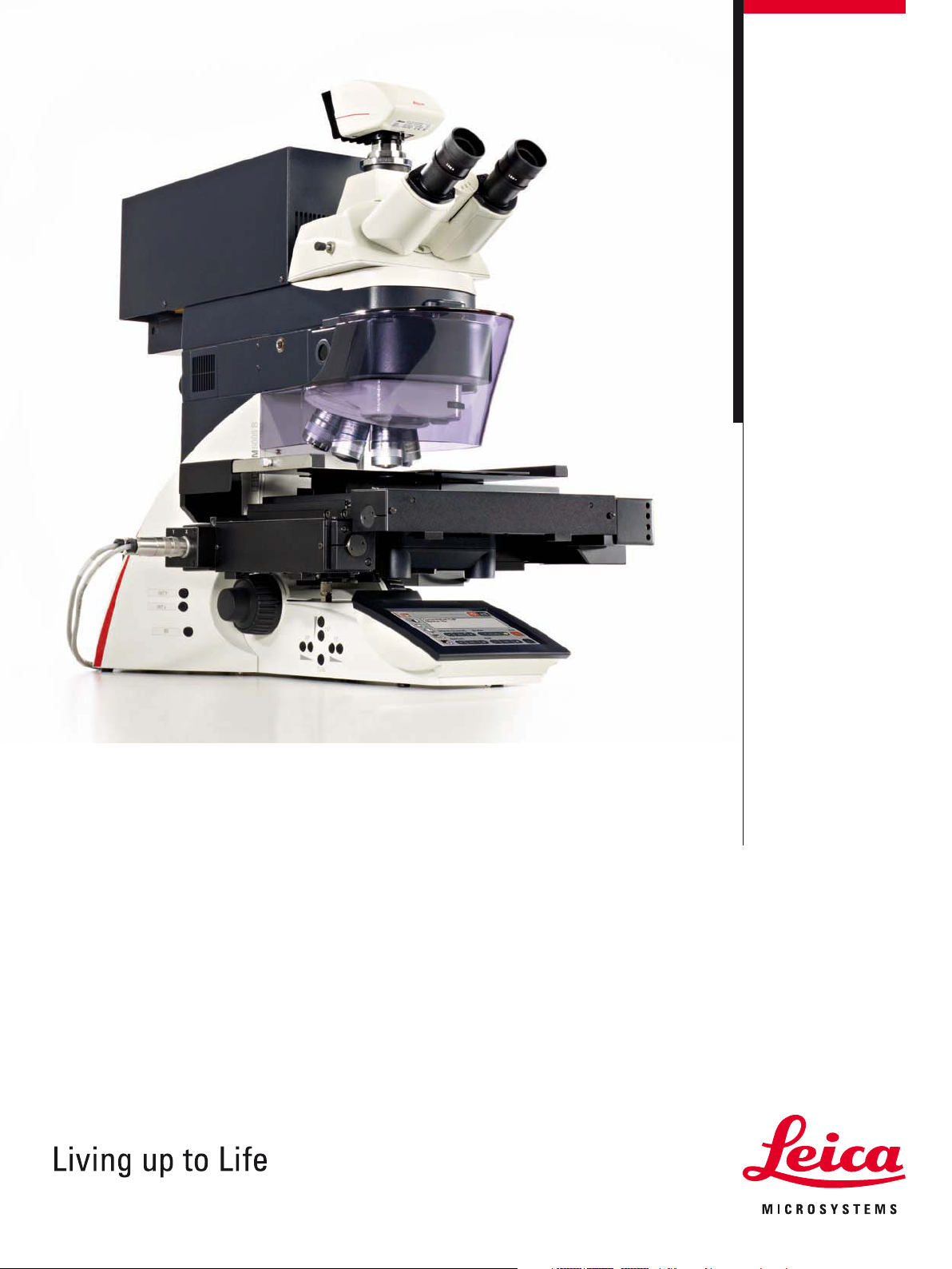
Leica LMD6500
Leica LMD7000
Laser Microdissection Systems
Page 2
Ultimate Perfection!
Laser microdissection (LMD) is the ultimate tool for perfection.
LMD makes it possible to obtain homogenous, ultrapure samples
from heterogenous starting material. A researcher can selectively
and routinely analyze regions of interest down to single cells to
obtain results that are reproducible, and specific.
Laser microdissection uses a microscope to visualize individual
cells or cell clusters. Regions of interest are selected by software,
excised from the surrounding tissue by a laser, and collected by
gravity into specialized devices for analysis.
There has been enormous progress in the development of laser
microdissection instrumentation in recent years. The driving force
in this area is the development of laser microdissection systems
by Leica Microsystems.
2
Page 3
At the forefront of laser microdissection technology,
only the Leica LMD6500 and Leica LMD7000 offer:
t Laser beam movement via optics – fast and precise cuts
t Specimen collection by gravity – contact- and contamination-free
t Dedicated objectives for LMD – highest possible laser power
t Adjustable laser – for thick, thin, soft, and hard tissue
3
Page 4
Specimen Collection
by Gravity
Benefits:
t Contact- and contamination-free collection
t Fast and reliable specimen collection
t Dissect all specimen shapes and sizes
t Pool unlimited amounts of specimen
t Use standard consumables
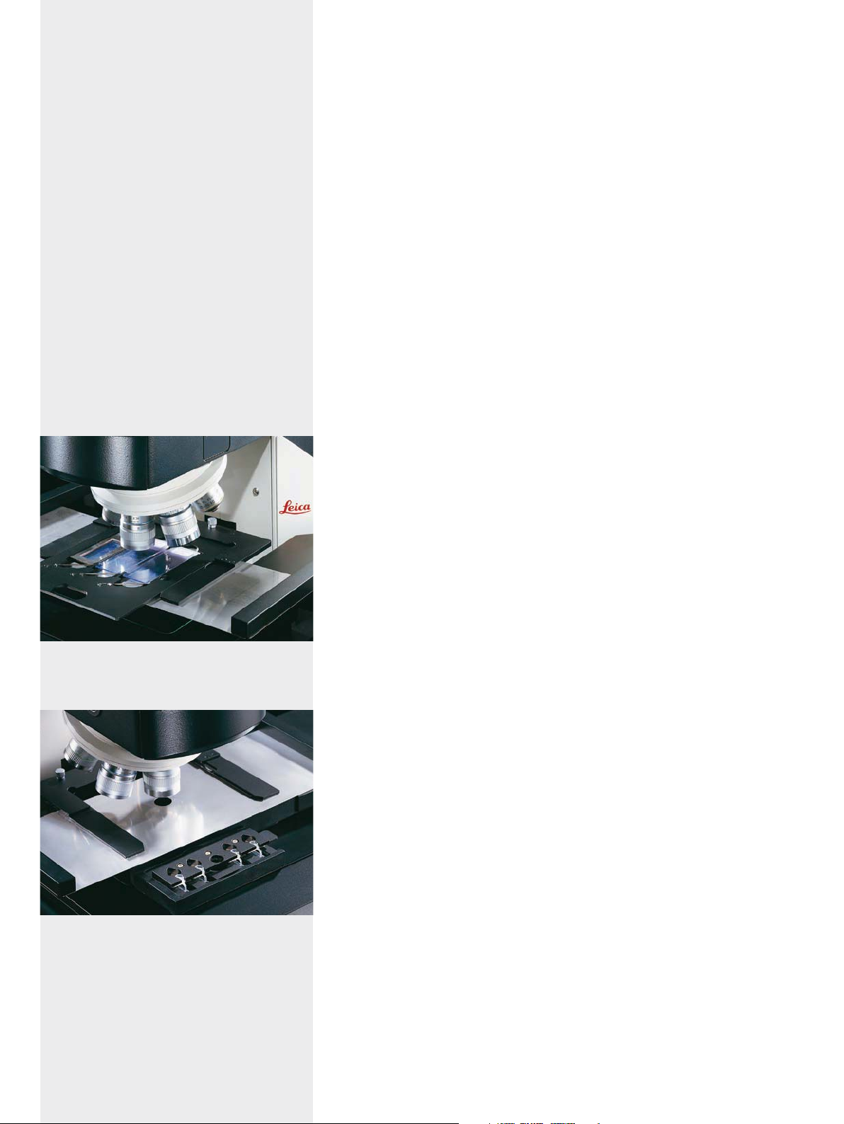
The three slide holder allows simultaneous work on up
to three slides. Several holders for different applications
are available.
LMD uses a UV laser to isolate microscopic regions from samples.
Leica Microsystems applies the most gentle technique for specimen collection – gravity.
Contact- and contamination-free
Gravity is the most sensible method of specimen collection. Once
the region of interest is excised, it gently falls into the collection
vessel. No additional steps or complicated methods are necessary. Most importantly, the specimens are not touched. This makes
specimen collection by gravity a contamination-free procedure.
Transfer dissectates of any size or shape
Gravity is the ideal tool to collect dissectates of any size or shape –
round and compact, or long and thin – it simply falls into the collection device. Large areas up to several mm2 are excised in one
step. No subdivision into smaller transportable sections is required. This ensures the complete recovery of the sample without
the loss of tissue due to additional cut lines.
Collect directly into reaction buffer
Collection by gravity is the fastest way to progress from tissue
section to reaction buffer. The collection vessel may contain
medium or buffer. Additionally, unlimited amounts of specimen
can be pooled.
Most comprehensive method
Leica Microsystems offers a wide range of specially designed
membrane systems for the best specimen preparations from tissue sections, over cell cultures, bacteria suspensions, plant materials, and many more. Samples and membranes are simultaneously cut by the laser. Specimen preparation has to be adapted to
the experimental needs, and with gravity as the motor for collection, the indispensable flexibility is provided.
Convenient handling of the collectors due to the automatically movable tray. The tray takes customized devices as
well.
4
Page 5
Laser Beam Movement
via Optics
When a high-energy laser pulse hits the sample a fast reaction
limited to the focus of the laser light called “cold ablation” occurs.
Surrounding tissue is not impaired or heated.
Only Leica Microsystems uses high-precision optics to direct the
laser beam along the desired cutting line.
Highest precision
Precision is the prerequisite to obtain homogenous material for
downstream analysis and reliable results. The unique laser beam
movement of the Leica LMD7000 and LMD6500 is controlled by
patented* high precision optics, while the microscope stage and
the sample are stationary. The cutting precision is unmatched and
far superior to using a fixed laser beam and moving the sample.
Maximal speed
Quick and reliable material collection for downstream analysis is
of highest importance. By moving only small optical parts during
the cutting process, Leica Microsystems’ approach allows precise cutting at high magnifications, and fast cutting speeds at low
magnifications. This technique ensures rapid capture, minimizing
degradation of the specimen.
Real-time laser cutting
A smart software tool allows live, on-the-fly dissections. The
unique function is especially beneficial for specimens that are difficult to cut. This tool can also be used for real-time ablation of
tissue. An important effect of laser beam movement is the ability
to conveniently document the dissection with movies. The image
remains fixed and only the laser beam moves – just the way our
eyes are used to.
Benefits:
t Highest possible precision
t Maximal speed
t Real-time laser cutting
t Convenient movie documentation
t Maintenance free
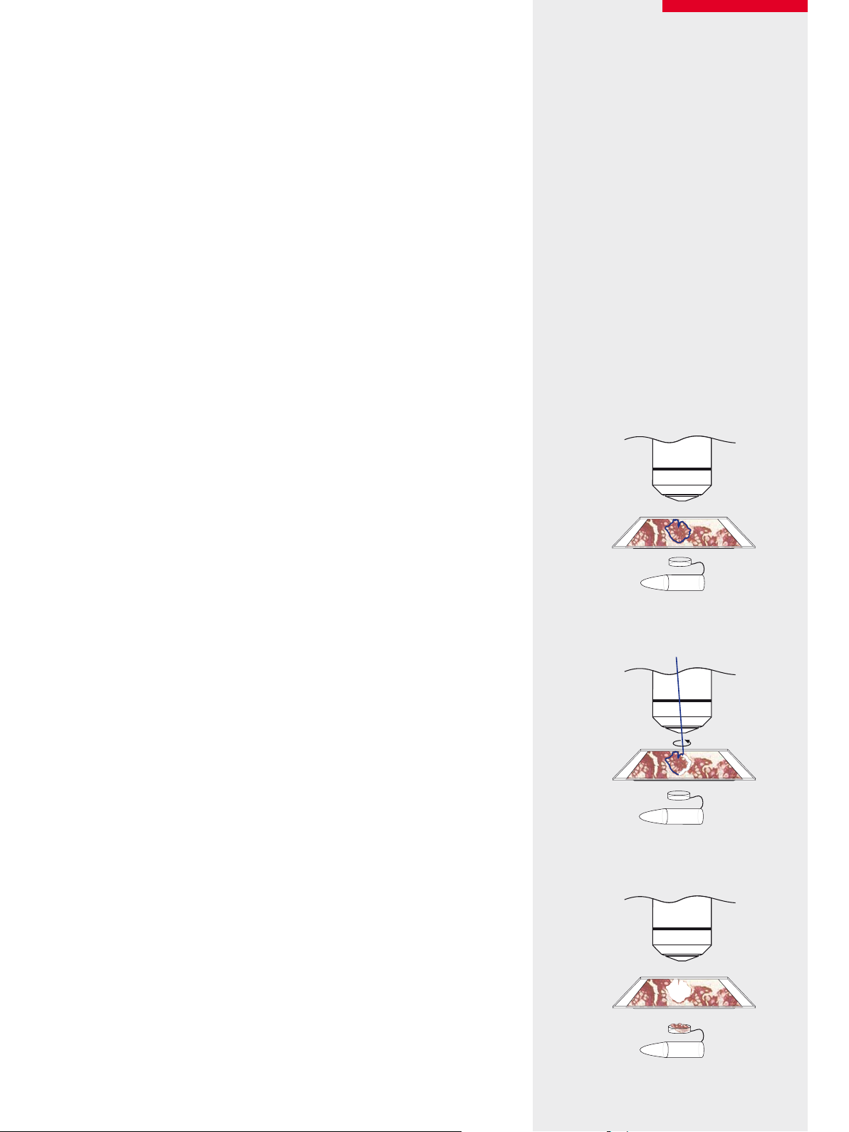
Step 1: Define region of interest
➟
Step 2: Laser beam steered by optics along the cut line
* Patented EP 1276586 B1, US 7035004 B2, JP 3996773 B2, TW 486566 B
➟
Step 3: Specimen collection by gravity
5
Page 6

➊
➋
6
➌
➍
Page 7

Leading-edge
Laser Technology
The laser is the core part of a laser microdissection system. The
two key features of a laser are pulse energy and pulse frequency.
The combination of these features within a LMD system was realized by Leica Microsystems for the first time.
Adapt the laser to the specimen
The Leica LMD7000 allows the control of laser pulse energy and
frequency. Adjust the laser according to the specimen and make
narrow, powerful, and fast cuts within a single system. Leica
Microsystems’ LMD systems provide the flexibility to align the UV
offset with respect to the focus of the visible light for the most precise cutting performance, for thick or thin samples, and ablation.
Control the thickness of the cutting line
The aperture control enables the adjustment of the width of the
cutting line. Combined with the high repetition rate, the narrowest cuts are possible. The laser aperture settings can be stored
individually for each objective.
Longlife, solid state laser
Leica Microsystems uses diode-pumped, solid state lasers. These
state-of-the-art lasers are maintenance free and reliable, ensuring
long lifetimes.
Choose the laser according to your needs: the Leica LMD6500 offers a pulse energy of 50 μJ and a fixed repetition rate, whereas
the Leica LMD7000 provides a pulse energy of up to 120 μJ and the
unique flexibility to adjust power and frequency.
Benefits:
t Control the pulse energy and frequency
(for Leica LMD7000)
t Adjust the width of the cutting line
t Control the UV offset
t Maintenance free solid state laser
LMD Laser Systems Leica LMD7000 Leica LMD6500
Max. pulse energy 120 μJ 50 μJ
Repetition rate Single pulse to
5000 Hz
Adjustable repetition
rate
Wavelength 349 nm 355 nm
Laser aperture
control
Yes No
Yes Yes
80 Hz
Please visit www.leica-microsystems.com/lmd
for more images and movies, application letters, etc.
Fig. 1: Brain, frozen section of GFAP-immunopositive astrocytes. Courtesy of G.J.
Burbach, MD, and T. Deller, MD, Institute of Clinical Neuroanatomy, J.W. Goethe
University, Frankfurt, Germany.
Fig. 2: Primary cell culture from pancreatic adenocarcinoma (PDAC) stained with
hematoxylin, objective 10x (before cutting, after cutting and inspection). Courtesy
of N. Funel, Department of Oncology, University of Pisa, Italy
Fig. 3: Maize root meristem. Courtesy of L. Feldman, University of California,
Berkeley, USA
Fig. 4: Brain section, stained with thionin. Courtesy of Axaron Bioscience AG,
Heidelberg, Germany
7
Page 8

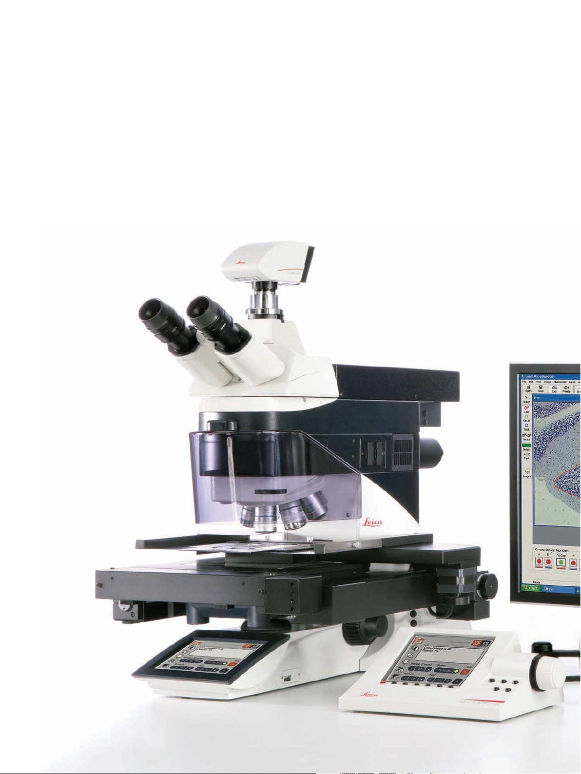
System Integration
Benefi ts:
t Fully automated DIC
t Motorized fl uorescence axis with
5- or 8-position fi lter cube turret
t Fluorescence intensity manager (FIM)
t Motorized Excitation Manager and fast
Internal Filter Wheel (IFW)
t Convenient Leica SmartTouch touchscreen
for controlling the automated modules
Leica Microsystems offers a wide range of objectives
(from 5x–150x) dedicated for laser microdissection.
All components of the Leica LMD systems are fully integrated and
act in concert, building a winning team greater than the sum of its
components.
High-end Leica DM6000 B research microscope
The foundation of every Leica LMD system is the high-end Leica
DM6000 B upright research microscope, which is suitable for all
types of life science research. Excellent optical performance, intelligent automation, and user-friendly operation make this fully
automated microscope the perfect tool for microscopic research.
Specially designed optics for laser microdissection
Leica Microsystems’ dedicated LMD objectives feature the highest possible UV transmission and outstanding imaging performance – the Leica SmartCut series. With SmartCut objectives,
thicker tissue can be cut faster. The unique high magnifi cation
150x dry objective enables the dissection of even the smallest areas without using oil.
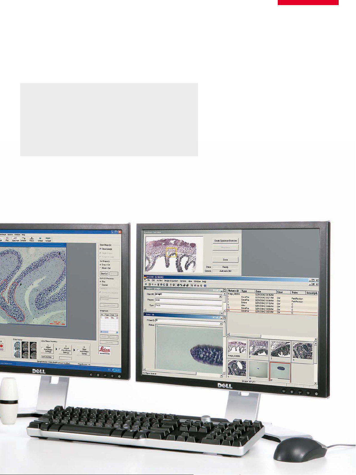
User-friendly software
Leica Microsystems’ LMD software is very easy to use, yet powerful without being complicated. A workfl ow guides the user
through the dissection process and intuitively provides all control elements where they are needed. The software contains all
features for dissection, such as serial section cutting, an optional
database, and fully integrated and automated cell recognition.
5x obj. for LMD
5x obj. Standard
The specially designed objectives for laser microdissection provide a much higher UV transmission than
standard objectives, and therefore, provide more laser
power on the sample.
8
With the Leica SmartTouch all automated functions are easily controlled.
Page 9

Application specifi c consumables for laser microdissection
Leica Microsystems offers four different types of metal frames,
glass slides and Petri dishes in different sizes. Whether the user
needs slides that show no autofl uorescence, or uses DIC contrast
to reveal the specimen areas – Leica Microsystems has the right
solution for any application.
a)
Benefi ts:
t High-end Leica DM6000 B
t Specially designed optics
t User-friendly software
t Application specifi c consumables
b)
d)
c)
a) 8-well strips building up a 96 well plate
b) Petri dishes
c) Big slides (50 mm x 76 mm)
d) Frame support
e) Membrane slides (PEN)
f) Frame slides (PET, POL, FLUO)
g) Microcentrifuge tubes
b)
e)
e)
f)
e)
g)
9
Page 10

200 μm
Solutions for
Advanced Applications
Cancer research
Cancer research requires visualization of morphologically altered
cell populations and their subsequent isolation.
t Contamination-free dissection by gravity
t Fast dissection of areas of any size or shape
t Collect directly into buffer
t Fully integrated database option for automated documentation
Single cells
The fastest possible dissection, the highest precision combined
with narrow cutting lines, are the prerequisite for expression
analysis of single cells.
t Laser movement via optics for fast and precise lasing
t Pen-screens for the most accurate drawing of cutting lines
t Leica SmartCut objectives with the highest UV transmission
t The unique 150x dry objective with the highest magnification
available for laser microdissection
200 μm
200 μm
Frozen section (10 μm) of a mouse aorta (whole vessel)
stained with cresyl violet on a POL frame slide. Courtesy
of K. Beuerlein, Rudolf-Buchheim-Institut für Pharmakologie, Justus-Liebig-Universität Giessen.
Proteomics
For proteomics analysis, either very sensitive analysis methods or
large amounts of protein are necessary for analysis.
t Auto vision control (AVC) for fast and fully automated
cell recognition and dissection
t Semi-automated cell recognition for assisted dissection
t Pool unlimited quantities of dissectates
Live cell cutting
Laser microdissection of living cells enables the separation of
single cells and cell clusters for recultivation or analysis.
t Unique, integrated consumables for sterile dissections
t Dissect non-adherent cells or bacteria
t Optional incubator
t Special sputtered GFP and BGR cubes for lasing in fluorescence
t Fully automated fluorescence axis with 5- or 8-position filter
cube turret
10
Page 11

Ablation and multi-dimensional imaging
Directly after the targeted ablation in a living structure, the imaging of the induced processes is important.
t Time lapse series with different fluorescence channels,
using Leica AF6000 (Advanced Fluorescence) software
and LMD at the same time
t Convenient movie documentation
t Real-time lasing for direct ablation
Specimen
Preparation
➟➟➟➟
Section and prepare
biological specimens
– Histological specimens
(formalin-fixed/
paraffin-embedded or
cryo sections)
– Living cells and
cell cultures
– Chromosome spreads
– Smears
– Cytospins
– Plant material
– Sperm and other
forensic preparations
Staining
Visualize regions of
interest
Brightfield:
– HE (hematoxylin-eosin)
– Cresyl violet
– Toluidin blue
– Thionin
– Immunohistochemistry
Fluorescence:
– Secondary antibodies
– Acridine-orange
– FISH
Microdissection
Selectively dissect
regions of interest
– Contact-free
– Contamination-free
– Any size from cell
compartments of a
2
few μm
to several mm
– Any shape
2
Extraction
Extract and prepare
important molecules
– DNA
– RNA
– Proteins
– Metabolites
– Biomarkers
Analysis
Obtain reproducible
and specific results
– PCR
– Quantitative real-time
PCR
– Microarrays
– Expression profiling
– Genetic fingerprinting
– LOH
– FISH
– LC-MS/MS
– 2-D PAGE
– SELDI
– MALDI
11
Page 12

4 5
3
11504133
HBO Lamp Adapter
11504117
1” fiber-optics
adapter
11888829
Condenser BF
mot. condenser head
11888830
Condenser DIC (suitable for BF, PH, DF, ICT)
mot. condenser head,
with mot. condenser disk (7 pos.),
with mot. polarizer
11504070
Lamp housing LH 106z
12 V 100 W halogen lamp
4-lens collector
0.55 m connection
11504114
Lamp housing
LH 106z Hg 100 W,
1” 6-lens collector
11504116
Liquid light guide, 2 m
ICT condenser prisms
(K1–K15)
11521505
Light ring set
11500325
Supply unit
Hg 100 W
11504115
External light
source EL6000
11505161
Tube attachment
with 1 camera port
C-Mount
HC 0.70x
11505223
Tube attachment
with 2 camera ports
selectable 100:100, manual
(LMD6500 only)
11505146
BDT 25+ V 100/50/0
Documentation tube
with documentation port
with variable beam splitting
12730203
Leica DFC400 FX
11547000
Leica DFC360 FX
11547002
Leica DFC310 FX
11501450
Hitachi HV-D20P
11505202
Imaging module
beam splitting: 100%:0%
with integrated and centrable
C-mount (0.7x)
(LMD7000 only)
11518146
Objective 5x/0.12
Microdissection
11518145
Objective 6.3x/0.13
Microdissection
11506208
Objective 40x/0.60 XT
Microdissection
11506222
Objective 63x/0.70 XT
Microdissection
11506214
Objective 150x/0.90 XT
Microdissection
Objective series BF
33
11888828
Stand top without fluorescence axis,
with 7-position objective turret M25, mot.
1
1
11600184
Holder for 1 slide
25 x 76 mm
11505214
Holder for big slide
50 x 76 mm
11505257
Holder for Petri dish
11505131
Lever for
4 x 0.2 ml PCR-tube
11505130
Lever for
4 x 0.5 ml PCR-tube
11505258
Lever for
1 x 8-well strips
11888826
Motorized Stage
1
4 4
11501255
Leica STP6000
SmartTouch Panel
11888831
Stand top with fluorescence axis,
with 5-position filter turret, mot.,
with 7-position objective turret M25, mot.
11888827
Scanning Stage
2
11505180
Remote Control
Smart Move
55 5
11888832
Stand top with fluorescence axis,
with 8-position filter turret, mot.,
with 7-position objective turret M25, mot.
2
2
11505226
Holder for 3 slides
25 x 76 mm
11505227
Holder for Petri dish and
big slide 50 x 76 mm
11505255
Holder for 18-well
double stack ibidi slide
11505229
Collector for
4 x 0.2 ml PCR-tube
11505228
Collector for
4 x 0.5 ml PCR-tube
11505230
Collector for
2 x 8-well strips
11888825
Leica LMD6500
max. 50 μJ laser
12
Basic stand without
stage and transmitted light axis
11888834
Leica LMD7000
max. 120 μJ laser
Basic stand without
stage and transmitted light axis
Page 13

Highlights of the Leica LMD Systems
Specimen collection by gravity
t Contact- and contamination-free specimen collection
t Collect all specimen shapes and sizes directly
into reaction buffer
t Pool unlimited amounts of specimen
t Use standard consumables for collection
t No additional laser pulses are needed for collection
Laser movement via optics
t Highest possible precision and speed
t Real-time laser cutting
t Convenient movie documentation
Leading-edge laser technology
t Control the pulse energy and frequency for thick, thin,
hard and soft tissues
t Control the width of the cutting line using the laser aperture
t Maintenance-free, longlife, solid state laser
t Control the UV offset for thicker samples or ablation
Specially designed optics for laser microdissection
t Wide range of LMD objectives, from 5x to 150x
t Higher UV transmission than standard objectives
t Use the unique 150x dry objective for the most
precise dissection
Lasing within fluorescence
t Special GFP and BGR cubes
t See three fluorescence channels (such as DAPI, FITC, TxRed)
at a glance through the eyepieces
t Use the internal filter wheel to visualize a single fluorochrome
t Non-fluorescent FLUO-frame slides
Intuitive software
t Workflow based and time saving
t Fully integrated database
t Overview images for navigation
t Serial section cutting
t Automated cell recognition
Fully automated high-end Leica DM6000 B
research microscope
t Fully automated DIC
t Unique Fluorescence Intensity Manager (FIM)
t Motorized excitation manager and fast internal filterwheel
t Constant color intensity control
Flexibility to use a variety of dissection devices
t Adapt the collection devices to specific research needs
t Use metal frames or glass slides, with or
without membrane (PEN, PET, POL, FLUO)
t Sterile dissection of living cells out of Petri dishes, stackable
membrane rings or 18-well membrane slides
t Use a sandwich of two metal frames for dissecting
non-adherent cells or bacteria
13
Page 14

Features and Specifications
Laser Leica LMD7000 Leica LMD6500
Type Diode pumped, solid state Diode pumped, solid state
Wavelength 349 nm 355 nm
Maximum pulse energy 120 μJ 50 μJ
Repetition rate Single pulse to 5000 Hz 80 Hz
Adjustable repetition rate Yes No
Laser aperture control Yes, continuously adjustable Yes, continuously adjustable
Free intensity control 1-100% 1-100%
UV offset freely adjustable and specific objective saved Yes Yes
Laser beam movement Via optics
Microscope
Transmitted light axis Contrast methods BF, optional PH, DF, POL, DIC (fully automated)
Illumination 12 V 100 W halogen lamp
Automation Automated illumination manager
Automated contrast manager
Constant color intensity control (CCIC)
Condenser Condenser head S28, 0.55 NA
Motorized 7x condenser disk
Motorized polarizer
Fluorescence axis Filter cube turret Motorized 5x or 8x
Automation Fluorescence intensity manager (FIM) for brightness adjustment
Circular and rectangular field diaphragms for eyepiece or camera viewing
Internal filter wheel and motorized Excitation Manager
Illumination Leica EL6000 (120 W metal halide) or
100 W HBO
Cubes All cubes size k
Special cubes for simultaneous fluorescence and cutting e.g.
– LMD-BGR
– LMD-GFP
Operation Focus Motorized:
– 5 electronic ratios
– Includes parfocal function
Memory function for two z-positions
Objective turret Motorized 7x M25 thread including dry and immersion modes
Controls 6 programmable function buttons
Leica SmartMove
Controls for z (focus) movement and x, y (stage) movement
4 programmable function buttons
Optional Leica STP6000
Controls for z (coarse and fine focus) and x, y (stage) movement
11 programmable function buttons
Touchscreen with information and control panels
Stand Display With integrated touchscreen Leica SmartTouch
2
Interfaces 2 x USB 2.0, 2 x I
Dimensions With scanning stage:
649.6 mm height, 512.0 mm width, 596.5 mm depth
C
14
Page 15

Dissection Dissection and Collection Unit Based on Scanning Stage Dissection and Collection Unit Based on Motorized Stage
Specimen Collection Contact- and contamination-free
Dissection modes Draw & Cut
Move & Cut (direct online cutting)
Draw & Scan (dot dissection scan)
Serial section cutting Ye s No
Stage precision ± 2 μm > ± 5 μm
Holding devices 3x standard slides (25 mm x 76 mm)
Optional 1x big slide (50 mm x 76 mm)
Optional Petri dish (50 mm)
Optional 18-well slide stack
Collection devices 4x 0.2 ml standard PCR tubes
4x 0.5 ml standard PCR tubes
Petri dish (50 mm)
Optional 2x 8-well strips building up a 96-well plate
Power supply CTR6500 CTR6000
System software
Package includes Dissection Automated collection devices and positioning of the PCR tubes
Fully automated inspection mode
Multi-cutting over the entire slide
Save and load drawn shapes
User guidance Workflow based graphical user interface
Free scaling drawn shapes
Saving user profiles
Overview images in BF and Fluorescence
Control Full laser control
Control software for the microscope
Laser and illumination settings are linked to objectives
Interfaces Export of shape list data for Microsoft Excel or OpenOffice
Integrated database interface to transfer all relevant data (laser, microscope and camera; database itself as option)
Optional software packages Automated vision control (AVC) for automated cell recognition within field of view (standard version) or
fully automated or semi automated over freely defined area (professional version)
Database Leica IM500 or Leica IM1000
1x standard slide (25 mm x 76 mm)
Optional 1x big slide
Optional Petri dish (50 mm)
4x 0.2 ml standard PCR tubes
4x 0.5 ml standard PCR tubes
Optional 1x 8-well strips building up a 96-well plate
Camera Leica DFC310 FX Leica DFC360 FX Leica DFC400 Hitachi HV-D20P
Type High sensitivity digital color High sensitivity digital monochrome High sensitivity digital color High sensitivity analog 3 CCD color
Cooled Yes, -20°K to ambient Yes, -20°K to ambient No No
Resolution 1392 x 1040 1392 x 1040 1392 x 1040 795 x 596 (x 3)
Pixel size 6.45 μm x 6.45 μm 6.45 μm x 6.45 μm 4.65 μm x 4.65 μm 8.00 μm x 8.00 μm
Speed 20 fps at 1392 x 1040
71 fps at 348 x 260
Electronic interface Single FireWire b cable
(IEEE1394b)
20 fps at 1392 x 1040
39 fps at 696 x 520
Single FireWire b cable
(IEEE1394b)
20 fps at 1392 x 1040
39 fps at 696 x 520
Single FireWire b cable
(IEEE1394b)
25 fps
PCI board
15
Page 16

“With the user, for the user”
Leica Microsystems
Leica Microsystems operates globally in four divi sions,
where we rank with the market leaders.
Life Science Division
•
The Leica Microsystems Life Science Division supports the
imaging needs of the scientific community with advanced
innovation and technical expertise for the visualization,
measurement, and analysis of microstructures. Our strong
focus on understanding scientific applications puts Leica
Microsystems’ customers at the leading edge of science.
Industry Division
•
The Leica Microsystems Industry Division’s focus is to
support customers’ pursuit of the highest quality end result.
Leica Microsystems provide the best and most innovative
imaging systems to see, measure, and analyze the microstructures in routine and research industrial applications,
materials science, quality control, forensic science investigation, and educational applications.
Biosystems Division
•
The Leica Microsystems Biosystems Division brings histopathology labs and researchers the highest-quality,
most comprehensive product range. From patient to pathologist, the range includes the ideal product for each
histology step and high-productivity workflow solutions
for the entire lab. With complete histology systems featuring innovative automation and Novocastra™ reagents,
Leica Microsystems creates better patient care through
rapid turnaround, diagnostic confidence, and close customer collaboration.
Surgical Division
•
The Leica Microsystems Surgical Division’s focus is to
partner with and support surgeons and their care of patients with the highest-quality, most innovative surgi cal
microscope technology today and into the future.
The statement by Ernst Leitz in 1907, “with the user, for the user,” describes the fruitful collaboration
with end users and driving force of innovation at Leica Microsystems. We have developed five
brand values to live up to this tradition: Pioneering, High-end Quality, Team Spirit, Dedication to
Science, and Continuous Improvement. For us, living up to these values means: Living up to Life.
Active worldwide
Australia: North Ryde Tel. +61 2 8870 3500 Fax +61 2 9878 1055
Austria: Vienna Tel. +43 1 486 80 50 0 Fax +43 1 486 80 50 30
Belgium: Groot Bijgaarden Tel. +32 2 790 98 50 Fax +32 2 790 98 68
Canada: Richmond Hill/Ontario Tel. +1 905 762 2000 Fax +1 905 762 8937
Denmark: Herlev Tel. +45 4454 0101 Fax +45 4454 0111
France: Nanterre Cedex Tel. +33 811 000 664 Fax +33 1 56 05 23 23
Germany: Wetzlar Tel. +49 64 41 29 40 00 Fax +49 64 41 29 41 55
Italy: Milan Tel. +39 02 574 861 Fax +39 02 574 03392
Japan: Tokyo Tel. +81 3 5421 2800 Fax +81 3 5421 2896
Korea: Seoul Tel. +82 2 514 65 43 Fax +82 2 514 65 48
Netherlands: Rijswijk Tel. +31 70 4132 100 Fax +31 70 4132 109
People’s Rep. of China: Hong Kong Tel. +852 2564 6699 Fax +852 2564 4163
Portugal: Lisbon Tel. +351 21 388 9112 Fax +351 21 385 4668
Singapore Tel. +65 6779 7823 Fax +65 6773 0628
Spain: Barcelona Tel. +34 93 494 95 30 Fax +34 93 494 95 32
Sweden: Kista Tel. +46 8 625 45 45 Fax +46 8 625 45 10
Switzerland: Heerbrugg Tel. +41 71 726 34 34 Fax +41 71 726 34 44
United Kingdom: Milton Keynes Tel. +44 1908 246 246 Fax +44 1908 609 992
USA: Bannockburn/lllinois Tel. +1 847 405 0123 Fax +1 847 405 0164
and representatives in more than 100 countries
Order nos. of the edition in: English 914 689 t VI/09/AX/Br.H.
LEICA and the Leica Logo are registered trademarks of Leica Microsystems IR GmbH.
www.leica-microsystems.com
 Loading...