

GE Healthcare Valve Planning Protocol Brochure
Visualization and quantification of
cardiac anatomy for effective
treatment strategies.
GE Healthcare
Overview
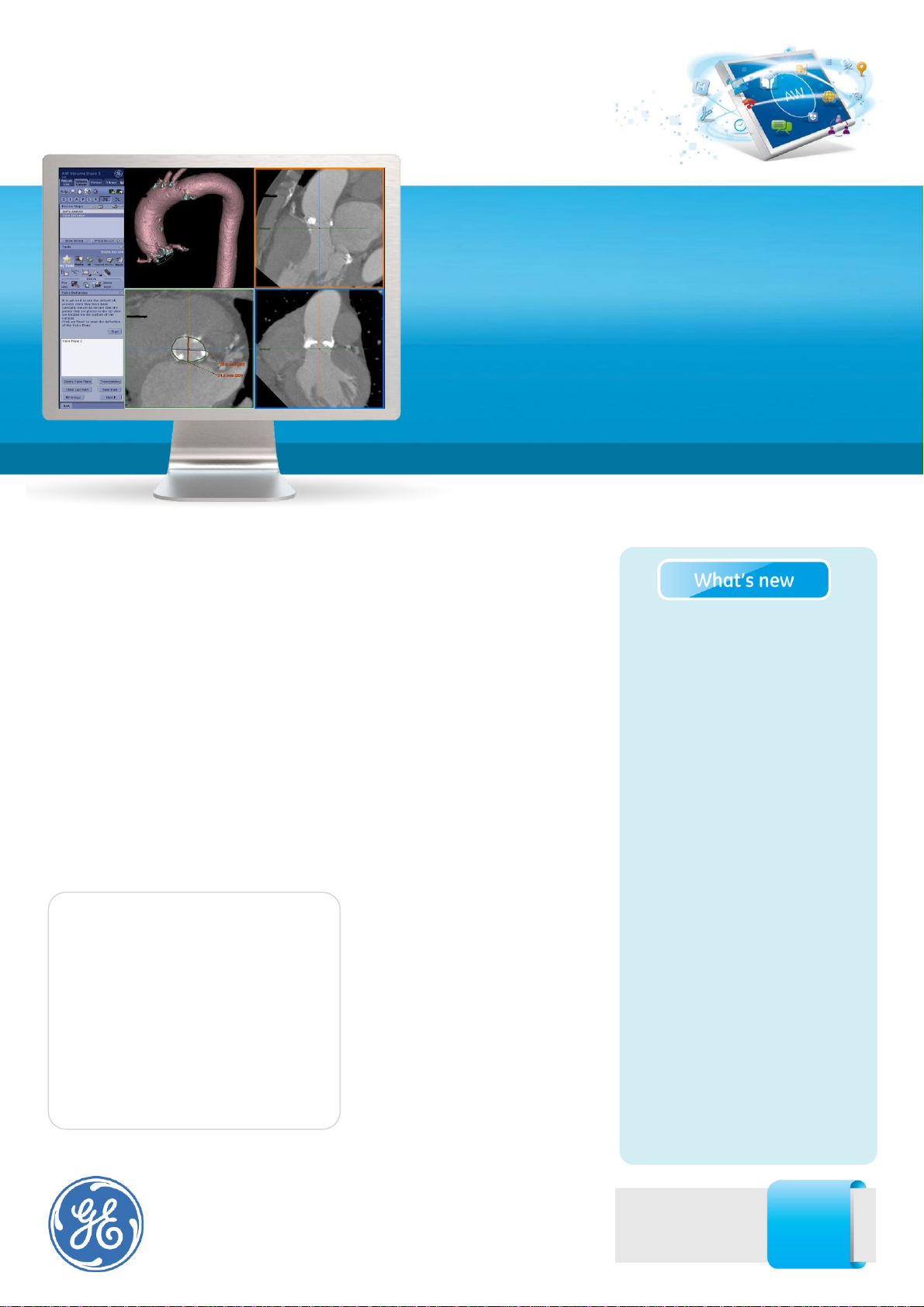
The Valve Planning protocol in the
Vessel IQ Xpress application lets you
visualize cardiac anatomy with the
degree of detail required to evaluate
the presentation of the aortic valve.
Armed with this information, you
can develop a preproceduralTAVR/TAVI plan to
establish a therapy strategy for the
Transcatheter Aortic Valve Replacement/Implantation
(TAVR/TAVI) demands meticulous, detailed planning to be
successful. Providing you with the necessary information,
confirmation of valve stenosis, viable access approach, distances
to ostia, elliptical aortic root dimensions, identification of the valve
plane and appropriate angle for valve deployment in the cathlab,
for example; is vital to your planning. You need a protocol that
helps process imaging data into 3D models to help you better
understand a particular patient’s cardiac anatomy and aid in
developing an appropriate therapy path and strategy.
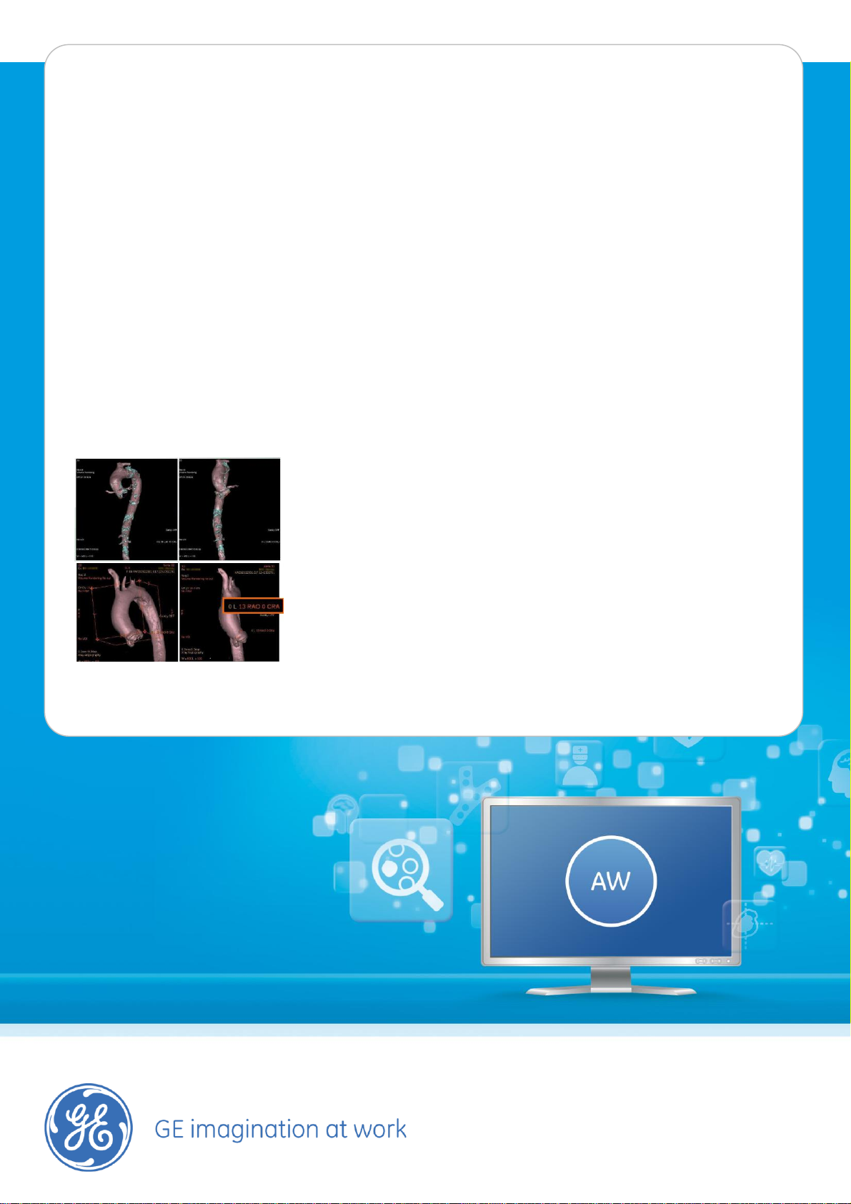
• Lets you segment the aorta
from iliacs to aortic root.
• Let’s you select the access
route.
• Let’s you see vessel tortuosity
and 3D visualization of
calcifications.
• Lets you measure the aortic
annulus and define valve plane
to help you select the
appropriate device to implant.
• Lets you measure the distance
between the leaflets and
coronary ostia for planning
valve deployment
• Automatically gives you the
optimized angle for the
intervention.
Valve Planning
Protocol
Visit us:
www.gehealthcare.com/aw/
applications/
Features
• Lets you visualize vessel tortuosity.
• Enables you to determine
calcification load to validate left or
right vascular pathways.
• Lets you segment the aorta for
better visualization, and to
ascertain aorta diameter and
valve-to-ostia measurement.
• Automatically segments
calcifications.
• Lets you visualize both iliac artery
diameter and profile.
•
• Lets you define the valve plane by
contouring the aortic annulus and
deposit 3D marks on the 3D
volume to locate coronary ostia or
other areas of interest..
• Gives you automatic C-Arm
angulation perpendicular to the
valve plane.
for the purpose of TAVR/TAVI
planning This software is designed to
support the physician in assessment
of vessel analysis, pre/post stent
planning and directional vessel
tortuosity visualization.
VesselIQ Xpress automatic
visualization tools provide the users
with the capabilities to facilitate
segmentation of bony structures for
accurate identification of the vessels.
Once vessels are visualized, tools are
available for sizing the vessel, the
valve annulus, and visualize the
calcifications
Regulatory Compliance
This product complies with the
European CE Marking regulation for
Medical Devices Directive: Directive
93/42/EEC, dated 14 June 1993.
• Lets you export the 3D CT images
to the cathlab through Innova
HeartVision for real time fluoro
overlays.
• With the AW Workstation, access
to 3D models based on CT or
angiography dataset is available in
the cath lab.
• Dedicated cath lab user interface
allows tableside control of 3D
images.
System Requirements
AW Workstation
VolumeShare 5 (voxtool.11.3) or
higher
VesselIQ Xpress.
Indications for Use
Valve Planning protocol of VesselIQ
Xpress is intended to provide an
optimized non–invasive application
to analyze vascular anatomy and
pathology and aid in determining
treatment paths (TA, TF, TAo,
Subclavian) from a set of Computed
Tomography (CT) Angiographic
images.
Valve Planning protocol of VesselIQ
Xpress is a post processing
application option for the Advantage
Workstation (AW) platform, CT
Scanner or PACS stations, which can
be used in the analysis of 2D and 3D
CT Angiography images/data derived
from DICOM 3.0 compliant CT scans
© 2012 General Electric Company.
All rights reserved. Data subject to change.
GE and GE Monogram are trademarks of General Electric Company.
* Trademark of General Electric Company.
 Loading...