

GE Healthcare Signa HDxt 1.5T Brochure
GE Healthcare
Signa* HDxt 1.5T
Optima* Edition 16.0
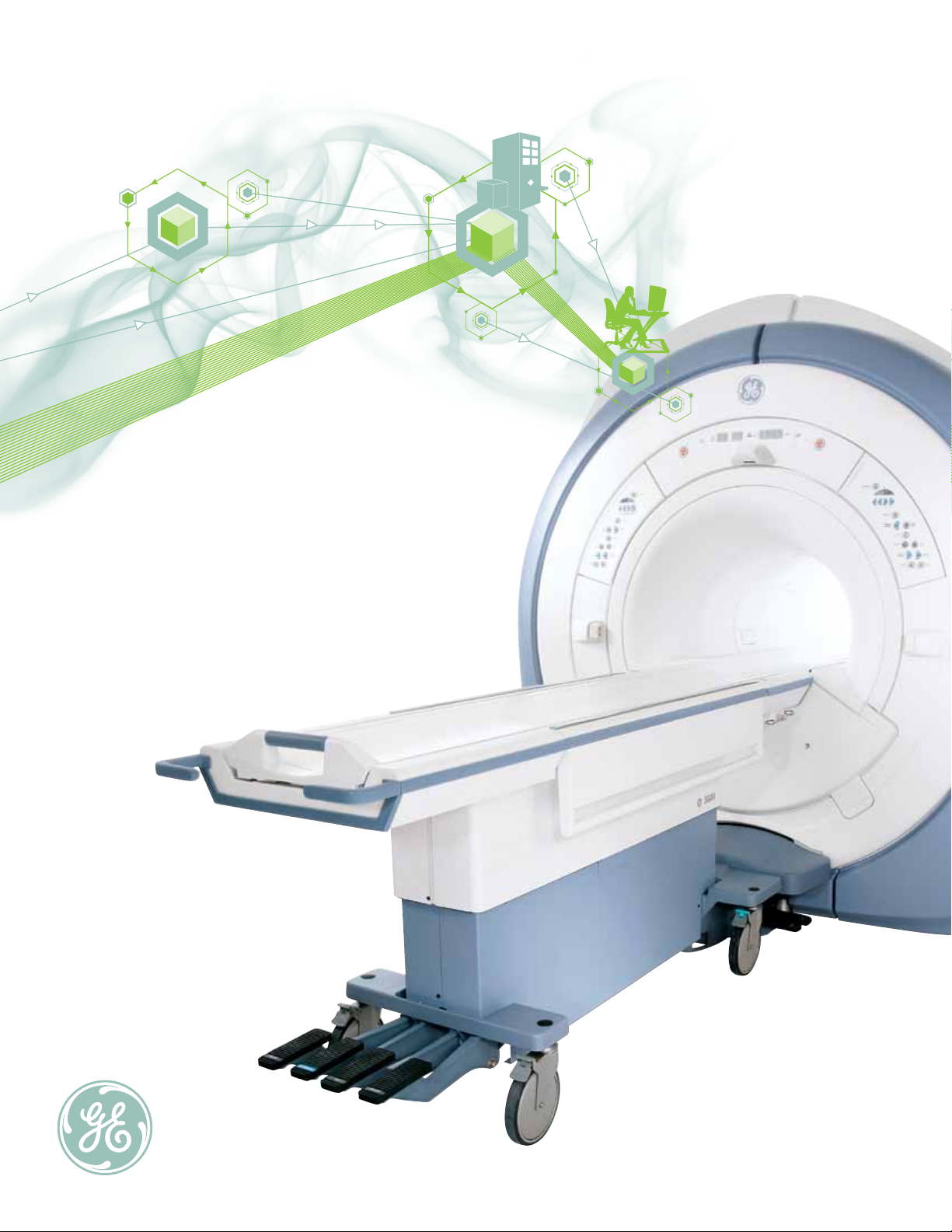
See More, Do More…
The next generation in High-Definition MR.
2 GE Healthcare Signa HDxt 1.5T Optima Edition

Expect More
You’ve been heard. When you want more out of your MRI
scanner, GE listens. And when you demand more
accuracy, more productivity, and more support, GE
delivers. Built on the high definition platform you know and
trust, Signa HDxt offers an MR System that allows you to see
more, do more, and expect more than ever before.
Introducing Signa HDxt 1.5T with Optima
Edition package,the next generation in
High-Definition MR.
GE Healthcare Signa HDxt 1.5T Optima Edition 3
Engineered for high definition
so you can see more.
See more with truly high-definition,
anatomically optimized imaging.
Engineered for enhanced image contrast,
reduced blurring, smaller field-of-view
prescriptions, and reduced artifacts.
4 GE Healthcare Signa HDxt 1.5T Optima Edition

Signa HDxt 1.5T
Designed for productivity
so you can do more.
Do more with consistent high-quality
imaging through can’t-miss applications.
Designed for optimal fat suppression,
tissue characterization, and artifact
reduction, every time.
Clinically proven to give you more
on every exam, every day.
GE was the first to introduce 1.5T MR technology.
Today, we have the world’s largest installed base of 1.5T
scanners . And we’re the only MR manufacturer celebrating
more than a quarter of a century of upgradeability. In fact,
HDxt is available as a new system — as well as an upgrade
to our current installed base customers.
Signa HDxt 1.5T: Highly rated service.
The peace of mind you’re looking for.
At 1.5T, the only choice is GE.
Built for longevity
so you can expect more.
Expect more with over 25 years of proven
commitment to MR system longevity.
Built for upgradeability, uptime, and
investment protection — GE’s Continuum*
commitment allows you to expect
more from your scanner.
GE Healthcare Signa HDxt 1.5T Optima Edition 5
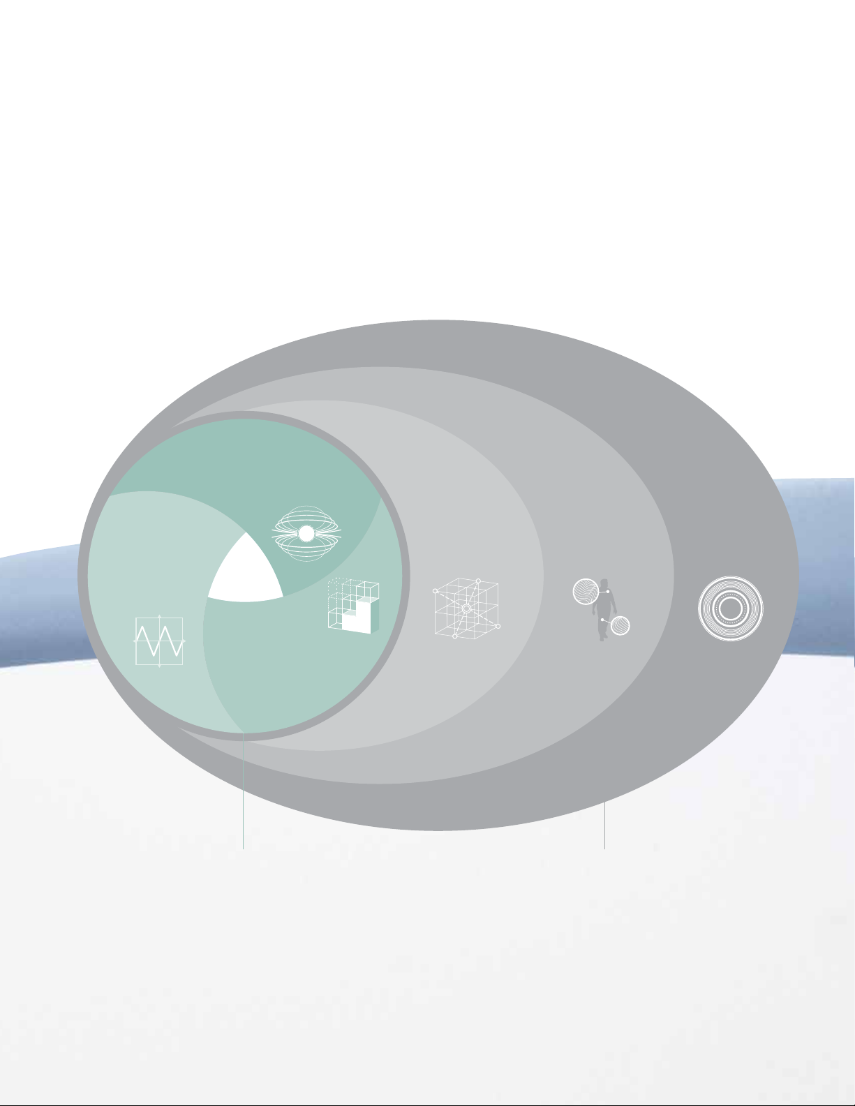
See More
Engineered for high-definition,
anatomically optimized imaging
The Signa HDxt 1.5T is engineered from end-to-end
to allow you to see more. With GE’s high-density coils, data
acceleration technology, and high-definition applications
optimized for each anatomical area, GE can deliver images
with the enhanced contrast, clarity, and accuracy you need.
Premium Performance
Anatomical Imaging
Optimization
Signa HDxt 1.5T starts with a foundation
of high-performance components, whose
integration enables superior imaging
capabilities that aim to bring you the
clearest, crispest images possible.
High Definition MR is more than just pulse
sequences, it is the sum total of our system.
And the best part? GE has done the
tweaking for you. Every component has been
specifically designed to deliver more detail and
more clarity, without compromise.
6 GE Healthcare Signa HDxt 1.5T Optima Edition
Signa HD Magnet
Engineered to deliver
the highest homogeneity
HD Gradients
Engineered for high-
fidelity to produce high
accuracy waveforms
The Signa HDxt MR Imaging Model
and stability
HD Reconstruction
Engineered for real-
time, high-performance
image generation
Data
Acceleration
Techniques
Engineered for
specific
applications
High-Definition
Applications
Engineered for
specific anatomical
challenges
High-Density
Coils
Engineered to
optimize the
specific exam
Anatomical Imaging OptimizationPremium Performance
GE Healthcare Signa HDxt 1.5T Optima Edition 7
See more in
Neuro Imaging
GE’s advancements in neuro imaging
continue with the delivery of Optima Edition
enhancements. In short, Signa HDxt 1.5T
Optima Edition is a natural for neuro.
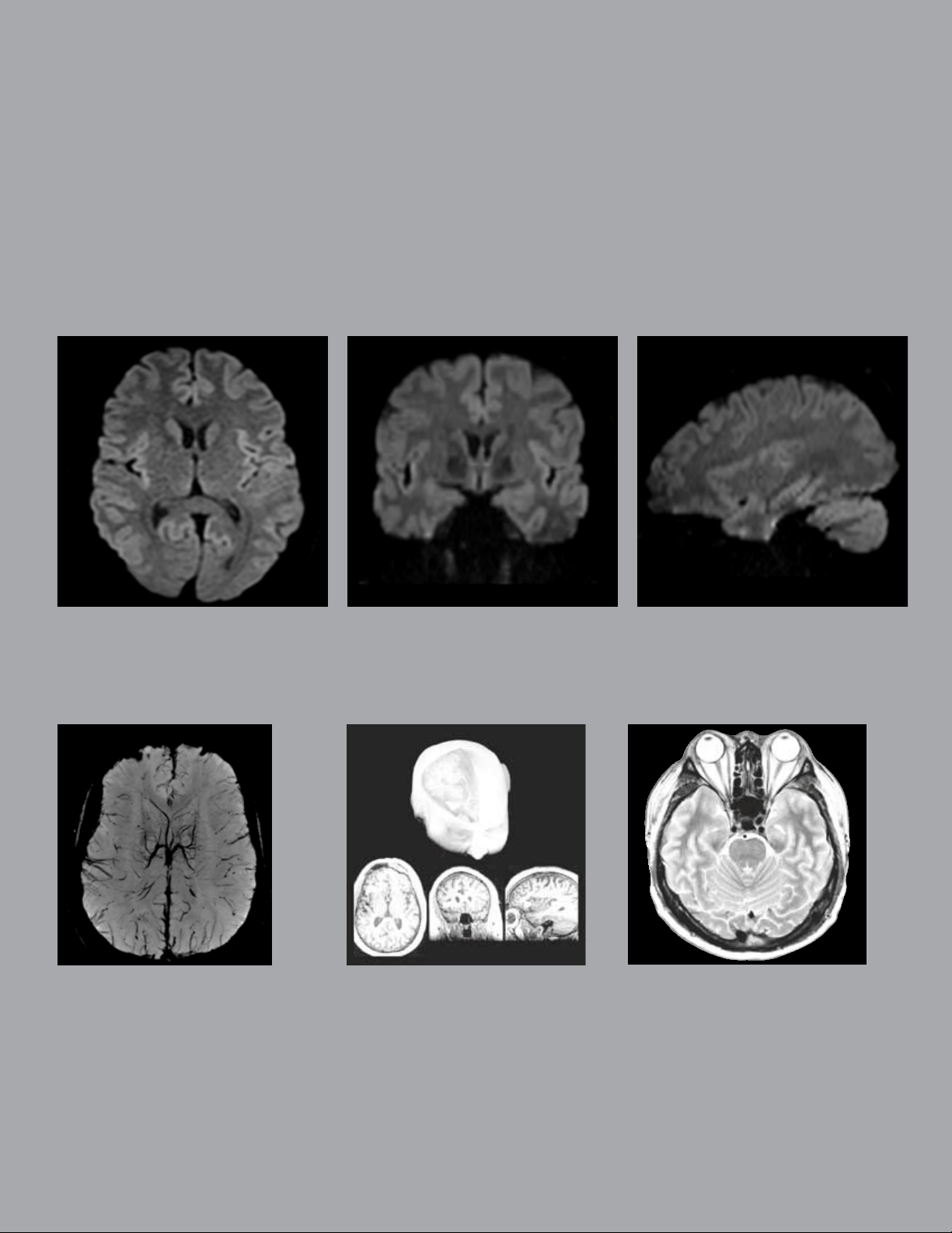
Enhanced DWI
NEW
The enhanced DWI technique supports multiple b-values in one acquisition with flexible control of NEX for each b-value. Novel diffusion techniques
“3-in-1” and “tetrahedral” allow applying gradients in multiple directions simultaneously to improve scan efficiency and signal-to-noise ratio.
SWAN
SWAN is a multi-echo 3D imaging
technique that helps visualize
and clearly delineate small vessels
and microbleeds, as well as large
vascular structures, and iron or
calcium deposits in the brain. This
image of the entire brain with the
0.5 x 0.6 x 2 mm voxel was
acquired in 4:50 min.
BrainWave
A suite of applications for functional brain
mapping. Includes a robust acquisition
sequence, easy-to-administer paradigms and
complete post-processing and visualization
tools. BrainWave Fusion integrates an
eloquent cortex map and DTI white-matter
trajectories with a high-resolution 3D anatomy
data set.
PROPELLER
Correct for motion artifacts and enhance
tissue contrast without compromising image
resolution or prolonging scan time — and reduce
susceptibility artifacts to clearly visualize small
or subtle lesions. Generate consistently
excellent images with less retakes, even on
restless children, or patients with tremors.
*
8 GE Healthcare Signa HDxt 1.5T Optima Edition
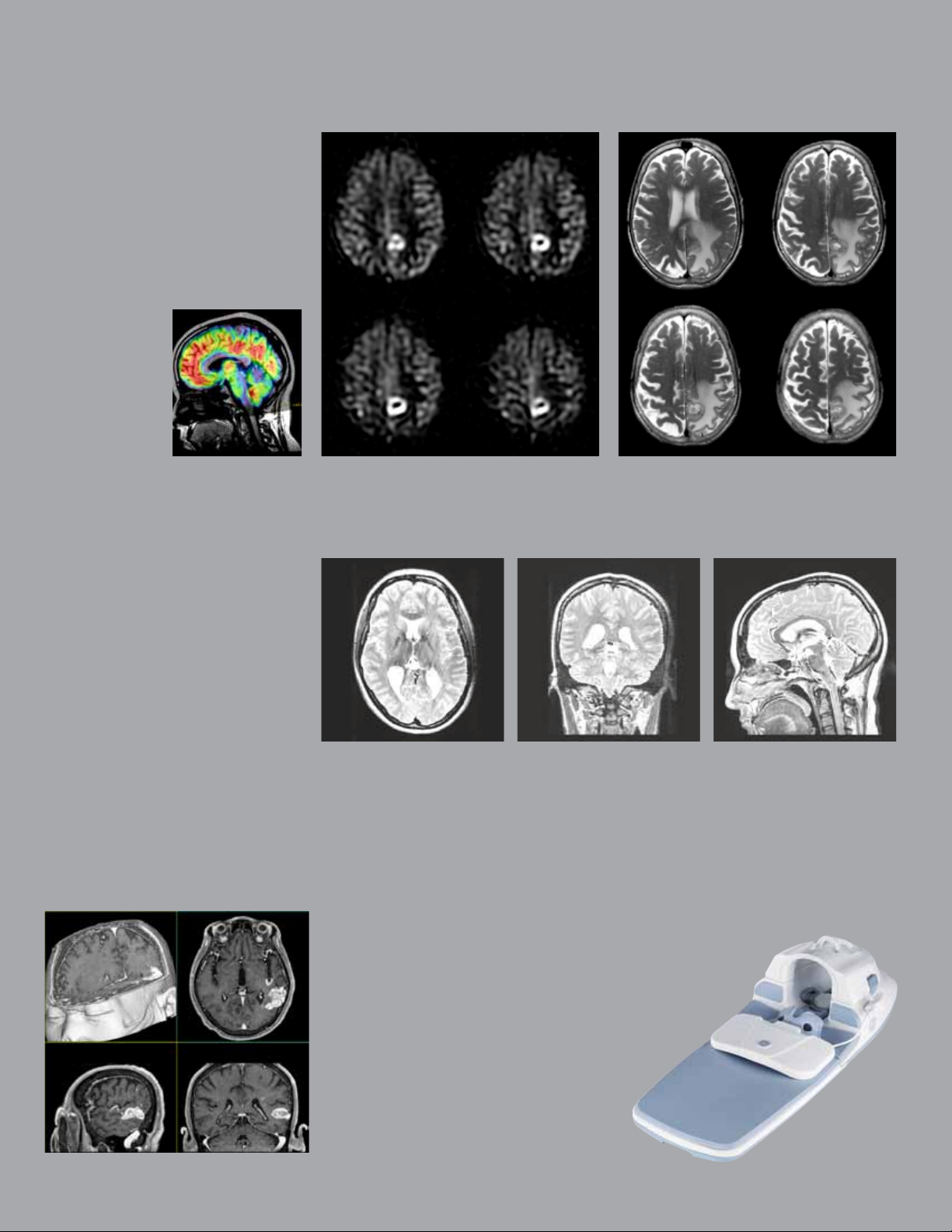
3D ASL
NEW
3D ASL is a robust non-contrast tissue perfusion
imaging technique that provides quantitative
assessment of cerebral blood flow (CBF)
and color perfusion maps (bottom). The
hyperintense signal in the left parietal occipital
lobe on this axial T2 image (far right) correlates
well with clearly depicted hyperperfusion on
the 3D ASL image (right). Note excellent SNR
throughout the entire anatomy.
Cube
Cube replaces several plane-after-plane 2D
acquisitions with a single 3D volume scan —
providing you with T2, T2 FLAIR, or PD contrast.
Easily reformat sub-millimeter isotropic volume
data from a single acquisition into any plane
without gaps — and with the same resolution as
the original plane.
ARC is an innovative, auto-calibrating, datadriven, parallel imaging method designed to
reduce scan time and streamline reconstruction
with high accuracy. In addition, it enables small
fields of view during prescription.
*
BRAVO
A 3D inversion-prepared
SPGR technique, BRAVO
provides high resolution
T1-weighted brain images
with optimized grey-white
matter contrast, using GE’s
innovative self-calibrating
ARC parallel imaging
technique. This 3D image
of a patient with brain
metastasis was acquired
with 1 mm
3
voxel.
High-Density
Head-Neck-Spine Array
Image the head, neck, and spine
without changing arrays or
repositioning the patient.
And, do it with high definition.
GE Healthcare Signa HDxt 1.5T Optima Edition 9
 Loading...