

Page 1
R04
(1000523)
Page 2

2
Page 3

®
The Animal CellEnglish
Introduction
Cells in animal multicellular organisms principally only occur in groups of similar cells or together with
other differentiated cells, or embedded in the intercellular substrate (intercellular substance, extracellular
matrix). The surrounding environment of the unicellular and primitive multicellular organisms (the “primordial soup“, so to speak) also surrounds the cells of more complex highly organized animal (human)
organisms, and ensures its nutrition via the blood vessels that penetrate throughout the tissues (down to
the capillaries).
The following basic characteristics distinguish the cells of living organisms: they possess higher complexity
of organization than their surroundings, they can react to stimuli from within and from their environment,
and they have the ability to reproduce (reduplication).
Overview of the construction and function of cells
The cell membrane (plasma membrane) encloses the cell and also provides a barrier to the external environment allowing the maintenance of its own internal environment. Within the cell, certain structures and
small organs (organelles, see list below) are also enclosed by a plasma membrane. The plasma membrane
itself consists of polar lipids that form a semi-permeable membrane. Thus the individual compartments
and organelles are separated from one another and from the specific molecules and ions they contain.
The plasma membrane is also connected to a fine framework of structural proteins, the filaments of the
cell skeleton (cytoskeleton). This cytoskeleton consists of fine actin filaments (7 nm diameter), hollow
microtubules (25 nm diameter) and, lying in between in diameter, the intermediary filaments. The microtubules develop from an organization centre, usually the centriole. They are also responsible for transport
processes along their length, to and from the organization centre (directional active transport, which also
occurs in the axons of nerve cells). The centriole itself is an organelle consisting of two groups of tubes
perpendicular to one another, from which the microtubules extend – this also occurs in newly formed cells.
During cell division the separation of the chromosomes is carried out by the “marionette threads”, the
microtubules emanating from the centriole.
As the name implies, the cytoskeleton ensures overall stability for the cell along with a corresponding
degree of flexibility. Furthermore the cytoskeleton enables extreme versatility in the active movements of
the cell: from stretching out foot-like appendages (e.g. filopodia) to make major changes in shape of the
entire cell (also the basis of active muscle contraction for example) to active movement of the cell (cell
migration). Moreover, the elements of the cytoskeleton propagate the tension lines within a cell via the
so-called cell-cell connections (e.g. desmosomes, see below) to the neighbouring cells and so mechanically
connect different areas of cells e.g. in the epidermis of the skin – particularly clear in the prickle cells.
Within the cell-cell connections (intercellular contact) structures with predominantly mechanical function
(contact adhesion: zonula; punctum; fascia adhaerens; macula adhaerens = desmosome) can be distinguished from those with an active metabolic and electro-coupling function (nexus, macula communicans = gap
junction; synapse). Finally, there are cell connections that seal off the intercellular area (contact barrier:
zonula occludens). Connections to the extracellular membrane form focal contacts and to the basal membrane the hemidesmosome.
All proteins, which make up the components of the cytoskeleton, are made by the “sewing machine“ of
the proteins, the ribosomes. These can be suspended in the cytoplasm or may be bound onto the vacuole
system of the rough endoplasmic reticulum (rough ER). Information is carried to the ribosomes from the
cell nucleus, where genetic information is stored on the chromosomes by means of the mRNA. The ribosome couples amino acid to amino acid to order and “sews” them onto a peptide or protein. Peptides and
proteins are further modified by auxiliary proteins within the ER, e.g. sugar groups may be added to the
protein (glycosylation). The smooth ER can synthesize lipids (cholesterol, triglycerides, steroid hormones),
lipoproteins and phospholipids. Furthermore the smooth ER makes fat-soluble compounds water-soluble
3
Page 4

®
The Animal Cell
and thus detoxifies them. In certain types of cell (e.g. muscle cells) the smooth ER also serves as a store for
calcium ions.
The Golgi apparatus (GA) consists of groups of flat sac-like membranes (sacculi) und numerous small vesicles. Here proteins newly synthesized in the ER are further modified. Specific sugar groups are synthesized
and coupled onto proteins, and also some proteins are cleaved. In addition polysaccharides with appropriate sulfatization are synthesized and glycolipids are synthesized and joined together. These mature proteins
are packed into vesicles in the GA and made ready for transport. Thus the GA plays an important role in the
sorting and delivery of proteins (“Post Office of the cell“).
The GA is also connected with exocytotic events (release of vesicles) and fagocytosis (protrusion of the
plasma membrane with subsequent “pinching off” of the membrane together with the components that it
surrounds). Vesicles can also be actively enveloped and captured (endocytosis) and also receptors acting as
mediators can be brought in (pinocytosis). Membrane protrusions such as the microvilli (small finger-like
protrusions with little mobility), stereocilia (larger protrusions with relatively little mobility) and kinocilia
(flagellum-shaped protrusions for active cilia movement) will only be discussed in the course reading.
Vacuoles can be formed both endocytotically and by cell components that digest the contents of vesicles
(lysosomes).
Alongside the lysosomes in a cell are small organelles (peroxisomes), which principally oxidize fatty acids,
amino acids and uric acid and can detoxify potentially cytotoxic organic compounds.
Mitochondria are bacteria that entered unicellular organisms in primeval times and developed into socalled symbionts. They possess their own genetic material and also ribosomes for the synthesis of their own
proteins. They have however become so intertwined with the genome of the host cell that they can no longer live independently. The benefit of this symbiosis is that the mitochondria can utilize oxygen (a principle
toxic substance for living cells, that first arose in the primeval atmosphere from the evolution of plants).
Thus carbohydrates, fatty acids and amino acids are oxidized to CO2 and H2O by consumption of elemental
oxygen. In this way ATP, the universal energy carrier, is obtained for the cell. The mitochondrion possesses
a double membrane wall. ATP synthesis and the processes of the respiratory chain take place on the internal wall itself. Fatty acid oxidation and the citric acid cycle take place within the mitochondria matrix. Thus
the mitochondria can be described as the powerhouse of the cell.
English
The cell nucleus is the information centre for the cell. The information itself is distributed over 46 deoxyribonucleic acid molecules (DNA). They are accommodated in the cell nucleus together with the histones
(capping proteins). The nucleus is typically more densely packed than the cytoplasm and is surrounded by
a nuclear double membrane (cistern of the ER) with defined channels (nuclear pores). The information for
protein synthesis is taken to the ribosomes by the messenger ribonucleic acid (mRNA), which is synthesized
on the gene segments of the DNA. This process is called transcription and produces copies of the DNA. The
ribosomal RNA is synthesized on specialized segments in the nucleoli (aggregations in the cell nucleus). In
addition there is close communication between the cytoplasm and the membrane receptors, so the cell
nucleus represents the central information and control unit of the cell.
Author: Prof. Dr. R.H.W. Funk, Institute of Anatomy, Dresden University of Technology
4
Page 5

®
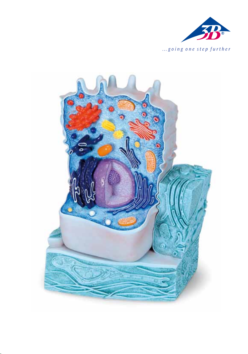
English The Animal Cell
1 Cell nucleus
2 Nucleolus
3 Mitochondrion
4 Smooth endoplasmic reticulum (ER)
5 Desmosome (Macula adhaerens)
6 Basal membrane
7 Hemidesmosome
8 Collagen fibres
9 Fibroblast
10 Peroxisome
11 Lysosome
12 Rough endoplasmic reticulum (ER)
13 Mitochondrion
14 Smooth endoplasmic reticulum (ER)
15 Golgi Apparatus
16 Centriole
17 Cytosol with embedded filament of the cytoskeleton
18 Microvilli
19 Secretion vesicle
20 Golgi Apparatus
21 Lysosome
22 Zonula occludens
23 Zonula adhaerens
24 Desmosome (Macula adhaerens)
25 Microplica
5
Page 6

®
DeutschDie tierische Zelle
Einleitung
Im tierischen mehrzelligen Organismus kommen Zellen grundsätzlich nur im Verbund von vielen gleichartigen bzw. unterschiedlich differenzierten Zellen, bzw. eingebettet in der zwischenzelligen Substanz
(Interzellularsubstanz, extrazelluläre Matrix) vor. Das Umgebungsmilieu der Einzeller und primitiven
Mehrzeller (gleichsam das „Urmeer“) umgibt so auch die Zellen in komplexen tierischen (menschlichen)
Organismen, wobei über die allseits das Gewebe durchdringenden Blutgefäße (bis herunter zu den
Haargefäßen, Kapillaren) die Ernährung gewährleistet wird.
Folgende Grundcharakteristiken zeichnen Zellen lebendiger Organismen aus: Sie sind höher komplex organisiert als ihre Umgebung, sie können auf Reize aus ihrem Inneren und ihrer Umgebung reagieren, schließlich haben sie die Fähigkeit sich zu vermehren (Reduplikation).
Übersicht über Bau und Funktion der Zelle
Die Umhüllung der Zelle und auch die Barriere zur Umwelt, zur Aufrechterhaltung eines eigenen inneren Milieus, wird durch die Zellmembran (Plasmamembran) erreicht. Auch innerhalb der Zelle werden
bestimmte Strukturen und kleine Organe (Organellen, siehe Aufzählung unten) durch die Plasmamembran
umhüllt. Die Plasmamembran selbst besteht aus polaren Lipiden, die eine semipermeable Membran
bilden. Daher können sich die einzelnen Kompartimente und Organellen voneinander bezüglich des
Gehalts bestimmter Moleküle und Ionen abgrenzen. Die Plasmamembran nimmt auch Verbindungen
mit einem feinen Gerüst von Strukturproteinen, den Filamenten des Zellskeletts (Zytoskelett) auf. Hier
findet man feine Aktinfäden (7 nm Durchmesser), röhrenförmige Mikrotubuli (25 nm Durchmesser) und
die im Durchmesser dazwischen (intermediär) gelegenen Intermediärfilamente. Die Mikrotubuli wachsen von einem Organisationszentrum, meist dem Zentriol aus. Dabei sind sie auch verantwortlich für
Transportvorgänge an ihnen entlang, zu und vom Organisationszentrum weg (gerichteter aktiver Transport
– auch in Axonen von Nervenzellen). Das Zentriol selbst ist ein Organell aus zwei senkrecht aufeinander
stehenden Gruppen von Röhren, von denen aus die Mikrotubuli ausstrahlen – auch bei neu entstehenden
Zellen. Bei der Zellteilung wird die Trennung der Chromosomen von den „Marionettenfäden“, der von den
Zentriolen ausgehenden Mikrotubuli, ausgeführt.
Wie der Name „Zytoskelett“ schon sagt, erfüllt dieses die Aufgaben, der Zelle insgesamt Stabilität und eine
entsprechende Flexibilität zu gewährleisten. Darüber hinaus ermöglicht das Zytoskelett die vielfältigsten
aktiven Bewegungen der Zelle: Vom Ausstrecken von füßchenförmigen Fortsätzen (z. B. Filopodien) über
starke Formveränderungen der gesamten Zelle (Grundlage z. B. auch der aktiven Muskelverkürzung) bis
hin zur aktiven Wanderung (Migration) der Zelle. Des Weiteren führen die Elemente des Zytoskeletts die
Spannungslinien innerhalb einer Zelle über die so genannten Zell-Zellverbindungen (z. B. Desmosomen,
s. u.) weiter an die Nachbarzellen und bilden so einen mechanischen Verbund von Zellarealen (z. B. in der
Epidermis der Haut – besonders deutlich bei den Stachelzellen).
Innerhalb der Zell-Zellverbindungen (Interzellulärkontakte) lassen sich funktionell solche, mit überwiegend mechanischer Funktion (Adhäsionskontakte: Zonula; Punctum; Fascia adhaerens; Macula
adhaerens = Desmosom) von solchen mit stoffwechselaktiver (metabolischer) und elektrisch-koppelnder
Funktion (Nexus, Macula communicans = Gap junction; Synapsen) unterscheiden. Schließlich existieren noch Zellverbindungen, die den Interzellularraum abdichten (Barrierenkontake: Zonula occludens). Verbindungen zur extrazellulären Matrix bilden fokale Kontakte bzw. zur Basalmembran das
Hemidesmosom.
Alle Proteine, die die Bestandteile des Zytoskeletts ausmachen, werden von den „Nähmaschinen“ der
Proteine, den Ribosomen, geliefert. Diese können frei im Zytoplasma liegen oder an das Vakuolensystem
des rauhen Endoplasmatischen Retikulums (rauhes ER) gebunden sein. Mit der Information, die aus
dem Zellkern (dieser speichert die Erbinformation über die Chromosomen) über die mRNA übermittelt
wird, koppelt das Ribosom nun „nach Anleitung“ Aminosäure an Aminosäure und „näht“ diese zu einem
6
Page 7

®
Die tierische ZelleDeutsch
Peptid bzw. Protein zusammen. Im Kompartiment des ER werden Peptide und Proteine weiter durch
Helferproteine modifiziert, so dass z. B. Zuckergruppen auf das Protein übertragen werden können
(Glykosylierung). Das glatte ER kann Lipide (Cholesterol, Triglyceride, Steroidhormone) und Lipoproteine
sowie Phospholipide synthetisieren. Darüber hinaus macht das glatte ER lipidlösliche Verbindungen wasserlöslich und entgiftet sie dadurch. In bestimmten Zellarten (z. B. Muskelzellen) dient dem glatten ER auch
als Speicher für Kalziumionen.
Der Golgi-Apparat (GA) besteht aus Gruppen flacher sackförmiger Membranen (Sacculi) und zahlreichen
kleinen Vesikeln. In diesem System werden die im ER synthetisierten noch unreifen Proteine weiter verändert. Es werden bestimmte Zuckergruppen synthetisiert und an Proteine angekoppelt, auch manche
Proteine gespalten. Des Weiteren werden Polysaccharide mit entsprechender Sulfatierung synthetisiert
sowie Glycolipide synthetisiert und angeknüpft. Die reifen Proteine werden im GA in Vesikel verpackt
und transportfähig gemacht. So spielt der GA eine wichtige Rolle bei der Sortierung und Auslieferung von
Proteinen („Postamt der Zelle“).
Hier steht der GA auch mit den Prozessen der Exozytose (Ausschleusung von Vesikeln) und Apozytose
(Ausbuchtungen der Plasmamembran, die mit den von ihnen umschlossenen Komponenten abgeschnürt
werden) in Verbindung. Darüber hinaus können Vesikel aktiv eingeschnürt werden (Endozytose) und auch
Rezeptor vermittelt eingebracht werden (Pinozytose). Nur kursorisch soll auf die Membranausstülpungen
wie Mikrovilli (kleine fingerförmige Ausstülpungen mit geringer Beweglichkeit), Stereozilien (größere
Ausstülpungen mit relativ geringer Beweglichkeit), Kinozilien (geißelförmige Ausstülpungen für aktiven
Flimmerschlag) eingegangen werden.
Sowohl endozytotisch als auch über Zellbestandteile selbst können Vakuolen gebildet werden, die die
abgeschnürten Inhalte verdauen (Lysosomen).
Neben den Lysosomen liegen in einer Zelle kleine Organellen (Peroxisomen), die vor allem Fettsäuren,
Aminosäuren und Harnsäure oxidieren und potentiell zelltoxische organische Verbindungen entgiften können.
Mitochondrien sind vor Urzeiten in Einzeller eingewanderte Bakterien, die sich zu so genannten
Symbionten entwickelten. Sie besitzen eine eigene Erbinformation und auch Ribosomen für die Synthese
von eigenen Proteinen. Sie haben sich jedoch so intensiv mit dem Genom der Wirtszellen verflochten, dass sie nicht mehr eigenständig lebensfähig sind. Der Nutzen dieser Symbiose besteht darin,
dass Mitochondrien Sauerstoff (eine für die lebenden Zellen prinzipiell giftige Substanz, die erst in der
Uratmosphäre durch die Evolution der Pflanzen entstanden ist) verbrennen können, d. h. Kohlenhydrate,
Fettsäuren und Aminosäuren werden unter Verbrauch von elementarem Sauerstoff zu CO2 und H2O
oxidiert. Dadurch wird der universelle Energieträger ATP für die Zelle gewonnen. Das Mitochondrium
besitzt eine doppelte Membranwand. An der inneren Membran selbst ist die Atmungskette und die ATPSynthesekette untergebracht. Im Inneren der Mitochondrienmatrix laufen die Fettsäureoxidation und der
Zitratzyklus ab. Damit stellen die Mitochondrien die „Kraftwerke“ der Zelle dar.
Der Zellkern ist das Informationszentrum für die Zelle. Die Information selbst ist auf 46
Desoxyribonukleinsäuremolekülen (DNA) verteilt. Sie sind zusammen mit den Histonen (Halteproteinen)
im Zellkern untergebracht. Der Kern, der insgesamt dichter gepackt ist als das Zytoplasma, wird von
einer Kerndoppelmembran (Zisterne des ER) mit definierten Kanälen (Kernporen) umgeben. Durch die
Boten-Ribonukleinsäure (mRNA), die an den Genabschnitten der DNA systhetisiert wird (Transkription)
und dann Kopien der DNA enthält, wird die Information zur Proteinsynthese zu den Ribosomen gebracht.
In den Nucleoli (Verdichtungen im Zellkern) werden an speziellen Abschnitten die ribosomale RNA
(rRNA) synthetisiert. Darüber hinaus besteht eine enge Kommunikation mit dem Zytoplasma und den
Membranrezeptoren, so dass der Zellkern die zentrale Informations- und Steuereinheit der Zelle darstellt.
Autor: Prof. Dr. R.H.W. Funk, Institut für Anatomie T.U. Dresden
7
Page 8

®
1 Zellkern (Nucleus)
2 Nucleolus
3 Mitochondrium
4 Glattes Endoplasmatisches Retikulum (ER)
5 Desmosom (Macula adhaerens)
6 Basalmembran
7 Hemidesmosom
8 Kollagene Fasern
9 Fibroblast
10 Peroxisom
11 Lysosom
12 Rauhes Endoplasmatisches Retikulum (ER)
13 Mitochondrium
14 Glattes Endoplasmatisches Retikulum (ER)
15 Golgi Apparat
16 Zentriol
17 Zytosol mit eingebetteten Filamenten des Zytoskeletts
18 Mikrovilli
19 Sekretvesikel
20 Golgi Apparat
21 Lysosom
22 Zonula occludens
23 Zonula adhaerens
24 Desmosom (Macula adhaerens)
25 Mikroplica
DeutschDie tierische Zelle
8
Page 9

®
La Célula AnimalEspañol
Introducción
En los organismos pluricelulares, básicamente, las células se encuentran sólo en conjunto con muchas
otras células del mismo tipo, o de un tipo diferenciado, o incrustadas en la sustancia intercelular (matriz
extracelular). El medio que rodea a los unicelulares y los pluricelulares primitivos (algo así como un mar
originario) rodea también las células de organismos animales complejos (humanos), en donde, sobre todo,
los vasos sanguíneos que atraviesan el tejido (hasta llegar a los capilares) garantizan la nutrición.
Las células de los organismos vivos presentan las siguientes características básicas: Están organizadas de
forma más elevada y compleja que su entorno, pueden reaccionar a sus propios estímulos internos y a los
de su entorno. Por último, tienen la capacidad de reproducirse (reduplicación).
Nociones generales sobre la estructura y la función de la célula
La membrana celular (membrana plasmática) le proporciona una cubierta a la célula y forma una barrera
con su entorno, que le permite mantener aislado su propio medio interno. En el interior de la célula también hay determinadas estructuras y órganos pequeños (organelos, mirar la enumeración abajo) que se
encuentran recubiertos por la membrana plasmática. La membrana plasmática está compuesta por lípidos
polares que forman una membrana semipermeable. Debido a esto, los compartimentos individuales y los
organelos pueden delimitar entre sí mismos el contenido de determinadas moléculas e iones. La membrana plasmática también tiene contactos con un fino andamio de proteínas estructurales y filamentos del
citoesqueleto. Aquí se encuentran los finos hilos de actina (7 nm de diámetro), los microtúbulos, llamados
así por su forma (25 nm de diámetro), y los filamentos intermedios que se encuentran en diámetros intermedios. Los microtúbulos crecen a partir de un centro de organización que, por lo general, es el centriolo.
También son responsables de los procesos de transporte que, a través de ellos, salen desde el centro de
organización o se dirigen hacia él (transporte activo y dirigido, también presente en los axones de las células nerviosas).
El centriolo, en sí mismo, es un organelo que se compone de dos grupos de tubos ubicados verticalmente
uno frente al otro, de los cuales se desprenden los microtúbulos, lo cual ocurre también en las células
recién formadas. Durante la división celular, los cromosomas se separan de los “hilos de marioneta” de los
microtúbulos que provienen del centriolo.
Como el nombre lo indica, el citoesqueleto tiene la tarea de garantizarle a la célula una estabilidad general
y una flexibilidad adecuada. Por otra parte, el citoesqueleto posibilita la multiplicidad de los movimientos
activos de la célula: desde la extensión de algunos apéndices con forma de pies pequeños (por ejemplo,
los filopodios), fuertes cambios en la forma de toda la célula (lo que, por ejemplo, constituye la base de la
contracción muscular activa) hasta un desplazamiento activo de la célula misma (migración). Además, los
elementos del citoesqueleto dirigen las líneas de tensión interna de la célula hacia las células vecinas por
medio de las llamadas uniones celulares (por ejemplo, los desmosomas, ver abajo) y forman una unión
mecánica de áreas de células (por ejemplo, en la epidermis de la piel, especialmente en las células del
estrato espinoso).
Dentro de las uniones de las células (contactos intercelulares) se pueden diferenciar funcionalmente aquéllas que poseen una función mecánica (contactos de adhesión: zónula, punctum, fascia adherente, mácula
adherente = desmosoma) de aquéllas que tienen un metabolismo activo (metabólicos) al igual que de
aquéllas con una función de acoplamiento eléctrico (nexos, mácula comunicante = uniones comunicantes,
sinapsis). Por último, también existen uniones celulares que hermetizan el espacio intercelular (contactos
de barrera: Zonula occludens). Las uniones con la matriz extracelular forman contactos focales como el
hemidesmosoma con la membrana basal.
Todas las proteínas que forman parte de los componentes del citoesqueleto son suministradas por los
“fabricantes” de proteínas: los ribosomas. Estos pueden encontrarse libremente en el citoplasma o unidos
al sistema de vacuolas del retículo endoplasmático granular. Los ribosomas usan la información que proviene del núcleo celular (éste almacena la información hereditaria por medio de los cromosomas) y que es
transmitida a ellos por el mRNA (RNA mensajero) para acoplar “de acuerdo con las instrucciones” un aminoácido a otro aminoácido, hasta formar un péptido o una proteína. Las proteínas y péptidos son modifi-
9
Page 10

®
EspañolLa Célula Animal
cados en los compartimentos del retículo endoplasmático por medio de proteínas auxiliares, de forma tal
que, por ejemplo, los grupos de azúcar puedan transmitirse a las proteínas (glicosilación). El retículo endoplasmático liso puede sintetizar lípidos (colesterol, triglicéridos, hormonas esteroides) y lipoproteínas como
los fosfolípidos. Además, trasforma en solubles en agua los compuestos solubles en lípidos, volviéndolos
no venenosas. En determinados tipos de células el retículo endoplasmático liso también puede almacenar
iones de calcio (por ejemplo, en las células musculares).
El aparato de Golgi se compone de grupos de membranas planas y en forma de saco (sacculi) y de muchas
vesículas pequeñas. En este sistema se modifican nuevamente las proteínas inmaduras que han sido sintetizadas en el retículo endoplasmático. Aquí se crean determinados grupos de azúcar y se acoplan a las proteínas; también se dividen algunas proteínas. Por otra parte, se sintetizan los polisacáridos con la sulfatación respectiva, y también los glicolípidos se sintetizan y acoplan. Las proteínas ya maduras son empacadas
en el aparato de Golgi volviéndose aptas para el transporte. El aparato de Golgi juega un papel importante
en la clasificación y suministro de proteínas (como si fuera una “oficina de correos de la célula”).
El aparato de Golgi está ligado a los procesos de exocitosis (exclusión de las vesículas) y fagocitosis (una prominencia de la membrana plasmática rodea una sustancia y la introduce al interior de la célula). Las vesículas pueden verse atrapadas activamente (endocitosis) y los receptores absorbidos también activamente
(pinocitosis).
Solo se abordarán de forma esquemática las extensiones de la membrana como las microvellocidades
(pequeñas prominencias con forma de dedo y movilidad mínima), estereocilios (grandes prominencias con
relativamente mínima movilidad), quinocilios (prominencias en forma de un cilio para una vibración activa
en forma de golpe).
Por medio de la endocitosis, o con partes de la célula misma, se pueden formar las vacuolas, que son
capaces de digerir el contenido introducido (lisosomas). Además de los lisosomas, hay en las células pequeños organelos (peroxisomas) que permiten la oxidación de ácidos grasos, aminoácidos y ácidos úricos, y
eliminan el veneno de las formaciones orgánicas potencialmente tóxicas para la célula.
Las mitocondrias son bacterias que, en tiempos inmemoriales, migraron al interior de los seres unicelulares convirtiéndose en lo que conocemos como simbiontes. Poseen información hereditaria propia, al
igual que ribosomas para la síntesis de las proteínas también propias. Se mezclaron tan profundamente
con el genoma de su huésped que ya no son capaces de sobrevivir independientemente. La utilidad de esta
simbiosis se basa en el hecho de que las mitocondrias pueden quemar oxígeno (una sustancia, en principio, venenosa para la célula, que sólo surgió en la atmósfera primitiva gracias a la evolución de las plantas), o sea, los carbohidratos, ácidos grasos y aminoácidos se oxidan en CO2 y H2O por medio del oxígeno
elemental. Así se genera para la célula el ATP, el transportador universal de energía. La mitocondria posee
una membrana con pared doble. En la propia membrana interna se encuentra la cadena respiratoria y la
cadena sintetizadora de ATP. En el interior de la matriz de la mitocondria se produce la oxidación de los
ácidos grasos y el ciclo de los citratos. De esta manera, las mitocondrias pueden considerarse como el “taller de energía de la célula”.
El núcleo celular es el centro de información de la célula. La información se encuentra repartida en 46
moléculas de ácido desoxirribonucléico (ADN). Ellas se alojan junto con las histonas (proteínas de sostén)
en el núcleo celular. El núcleo es en general más denso que el citoplasma y está rodeado por una membrana nuclear doble (cisterna del retículo endoplasmático) con canales definidos (poros nucleares). La
información sobre la síntesis de las proteínas se transmite a los ribosomas a través de los ácidos ribonucléicos mensajeros (mRNA) que se sintetizan en algunos sectores del gen del ADN (transcripción) y que luego
contienen copias de ADN. Los ácidos ribonucléicos mensajeros (mARN) se sintetizan en algunos sectores del
gen del AND (transcripción) y contienen una copia del ADN, por medio de éste llega la información de la
síntesis de las proteínas a los ribosomas. En los nucleolos (masa del núcleo celular), en algunas zonas especiales, se sintetiza el ARN ribosomal (rARN). Además, existe una estrecha comunicación entre el citoplasma
10
Page 11

®
Español La Célula Animal
y los receptores membranales, de tal forma que el núcleo representa el centro de información y dirección
de la célula.
Autor: Prof. Dr. R.H.W. Funk, Instituto de Anatomía de la U. T. de Dresden
1 Núcleo celular
2 Nucleolo
3 Mitocondria
4 Retículo endoplasmático liso (RE)
5 Desmosoma (mácula adherente)
6 Membrana basal
7 Hemidesmosoma
8 Fibras colágenas
9 Fibroblasto
10 Peroxisoma
11 Lisosoma
12 Retículo endoplasmático granular (RE)
13 Mitocondria
14 Retículo endoplasmático granular liso (RE)
15 Aparato de Golgi
16 Centriolo
17 Citosol con filamentos incrustados del citoesqueleto
18 Microvellocidades
19 Vesícula secretora
20 Aparato de Golgi
21 Lisosoma
22 Zónula ocludente
23 Zónula adherente
24 Desmosoma (mácula adherente)
25 Micropliegue
11
Page 12

®
FrançaisLa cellule animale
Introduction
Dans l’organisme animal pluricellulaire, les cellules ne se présentent en principe que sous forme d’une
liaison de plusieurs cellules de même type ou sous forme de cellules à différenciation diverse. Les cellules
peuvent être également incluses dans la substance intercellulaire (substance intercellulaire, matrice extracellulaire). Le milieu environnemental des organismes unicellulaires et des organismes pluricellulaires
primitifs (la « soupe primitive » pour ainsi dire) enveloppe également les cellules des organismes animaux
(humains) complexes. Leur alimentation est assurée par les vaisseaux sanguins se chargeant d’une irrigation générale des tissus (jusqu’aux artères ciliaires, capillaires).
Les caractéristiques de base suivantes définissent les cellules d’organismes vivants : L’organisation de ces
cellules est plus complexe que celle de leur environnement. Elles peuvent réagir à des stimuli venant de
leur milieu interne ou de leur environnement, et elles disposent enfin de la faculté de se reproduire (réplication).
Aperçu sur la structure et le fonctionnement de la cellule
La membrane cellulaire (membrane plasmique) forme l’enveloppe de la cellule et constitue également
une barrière contre l’environnement qui permet de préserver l’intégrité du milieu interne cellulaire. À
l’intérieur de la cellule elle-même, certaines structures et des petits organes intracellulaires (organelles, cf.
énumération ci-dessous) sont également enveloppés d’une membrane plasmique. La membrane plasmique
elle-même est composée de lipides polaires, formant une membrane semi-perméable. Les divers compartiments et organelles peuvent donc s’isoler les uns par rapport aux autres quant à la teneur de certains
ions et molécules. La membrane plasmique présente également des jonctions avec une ossature fine de
protéines structurales, les filaments du squelette cellulaire (cytosquelette). Nous y trouvons des filaments
fins d’actine (diamètre de 7 nm), des microtubules à structure tubulaire (diamètre de 25 nm) ainsi que les
filaments intermédiaires dont le diamètre présente une valeur intermédiaire. Les microtubules « poussent
» à partir d’un centre d’organisation, généralement depuis le centriole. Ils assument en outre les processus
de transport sur toute leur propre longueur, vers et depuis le centre d’organisation (transport directionnel
actif – également dans les axones de cellules nerveuses). Le centriole lui-même est un organelle composé
de deux groupements de tubes perpendiculaires les uns aux autres, à partir desquels les microtubules
rayonnent – également lors de la formation de nouvelles cellules. Lors de la division cellulaire, la séparation des chromosomes se fera au moyen des « ficelles de marionnette », les microtubules partant des
centrioles.
Comme son nom l’indique, les tâches assumées par le « cytosquelette » permettront une plus grande
stabilité générale de la cellule tout en conférant à cette dernière la flexibilité requise. Le cytosquelette
permet en outre les mouvements actifs les plus variés de la cellule : depuis l’extension des prolongements
pédiformes (tels que filopodes) en passant par des modifications importantes de l’ensemble de la forme
cellulaire (sur lesquelles se base par exemple le raccourcissement musculaire) jusqu’à la migration active
de la cellule. Par ailleurs, les éléments du cytosquelette transmettent les lignes de contrainte se présentant
à l’intérieur d’une cellule aux cellules voisines, via les jonctions cellule-cellule (telles que les desmosomes,
cf. ci-dessous), en formant une liaison mécanique, composée de zones cellulaires (telles que celles présentes dans l’épiderme de la peau – ce qui se manifeste visiblement dans le cas de la couche des cellules
épineuses).
À l’intérieur des jonctions cellule-cellule (contacts intercellulaires), il est possible de faire une distinction
fonctionnelle entre d’une part les jonctions assumant une fonction essentiellement mécanique (contacts
d’ancrage : zonula ; punctum ; fascia adhaerens ; macula adhaerens = desmosome) et entre d’autre
part les jonctions assumant une fonction métabolique active et de jonction électrique (nexus, macula
communicante = jonction communicante ; synapses). Enfin, il existe encore des jonctions cellulaires
assurant une étanchéification de l’espace intercellulaire (contacts au niveau de la barrière : zonula occlu-
12
Page 13

®
Français La cellule animale
dens). Des contacts focaux forment des jonctions vers la matrice extracellulaire ou la membrane basale,
l’hémidesmosome.
Toutes les protéines, composant le cytosquelette, sont délivrées par les « machines à coudre » des protéines, les ribosomes. Ces derniers peuvent circuler librement dans le cytoplasme ou être reliés au système
vacuolaire du réticulum endoplasmique rugueux (RE rugueux). Au moyen des informations transmises
depuis le noyau cellulaire (ce dernier stocke l’information génétique dans les chromosomes) via l’ARNm,
le ribosome couple alors « conformément au mode d’emploi » acide aminé à acide aminé et les « coud »
ensemble pour en faire un peptide ou une protéine. Dans le compartiment du RE, des protéines auxiliaires
se chargeront de poursuivre la modification des peptides et des protéines afin que des groupements sucrés
puissent être par exemple transférés à la protéine (glycosylation). Le RE lisse peut réaliser la synthèse de
lipides (cholestérol, triglycérides, hormones stéroïdiennes) et de lipoprotéines ainsi que celle de phospholipides. Le RE lisse se chargera en outre de rendre composés liposolubles solubles dans l’eau, ce qui les
détoxiquera. Dans certains types de cellules (telles que cellules musculaires), le RE lisse joue également le
rôle d’un réservoir pour les ions calcium.
L’appareil de Golgi (AG) comprend des groupements de membranes plates en forme de sac (saccules) ainsi
que beaucoup de petites vacuoles. C’est au niveau de ce système que les protéines synthétisées dans le RE
et qui n’y sont pas encore arrivées à maturité subiront d’autres modifications. Certains groupements sucrés
seront synthétisés et couplés à des protéines, quelques protéines seront également scindées. Nous assistons
par ailleurs à une synthèse de polysaccharides avec une sulfatation correspondante ainsi qu’à une synthèse
et un attachement de glycolipides. Dans l’AG, les protéines arrivées à maturité seront emballées dans des
vacuoles et préparées au transport. L’AG joue donc un rôle important lors du tri et de la distribution des
protéines (« bureau de poste de la cellule »).
Dans ce cas, l’AG est également en rapport avec les processus de l’exocytose (rejet de vacuoles à l’extérieur)
et ceux de l’apocytose, le clivage de composants cellulaires (saillies de la membrane plasmique, étranglées
au moyen des composant les entourant). Des vacuoles peuvent en outre être activement étranglées (endocytose) et des récepteurs intermédiaires également mis en place (pinocytose). Nous ne voulons traiter ici
que brièvement les excroissances de la membrane sous forme de microvillosité (petites excroissances à
mobilité restreinte), les stéréocils (excroissances de plus grande taille à mobilité relativement restreinte) et
les kinocils (excroissances de forme flagellée permettant une action sur le battement ciliaire).
Des vacuoles, digérant le matériel étranglé (lysosomes), pourront se former aussi bien par endocytose que
par les composants cellulaires eux-mêmes.
Une cellule contient, outre les lysosomes, de petites organelles (péroxisomes) pouvant avant tout oxyder
des acides gras, des acides aminés et des acides uriques et pouvant potentiellement détoxiquer des composés organiques toxiques au niveau cellulaire.
Les mitochondries sont des bactéries, ayant émigré, il y a des millions d’années, dans des organismes
unicellulaires et s’étant transformées en symbiontes. Ces mitochondries disposent d’une information génétique propre et également de ribosomes leur permettant de synthétiser leurs propres protéines. Mais ces
bactéries se sont cependant alliées si étroitement au génome des cellules hôtes qu’elles ne sont plus autonomement viables. Cette symbiose présente l’avantage que les mitochondries peuvent brûler de l’oxygène
(une substance en principe toxique pour les cellules vivantes, apparue seulement à la suite de l’évolution
végétales dans l’atmosphère primaire), en d’autres mots, des hydrates de carbone, des acides gras et des
acides aminés seront oxydés pour être transformés en CO2 et H2O, en faisant appel à de l’oxygène élémentaire. Ce qui permettra de faire bénéficier la cellule de l’A.T.P, porteur universel d’énergie. La mitochondrie
présente une paroi membranaire double. La chaîne respiratoire permettant la synthèse de l’A.T.P se trouve
sur la membrane interne elle-même. L’oxydation des acides gras et le cycle de Krebs prennent place dans la
matrice des mitochondries. Les mitochondries représentent donc les « centrales électriques » de la cellule.
Le noyau cellulaire est le centre d’informations de la cellule. Les informations elles-mêmes sont réparties
13
Page 14

®
FrançaisLa cellule animale
sur 46 molécules d’acide désoxyribonucléique (ADN). Qui se trouvent avec les histones (protéines d’ancrage)
dans le noyau cellulaire. Le noyau, dont la densité est dans l’ensemble plus compacte que celle du cytoplasme, est enveloppé d’une membrane nucléaire double (citerne du RE) présentant des canaux définis
(pores nucléaires). Les informations concernant la synthèse protéinique seront transmises aux ribosomes
par l’acide ribonucléique messager (ARNm), synthétisé sur les segments génétiques de l’ADN (transcription)
et contenant ensuite des copies de l’ADN. Dans les nucléoles (condensations dans le noyau cellulaire), la
synthèse de l’ARN ribosomique (ARNr) se fera au niveau de segments spéciaux. Il existe en outre une communication étroite avec le cytoplasme et les récepteurs de la membrane, ce qui fait donc du noyau cellulaire l’unité centrale d’information et de commande de la cellule.
Auteur : Professeur Dr. R.H.W. Funk, Institut d’anatomie de l’Université Technique de Dresde
1 Noyau cellulaire (nucléus)
2 Nucléole
3 Mitochondrie
4 Réticulum endoplasmique lisse (RE)
5 Desmosome (Macula adhaerens)
6 Membrane basale
7 Hémidesmosome
8 Fibres collagènes
9 Fibroblaste
10 Péroxysome
11 Lysosome
12 Réticulum endoplasmique rugueux (RE)
13 Mitochondrie
14 Réticulum endoplasmique lisse (RE)
15 Appareil de golgi
16 Centriole
17 Cytosol avec filaments inclus du cytosquelette
18 Microvillosité
19 Vacuole de sécrétion
20 Appareil de golgi
21 Lysosome
22 Zonula occludens
23 Zonula adhaerens
24 Desmosome (Macula adhaerens)
25 Microplica
14
Page 15

®
Português A célula animal
Introdução
Nos organismos animais pluricelulares as células geralmente sempre se encontram em associação com
muitas outras do mesmo tipo ou diferenciadas em diversas formas, integradas dentro de uma substância
entre as células (substância intercelular, matriz extracelular). O meio ambiente dos organismos unicelulares
e dos pluricelulares primitivos (parecido ao „mar originário“) envolve assim as células nos organismos animais complexos (humanos), sendo que a alimentação é garantida pelos vasos sangüíneos que atravessam o
tecido por todo lado (até os capilares).
As seguintes caraterísticas básicas definem os organismos vivos: eles tem uma organização mais complexa
do que o seu entorno, podem reagir a excitações internas e oriundas do seu meio ambiente externo, e
finalmente, eles têm a habilidade de se reproduzir (reduplicação).
Noções gerais sobre a constituição e o funcionamento da célula
O envoltório da célula, que é também uma barreira frente ao meio ambiente para poder criar um meio
interno próprio, é constituído pela membrana celular (a membrana plasmática). Também dentro da célula
existem algumas estruturas e pequenos órgãos (orgânulos, veja lista abaixo) que são envoltos de membrana plasmática. A própria membrana plasmática é feita de lipídios polares que formam uma membrana
semipermeável. Por isso, cada compartimento e orgânulo fica separado do outro conforme o conteúdo em
certas moléculas e íons. A membrana plasmática também tem contatos por meio de uma fina construção
de proteínas estruturais, os filamentos do esqueleto da célula (o citoesqueleto). Aqui encontram-se finos
filamentos de actina (7 nm de diâmetro), pequenos microtúbulos em forma de tubo (25 nm de diâmetro), e
os filamentos intermediários, intermediários pelo tamanho do diâmetro. Os microtúbulos crescem à partir
de um centro de organização, em geral o centríolo. Estes são responsáveis pelos transportes ao longo deles,
para outros centros de organização e desde eles (Transporte dirigido ativo – também presente nas axonas
das células nervosas). O centríolo em si é um orgânulo feito de dois grupos de tubos perpendiculares uns
aos outros, do qual partem os microtúbulos, também em células ainda em formação. Durante a divisão
celular, a divisão dos cromossomas é realizada através dos microtúbulos que saem dos centríolos.
Como o nome „citoesqueleto“ já indica, ele cumpre a função de garantir a estabilidade geral da célula,
assim como a necessária flexibilidade. Além disso, o citoesqueleto possibilita os mais variados movimentos ativos da célula: desde a projeção de pontas em forma de pezinhos (por ex., os filopódios), ou fortes
alterações na aparência geral de toda a célula (a base, por ex., do encurtamento ativo dos músculos), até o
deslocamento ativo (migração) da célula. Pelo mais, os elementos do citoesqueleto conformam as linhas de
tensão dentro da célula e a conectam com outras células através da chamada comunicação célula a célula
(por ex., desmossomas), formando assim uma associação mecânica de zonas celulares (por ex., na epiderme cutânea – particularmente visível nas células espinhosas).
Dentro das conexões de célula a célula (contatos intercelulares) distingue-se funcionalmente entre as
que cumprem funções mecânicas (contatos por adesão: zônula; junção intermediária (Fascia adhaerens);
mácula de adesão = desmossoma) das que cumprem funções ativas ligadas a processos do metabolismo
(metabólicas) ou uma função de conexão elétrica (nexo, junção comunicante = gap junction; sinapses)
Finalmente, existem também conexões celulares que vedam o espaço intercelular (contatos de barreira:
zônula de oclusão). As conexões com a matriz extracelular são formadas por contatos focais, como o
hemidesmossoma com a membrana basal.
Todas as proteínas que formam os componentes do citoesqueleto são fornecidas pelas „máquinas de costura“ das proteínas, os ribossomas. Estes podem estar livres no citoplasma ou estar associados ao sistema de
vacúolos do retículo endoplasmático rugoso (RE rugoso). Com as informações contidas no núcleo da célula
(este armazena a informação hereditária por meio dos cromossomas) e transmitidas pelo mRNA, o ribossoma junta aminoácido com aminoácido conforme „as instruções“ e os „costura“ para formar um peptídeo
ou uma proteína. No compartimento do RE, os peptídeos e as proteínas são modificados por proteínas
15
Page 16

®
A célula animal Português
auxiliares de modo a que grupos de açúcares, por exemplo, possam ser transferidos para a proteína (glicuronidação). O RE liso pode sintetizar lipídios (colesterol, triglicerídeos, hormônios esteróides) e lipoproteínas, assim como fosfolipídios. Além disto, o RE liso torna associações solúveis em lipídios em solúveis em
água, desintoxicando-as assim. Em certos tipos de células (por ex., nas células musculares) o RE liso serve
de armazém para íons de cálcio.
O complexo de Golgi (CG) consiste num grupo de membranas em forma de sacos achatados (sáculos) e
numerosas pequenas vesículas. Neste sistema, as proteínas sintetizadas no RE e ainda não maduras são
transformadas. Grupos específicos de açúcares são sintetizados e associados a proteínas, algumas proteínas
também são divididas. A continuação, são sintetizados polissacáridos com a sulfatação correspondente,
assim como glicolipídios são sintetizados e acoplados. As proteínas maduras são embaladas no CG numa
vesícula e preparadas para o transporte. Assim, o CG cumpre um papel importante na seleção e distribuição de proteínas („o correio da célula“).
Aqui, o CG também está relacionado com os processos da exocitose (expulsão de vesículas) e da fagocitose
(invaginação da membrana que é logo fechada como um saco junto com os componentes assim abrangidos). Além disso, vesículas podem ser fechadas ativamente (endocitose) e receptores podem ser absorvidos
ativamente (pinocitose). Só se abordarão de forma esquemática as extensões da membrana como os microvilos (pequenas extensões em forma de dedo com pouca mobilidade), estereocílios (extensões maiores
com mobilidade relativamente pequena), cílios (extensões maiores em forma de chicote para vibrar ativamente).
Tanto por endocitose como também por componentes da própria célula, podem formar-se vacúolos que
digerem os conteúdos absorvidos (lisossomas).
Além dos lisossomas encontram-se numa célula pequenos orgânulos (os peroxissomas), os quais oxidam
principalmente ácidos grassos, aminoácidos e ácido úrico e assim podem decompor formações químicas
orgânicas tóxicas para a célula.
As mitocôndrias são bactérias que em tempos primordiais migraram para dentro de seres unicelulares
tornando-se ditos simbiontes. Elas possuem uma informação hereditária própria assim como ribossomas
para a síntese de proteínas próprias. Elas se mesclaram tão profundamente com o genoma do hóspede que
elas não são mais capazes de sobreviver independentemente. A utilidade desta simbiose consiste no fato
que as mitocôndrias podem queimar o oxigênio (uma substância, em princípio, venenosa para a célula
que só surgiu na atmosfera primitiva graças à evolução das plantas), ou seja, os carboidratos, ácidos grassos e aminoácidos são oxidados em CO2 e H2O utilizando oxigênio elementar. Assim é produzido para a
célula o transportador universal de energia ATP. A mitocôndria possui uma membrana com parede dupla.
Na própria membrana interna encontram-se a corrente respiratória e a corrente sintetizadora de ATP. No
interior da matriz da mitocôndria ocorrem a oxidação dos ácidos grassos e o ciclo dos citratos. Com isto, as
mitocôndrias são as „usinas de energia da célula“.
O núcleo celular é o centro de informações da célula. A informação em si encontra-se distribuída em 46
moléculas de ácido desoxiribonucléico (DNA). Elas se encontram no núcleo junto com os histonos (proteínas de suporte). O núcleo é geralmente mais denso do que o citoplasma, ele é envolto de uma membrana nucléica dupla (cisterna do RE) com canais definidos (poros nucléicos). Através dos ácidos ribonucléicos
mensageiros (mRNA) que são sintetizados nos trechos genéticos do DNA (transcrição) e que logo contêm
cópias do ADN, a informação sobre a síntese das proteínas é levada aos ribossomas. Nos nucléolos (densificações no núcleo) é sintetizado o RNA ribossomático (rRNA) em trechos especiais. Além disso, existe uma
comunicação próxima entre o citoplasma e os receptores da membrana, de modo que o núcleo representa
o centro de informações e a unidade de comando da célula.
Autor: Prof. Dr. R.H.W. Funk, Instituto de Anatomia da T.U. Dresden
16
Page 17

®
1 Núcleo
2 Nucléolo
3 Mitocôndria
4 Retículo endoplasmático liso (RE)
5 Desmossoma (mácula de adesão)
6 Membrana basal
7 Hemidesmossoma
8 Fibras colágenas
9 Fibroblasto
10 Peroxissoma
11 Lisossoma
12 Retículo endoplasmático rugoso (RE)
13 Mitocôndria
14 Retículo endoplasmático liso (RE)
15 Aparelho de Golgi
16 Centríolo
17 Citosol com filamento do citoesqueleto integrados
18 Microvilos
19 Vesícula secretora
20 Aparelho de Golgi
21 Lisossoma
22 Zônula de oclusão
23 Zônula de adesão
24 Desmossoma (mácula de adesão)
25 Microprega
A célula animalPortuguês
17
Page 18

15 16 17 18 19
20
14
13
12
11
10
21
3
1
2
4
5
18
689
Page 19

22
23
24
25
7
19
19
Page 20

La cellula animale Italiano
Introduzione
Generalmente, nell’organismo pluricellulare animale esistono solo insiemi di cellule dello stesso tipo o con
caratteristiche diverse o cellule situate nella sostanza intracellulare (sostanza intracellulare, matrice extracellulare). L’ambiente di vita di protozoi e organismi pluricellulari primitivi (il cosiddetto „mare preistorico“) circonda anche le cellule di organismi animali (umani) complessi, garantendo l’alimentazione tramite i
vasi sanguigni che permeano i tessuti (arrivando fino ai vasi capillari).
Le caratteristiche basilari seguenti contraddistinguono le cellule di organismi viventi: Sono caratterizzate
da una complessità più elevata rispetto all’ambiente in cui vivono e possono reagire a stimoli inviati ad essi
stessi o all’ambiente che li circonda. Infine, hanno la capacità di riprodursi (reduplicazione).
Panoramica della struttura e della funzione della cellula
Il rivestimento della cellula, ed anche la barriera nei confronti dell’ambiente extracellulare per il mantenimento del proprio ambiente interno, è costituito dalla membrana cellulare (membrana plasmatica). Anche
all’interno della cellula, la membrana plasmatica riveste determinate strutture e organi di piccole dimensioni (organuli, ved. elenco in basso). La membrana plasmatica stessa è costituita da lipidi polari che formano una membrana semipermeabile. Pertanto, i singoli compartimenti e organuli possono essere separati
l’uno dall’altro in funzione del contenuto di determinate molecole e ioni. La membrana plasmatica stabilisce collegamenti anche con una sottile struttura di proteine strutturali, i filamenti dello scheletro della
cellula (citoscheletro). In questo scheletro si possono trovare microfilamenti di actina (diametro di 7 nm),
microtubuli (diametro di 25 nm) e filamenti intermedi situati nel diametro. I microtubuli vengono sviluppati da un centro di organizzazione principalmente costituito da un centriolo e sono inoltre responsabili
dei processi di trasporto per tutta la loro lunghezza, da e verso il centro di organizzazione (trasporto attivo
direzionato, anche in assoni da cellule nervose). Il centriolo stesso è un organulo formato da due gruppi
di tubi situati verticalmente, dai quali partono i microtubuli, anche in caso di cellule nuove. Durante la
divisione cellulare, la separazione dei cromosomi viene eseguita dai „filamenti finti“ dei microtubuli che si
diffondono dai centrioli.
Come suggerisce la parola „citoscheletro“, questo elemento garantisce la stabilità ed una corrispondente
flessibilità della cellula. Inoltre, il citoscheletro consente alla cellula di eseguire i più svariati movimenti
attivi: dall’estroflessione di pseudopodi (ad es. filopodi) mediante forti variazioni di forma dell’intera cellula (base ad es. anche della contrazione muscolare) fino alla mutazione attiva (migrazione) della cellula.
Inoltre, gli elementi del citoscheletro inviano le linee di tensione all’interno di una cellula attraverso le
cosiddette adesioni cellula-cellula (ad es. desmosomi, ved. sotto.) alle cellule adiacenti formando quindi
un’unione meccanica di areali (ad es. nell’epidermide della pelle, particolarmente evidenti nelle cellule
spinose).
All’interno delle adesioni cellula-cellula (contatti intracellulari) è possibile distinguere in modo funzionale
le adesioni con funzione prevalentemente meccanica (contatti di adesione: Zonula; Punctum; Fascia
adhaerens; Macula adhaerens = desmosomi) da quelle attive a livello metabolico e quelle con funzione di
giunzione elettrica (Nexus, Macula communicans = Gap junction; sinapsi). Infine esistono anche legami cellulari che chiudono lo spazio intracellulare (contatti : Zonula occludens). Le giunzioni con la matrice extracellulare generano contatti focali, mentre i contatti con la membrana basale generano emidesmosomi.
Tutte le proteine che sono parte integrante del citoscheletro sono fornite dalle „macchine da cucire“ delle
proteine, i ribosomi. I ribosomi sono contenuti liberamente nel citoplasma oppure possono essere collegati al sistema vacuolare del reticolo endoplasmatico ruvido (RE ruvido). Con l’informazione trasmessa
dal nucleo cellulare (che immagazzina l’informazione ereditaria tramite i cromosomi) attraverso l’mRNA,
il ribosoma unisce „a comando“ due amminoacidi alla volta e li „cuce“ ad un peptide o ad una proteina.
Nello scompartimento del RE, i peptidi e le proteine vengono ulteriormente modificati dalle proteine
20
Page 21

La cellula animaleItaliano
coadiuvanti, in modo che i gruppi di zuccheri possano essere trasferiti alla proteina (glicolizzazione). Il RE
liscio è in grado di sintetizzare lipidi (colesterolo, trigliceridi, ormoni steroidi) e lipoproteine, nonché fosfolipidi. Inoltre, il RE liscio rende i legami liposolubili idrosolubili e li depura. In determinati tipi di cellule
(ad es. le cellule muscolari), il RE liscio funge anche da accumulatore per ioni calcio.
L’apparato di Golgi (AG) è costituito da gruppi di membrane a forma di sacco appiattito (Sacculi) e da numerose vescicole. In questo sistema, le proteine ancora impure sintetizzate nel RE vengono ulteriormente
modificate. Determinati gruppi di zuccheri vengono sintetizzati e legati a proteine, ed alcune proteine
vengono scisse. Inoltre, i polisaccaridi con solfatazione corrispondente vengono sintetizzati, mentre i glicolipidi vengono sintetizzati e collegati. Le proteine pure vengono impacchettate nella vescicola dell’AG e rese
trasportabili. Quindi, l’AG svolge un ruolo molto importante nella selezione e nella consegna delle proteine
(„ufficio postale della cellula“).
In questo caso, l’AG è legato anche ai processi della esocitosi (espulsione di vescicole) e della fagocitosi
(rigonfiamenti della membrana plasmatica, che vengono isolati dai componenti esclusi). Inoltre, le vescicole possono essere segmentate attivamente (endocitosi) ed anche inglobate mediante i recettori (pinocitosi).
Solo sommariamente si può ignorare le estroflessioni della membrana come i microvilli (microscopiche
estroflessioni bastoncellari con ridotta mobilità), le stereociglia (grandi estroflessioni con mobilità relativamente ridotta), chinocilia (estroflessioni a forma di frusta per uno sfarfallamento attivo).
I vacuoli, che assimilano i contenuti isolati (lisosomi), possono essere formati sia tramite processo endocitico sia tramite le parti integranti della cellula.
Oltre ai lisosomi, in una cellula sono presenti piccoli organuli (perossisomi), i quali ossidano principalmente acidi grassi, amminoacidi e acidi urici e possono purificare legami organici potenzialmente tossici
per la cellula.
Sin dall’origine dei tempi, i mitocondri sono batteri migrati in organismi unicellulari, che si sono successivamente trasformati nei cosiddetti simbionti. Possiedono una propria informazione ereditaria ed anche
ribosomi per la sintesi delle proprie proteine. Si sono intrecciati in modo così complicato con il genoma
delle cellule ospiti che non sono più in grado di vivere autonomamente. Questa simbiosi è sfruttata in
modo tale da consentire ai mitocondri di bruciare ossigeno (una sostanza particolarmente nociva per le
cellule viventi, generato nell’atmosfera originaria tramite l’evoluzione delle piante), ovvero carboidrati,
acidi grassi e amminoacidi vengono ossidati in CO2 e H2O mediante l’uso di ossigeno elementare. In questo
modo si ricava il vettore energetico universale ATP per la cellula. Il mitocondrio possiede una membrana
a doppia parete. Sulla membrana interna si trova la catena respiratoria e la catena per la sintesi dell’ATP.
All’interno della matrice del mitocondrio si svolgono l’ossidazione degli acidi grassi e il ciclo del citrato.
Quindi, i mitocondri rappresentano la „centrale motrice“ della cellula.
Il nucleo cellulare è il centro di informazioni della cellula. L’informazione è suddivisa in 46 molecole di
acido desossiribonucleico (DNA). Queste molecole sono tenute insieme da istoni (proteine leganti) nel
nucleo cellulare. Il nucleo, molto più compatto del citoplasma, è circondato da una membrana nucleare
doppia (cisterna del RE) con canali definiti (pori nucleari). Attraverso l’acido ribonucleico messaggero
(mRNA) che viene sintetizzato sulle sezioni genetiche del DNA (trascrizione) e contiene quindi copie del
DNA, l’informazione sulla sintesi proteica viene portata ai ribosomi. Nei nucleoli (concentrazioni nel nucleo
cellulare) viene sintetizzato l’RNA ribosomiale (rRNA) in sezioni speciali. Inoltre, è presente una stretta
comunicazione con il citoplasma e i recettori della membrana, in modo che il nucleo cellulare rappresenti
l’unità di informazione e gestione centrale della cellula.
Autore: Prof. Dr. R.H.W. Funk, Istituto di anatomia T.U. Dresda
21
Page 22

®
La cellula animale Italiano
1 Nucleo cellulare (nucleo)
2 Nucleolo
3 Mitocondrio
4 Reticolo endoplasmatico liscio (RE)
5 Desmosoma (Macula adhaerens)
6 Membrana basale
7 Emidesmosoma
8 Fibre collagene
9 Fibroblasto
10 Perossisoma
11 Lisosoma
12 Reticolo endoplasmatico ruvido (RE)
13 Mitocondrio
14 Reticolo endoplasmatico liscio (RE)
15 Apparato di Golgi
16 Centriolo
17 Citosolo con filamenti del citoscheletro avvolti
18 Microvilli
19 Vescicolo per secrezione
20 Apparato di Golgi
21 Lisosoma
22 Zonula occludens
23 Zonula adhaerens
24 Desmosoma (Macula adhaerens)
25 Micropieghe
22
Page 23

日本語
動物細胞モデル
はじめに
多細胞生物の動物細胞は大抵、機能が類似した細胞が集まった状態で、細胞間質(細胞外マトリックス)に囲
まれています。細胞の周囲には毛細血管を通して血漿が濾出することで作られる組織液があり、ここから栄養
分を得ています。
細胞の構造と機能
生体膜と細胞骨格
細胞膜(生体膜)は細胞を包み込み、外的環境への防壁を兼ねているので、自身の内的環境の維持管理を可能
にします。 細胞の内部では、ある特定の構造や小さい器官(細胞小器官,下記リスト参照)もまた生体膜で包
まれています。生体膜は極性脂質より成り、半透性を有しています(半透膜)。この様に、個々の細胞区画、細
胞小器官はお互い独立して存在し、それらは特定の分子やイオンを包含しています。 生体膜は、細胞の形態を
形成するタンパク質である細胞骨格に結びついています。この細胞骨格は、アクチンフィラメントなどからな
る微小繊維(直径
成ります。
微小管は通常、形成中心、中心小体から成長します。微小管は細胞骨格としてだけではなく細胞内輸送にも関
わっており、微小管に沿ってタンパク複合体が形成中心から、もしくは中心へ向かって移動します。(この能動
輸送は神経細胞の軸索でも起こります。)中心小体は、互いに垂直に並ぶ
ここから微小管が伸びています。微小管は新しく形成されたばかりの細胞にも見ることができます。細胞分裂
の際、染色体の分離は、中心小体から生じる微小管の集合である紡錘体によって行なわれます。
また細胞骨格とは、その名前が示唆するように、細胞の全体的形態の安定性と適度な柔軟性を確保します。
細胞骨格は細胞運動にも深く関わっています。たとえば、細胞全体の形に大きな変化をもたらす筋収縮、細胞
から化合物を分泌するエンドサイトーシス、原形質流動などの要として重要な役割を果たしています。
7 nm)、中空の微小管(直径 25 nm)、それらの中間の直径を持つ中間径フィラメントより
2 つの管で成り立つ細胞小器官であり、
さらに、細胞骨格の成分で細胞質の枠となっている中間径フィラメントは、隣接する細胞同士が付着する部位
(例えばデスモソーム,下記参照)で固定されています。またこのフィラメントはデスモソームを通して隣接す
る細胞同士を間接的に結合しています。この結合は特に上皮細胞でよく見られます。結合様式にはデスモソー
ム以外にも、神経細胞で見られるシナプス結合、細胞間連絡の場となるギャップ結合などがありますが、それ
ぞれ機能、組成が異なり区別されます。上皮細胞が基底膜とつながる箇所ではヘミデスモソームが形成されます。
リボソームと小胞体
細胞骨格の成分ともなっているタンパク質はすべて、リボソームというタンパク質の工場によって作られます。
リボソームは細胞質中を漂って、もしくは粗面小胞体(粗面
遺伝情報を転写した
アミノ酸を次々につなぎ、ペプチドあるいはタンパク質を合成します。ペプチドやタンパク質はさらに小胞体
内部で糖等が付加され修飾されることもあります。(グリコシル化)。
滑面小胞体は脂質(コレステロール、トリグリセリド)、ステロイドホルモン、リポタンパク、リン脂質の合成
に関わっています。さらに、滑面小胞体では脂溶性の化合物を酵素により水溶性にすることで解毒する作業も
行われます。筋細胞などある種の細胞では、滑面小胞体はカルシウムイオンの貯蔵所としての役目も果たします。
ゴルジ体とサイトーシス
ゴルジ体は平らな嚢状の槽であるゴルジ槽のグループと、多数の小嚢であるゴルジ小胞から成ります。ゴルジ
体では、小胞体で修飾を受けたタンパク質に糖類の付加、分割などの修飾がさらに加えられます。タンパク質
以外にも脂質を受け取り、糖脂質の合成も行っています。これらの修飾が終わったタンパク質や糖脂質は最終
mRNA が細胞核からリボソームに伝わります。リボソームは tRNA によって運ばれてきた
ER)と結合して細胞中に存在します。DNA から
23
Page 24

動物細胞モデル
的には輸送小胞に包まれた状態となり、運搬に備えます。このようにして、ゴルジ体はタンパク質の分類と運
搬を行い、細胞の郵便局ともいえる役割を果たしています。
ゴルジ体は、生成物を細胞外への分泌するエキソサイトーシス、反対に細胞膜が細胞外の物質を囲み取り込む
エンドサイトーシス(ファゴサイトーシスとピノサイトーシス)にも関わっています。微絨毛、不動毛、運動
毛は細胞表面に見られる細胞質の突起ですが(不動毛、運動毛は微絨毛の一種と捉えられる)、ある種の微絨毛
を持つ細胞ではエンドサイトーシスにより物質の取り込みが行われています。エンドサイトーシスで細胞内部
に取り込まれた物質は、細胞膜に囲まれ、エンドサイトーシス小胞として細胞内に存在します。これはリソソー
ムに運ばれ、取り込んだ物質をリソソームが含む酵素によって分解します。リソソームと同様に一重膜で包ま
れた小さい細胞小器官であるペルオキシソームは酸化酵素を持ち、主に脂肪酸、アミノ酸、尿酸を酸化します。
またカタラーゼを持つため、細胞にとって毒性を示す過酸化水素を分解します。
ミトコンドリア
ミトコンドリアは原始の時代に単細胞の生命体に取り込まれたバクテリアで、まるで共生生物のように進化し
ました。ミトコンドリアは独自の遺伝物質と、また自分達のタンパク質の合成のためのリボソームも持ってい
ます。しかし宿主細胞のゲノムと密接に結びついてしまったため、単独ではもはや生きられなくなりました。
この共生関係の利点は、嫌気性の生物が酸素を活用できるようになったことです。太古の時代、植物の繁栄によっ
てもたらされた大量の酸素は嫌気性の生物にとって有害な物質でした。しかし酸素を活用できるミトコンドリ
アを取り込むことで、酸素からエネルギーを作り出せるようになりました。酸素を利用して、炭水化物、脂肪酸、
アミノ酸などの有機物を酸化することで、
り出されています。
ミトコンドリアは二重膜構造を持っており、内部の腔部分であるマトリックス、内膜、外膜、外膜と内膜の間
である膜間部分に分けられます。内膜では
脂肪酸酸化酵素やクエン酸回路に関わる酵素が存在し、細胞のエネルギー生成の核となっています。そのため、
ミトコンドリアは細胞の発電所と表されます。
CO
、H
O、そして生命の共通したエネルギー担体である ATP が作
2
2
ATP 合成と呼吸鎖における酸化反応が行なわれ、マトリックスには
日本語
核
細胞核は細胞の情報センターです。遺伝情報を持つ
染色室は細胞周期の
は核孔をもつ二重膜、核膜に包まれています。他の小器官より大きいため容易に単離することもできます。
M 期には高次構造である染色体となり、ヒトの場合は 46 本の染色体が確認できます。核
DNA は核内でヒストンとともに染色質を形成しています。
DNA が持つタンパク質合成の情報は、mRNA に写し取られ、これがタンパク質の工場であるリボソームに運ば
れます。この
スを翻訳といいます。
核内にある核小体にはリボソーム
ムタンパクが付き、プロセシングを受けて大小のリボソームサブユニットがつくられます。サブユニットはそ
れぞれ核膜孔から出て、結びつきリボソームとなります。
DNA が mRNA に写し取られるプロセスを転写、mRNA の情報からペプチド鎖が作られるプロセ
RNA(rRNA)をコードする DNA があります。ここで作られた rRNA にリボソー
Author: Prof. Dr. R.H.W. Funk, Institute of Anatomy, Dresden University of Technology
24
Page 25

日本語
1 核
2 核小体
3 ミトコンドリア
4 滑面小胞体(滑面 ER)
5 デスモソーム
6 基底膜
7 ヘミデスモソーム
8 膠原繊維(コラーゲン繊維)
9 線維芽細胞
10 ペルオキシソーム
11 リソソーム
12 粗面小胞体(粗面 ER)
13 ミトコンドリア
14 滑面小胞体(滑面 ER)
15 ゴルジ体
16 中心小体
17 細胞質基質と繊維状の細胞骨格
18 細胞膜(突起部は微絨毛)
19 輸送小胞
20 ゴルジ体
21 リソソーム
22 閉鎖帯
23 接着帯(アドヘレンスジャンクション)
24 デスモソーム
25 Microplica(微細なヒダ状の構造)
動物細胞モデル
25
Page 26

®
РусскийЖивотная клетка
Предисловие
Клетки многоклеточных животных организмов в основном располагаются в виде групп сходных клеток
или вместе с другими дифференцированными клетками, либо встроены в межклеточное вещество
(промежуточное вещество, внеклеточный матрикс). Окружающая среда одноклеточных и примитивных
многоклеточных организмов (так называемый «первичный бульон») также окружает клетки более
сложных высокоорганизованных животных (человеческих) организмов и обеспечивает их питание через
кровеносные сосуды, которые проникают сквозь ткани (вплоть до капилляров).
Клетки живых организмов отличаются следующими основными характеристиками: они обладают более
сложной организацией, чем окружающая их среда, они могут реагировать на внутренние стимулы и
стимулы окружающей среды, и обладают способностью к размножению (редупликация).
Обзор строения и функции клеток
Клеточная мембрана (плазматическая мембрана) окружает клетку и образует барьер, ограждающий
клетку от наружной окружающей среды, для поддержания своей собственной внутренней среды. Внутри
клетки определенные структуры и маленькие органы (органеллы, см. перечень ниже) также окружены
плазматической мембраной. Сама плазматическая мембрана состоит из полярных липидов, которые
образуют полупроницаемую оболочку. Таким образом, отдельные камеры и органеллы отграничены
друг от друга и от содержащихся в них определенных молекул и ионов. Плазматическая мембрана
также связана с тонким каркасом структурных белков, филаментов клеточного скелета (цитоскелет). Этот
цитоскелет состоит из тонких филаментов актина (диаметром 7 нм), полых микротрубочек (диаметром 25
нм) и лежащих между ними промежуточных филаментов. Микротрубочки образуются в организационном
центре, обычно в центриоли. Они также отвечают за процессы транспортировки по всей их длине в
организационный центр и из него (направленный активный транспорт, который также осуществляется в
аксонах нервных клеток). Центриоль представляет собой органеллу, состоящую из двух групп трубочек,
расположенных перпендикулярно друг к другу, из которых исходят микротрубочки – этот процесс
также происходит во вновь образовавшихся клетках. Во время деления клетки разделение хромосом
обеспечивается «нитями марионетки» - микротрубочками, исходящими из центриоли.
Исходя из названия, цитоскелет обеспечивает общую стабильность клетки и соответствующий уровень
гибкости. Более того, цитоскелет обеспечивает чрезвычайную универсальность активных движений клетки:
от растяжения отростков в виде «ножек» (например, филоподий) для осуществления основных изменений
формы клетки в целом (например, при активных мышечных сокращениях) до активных движений клетки
(миграция клетки). Кроме того, элементы цитоскелета формируют линии натяжения внутри клетки
посредством так называемых межклеточных соединений (например, десмосом, см. ниже), направленные
к соседним клеткам, и, таким образом, механически связывают различные области клетки, например, в
эпидермисе кожи – особенно четко это видно в шиповатых клетках.
В межклеточных соединениях (межклеточный контакт) структуры с преимущественно механической
функцией (адгезионные соединения: зона, точка, полоса слипания, пятно сцепления = десмосома) могут
отличаться от таковых, несущих активную метаболическую и электропроводящей функцией (нексус,
соединительное пятно = щелевое соединение, синапс). Наконец, существуют клеточные соединения,
которые запирают межклеточную область (контактный барьер: плотное соединение (зона замыкания)
Соединения с внеклеточной мембраной образуют фокальные контакты и контакты с базальной
мембраной полудесмосомы.
Все белки, входящие в состав компонентов цитоскелета, сформированы «швейной машиной»
протеинов - рибосомами. Они могут быть подвешены в цитоплазме или соединены с системой вакуолей
шероховатого эндоплазматического ретикулума (шероховатого ЭР). Информация поступает в рибосомы
из клеточного ядра, где в хромосомах хранится генетическая информация в виде мРНК. Рибосома
связывает аминокислоты в определенной последовательности и «пришивает» их к пептиду или белку.
26
Page 27

®
Русский Животная клетка
Пептиды и белки далее изменяются с помощью вспомогательных белков внутри ЭР, например, к белку
могут присоединяться группы сахаров (гликозилирование). Гладкий ЭР может синтезировать липиды
(холестерин, триглицериды, стероидные гормоны), липопротеины и фосфолипиды. Кроме того, гладкий ЭР
делает жирорастворимые вещества водорастворимыми и, таким образом, обезвреживает их. В отдельных
типах клеток (например, в мышечных клетках) гладкий ЭР также служит для хранения ионов кальция.
Аппарат Гольджи состоит из групп горизонтальных мешковидных мембран (мешочки) и множества мелких
везикул. Здесь происходит дальнейшая модификация вновь синтезированных в ЭР белков. Специфические
сахарные группы синтезируются и связываются с белками, а также расщепляются некоторые белки.
Кроме того, синтезируются полисахариды с соответствующей сульфатизацией и гликолипиды и
соединяются вместе. Эти зрелые белки упакованы в везикулы в АГ и готовы к транспорту. Таким образом,
АГ играет важную роль в сортировке и доставке белков («почта клетки»).
АГ также связан с процессами экзоцитоза (высвобождение пузырьков) и фагоцитоза (выпячивание
плазматической мембраны с последующим «замыканием» мембраны вокруг захваченных ею
компонентов). Также могут активно обволакиваться и захватываться везикулы (эндоцитоз) и рецепторы,
выступающие в качестве медиаторов (пиноцитоз). Выпячивания мембраны, такие как микроворсинки
(маленькие малоподвижные пальцевидные выпячивания), стереоцилии (выпячивания большего размера
с относительно малой подвижностью) и киноцилии (выпячивания в форме жгутика, обеспечивающие
активное движение ресничек) будут только обсуждаться в рамках курса.
Вакуоли могут образовываться и путем эндоцитоза, и компонентами клетки, которые переваривают
содержимое везикул (лизосомы).
Вдоль лизосом в клетке расположены маленькие органеллы (пероксисомы), которые преимущественно
окисляют жирные кислоты, аминокислоты и мочевую кислоту и могут обезвреживать потенциально
цитотоксические органические вещества.
Митохондрии – это бактерии, которые попали в одноклеточные организмы в первобытные времена и
развились в так называемые симбионты. Они имеют свой собственный генетический материал, а также
содержат рибосомы для синтеза своих собственных белков. Однако они настолько тесно переплетены с
геномом клетки хозяина, что не могут существовать независимо. Польза подобного симбиоза заключается
в том, что митохондрии могут утилизировать кислород (принципиально токсическое вещество для живых
клеток, которое возникло впервые в первобытной атмосфере в ходе эволюции растений). Таким образом,
углеводы, жирные кислоты и аминокислоты окисляются до CO2 и H2O при потреблении элементарного
кислорода. Таким путем АТФ, универсальный переносчик энергии, поступает в клетку. Митохондрия
обладает двойной мембраной. Синтез АТФ и процессы дыхательной цепи происходят во внутренней
стенке. Окисление жирных кислот и цикл лимонной кислоты происходят внутри митохондриального
матрикса. Таким образом, митохондрии можно назвать «электростанцией» клетки.
Клеточное ядро – представляет собой информационный центр клетки. Информация распределена в
46 молекулах дезоксирибонуклеиновой кислоты (ДНК). Они расположены в клеточном ядре вместе
с гистонами (кэппирующие белки). Ядро, как правило, упаковано более плотно, чем цитоплазма, и
окружено двойной ядерной мембраной (цистерна ЭР) с определенными каналами (ядерные поры).
Информация для синтеза белков передается в рибосомы мессенджерными рибонуклеиновыми кислотами
(мРНК), которые синтезируются в генных сегментах ДНК. Этот процесс называется транскрипцией, при
этом образуются копии ДНК. Рибосомальная РНК синтезируется на специализированных сегментах
в нуклеолах (скопления в клеточном ядре). Кроме того, существует тесная связь между цитоплазмой и
рецепторами мембраны, таким образом, ядро клетки представляет собой информационный центр и
орган управления клетки.
Автор: проф. Р. Функ (Dr. R.H.W. Funk), Институт анатомии, Дрезденский технический университет
27
Page 28

®
Животная клетка
1 Ядро клетки
2 Ядрышко
3 Митохондрия
4 Гладкий эндоплазматический ретикулум (ЭР)
5 Десмосома (пятно сцепления)
6 Базальная мембрана
7 Полудесмосома
8 Волокна коллагена
9 Фибробласт
10 Пероксисома
11 Лизосома
12 Шероховатый эндоплазматический ретикулум (ЭР)
13 Митохондрия
14 Гладкий эндоплазматический ретикулум (ЭР)
15 Аппарат Гольджи
16 Центриоль
17 Цитозоль с встроенным филаментом цитоскелета
18 Микроворсинки
19 Секреторный пузырек
20 Аппарат Гольджи
21 Лизосома
22 Зона замыкания
23 Зона сцепления
24 Десмосома (пятно сцепления)
25 Микроскладка
Русский
28
Page 29

®
中文 动物细胞
引言
动物的多细胞有机体中的细胞大多只以相同的细胞群组的形式出现或者以与其他分化的细胞混合在
一起的形式出现, 或者是嵌入在细胞基质中(胞内基质,胞外基质)。 单细胞和原始多细胞有机
体的周围环境(也称为“原始汤”)也包围着更为复杂和高级的有机化的动物(人类)有机体, 并
且通过渗透于整个组织(至毛细血管)的血管来保证其营养供应。
以下的主要特征用以区分生物的细胞: 它们拥有比它们的环绕物更为高级复杂的组织, 它们可以
对内部刺激和外部环境刺激发生反应, 它们具有再生(复制)的能力。
纵览细胞的结构和功能
细胞膜(质膜)将细胞裹覆并且也向外部环境提供了一个维持其自身内部环境的载体。 在细胞内
部,特定的构造和小的器官(细胞器,见下表)也被质膜所包覆。 质膜本身由形成半渗透膜的极性
类脂所构成。 这样一来,单独的区室和细胞器被互相分离开来,并且也与它们所含有的特殊的分子
和离子互相分离开来。质膜也被连接到结构蛋白的微细构造上,细胞骨胳的纤丝(细胞骨架)。 这
个细胞骨架由微细的肌动蛋白丝(直径7 nm ),空心微管(直径25 nm )和径向排列的中间纤丝构
成的。微管产生于一个有机体中心,通常称为中心粒。它们也负责沿着它们的长度方向从有机体中
心向外进行传输和向有机体中心进行传输的责任(也可以在神经细胞轴突中发生定向主动转运)。
中心粒本身是一个由两组相互垂直的管道构成的细胞器,微管经过这个细胞器进行延伸-这种情况
在新形成的细胞中也有发生。在细胞分裂过程中,染色体的分离是由“木偶控制线”-中心粒发出
的微管来进行的。
如同这个名字所表达的,细胞骨架确保了细胞的整体稳定性并使细胞具有相应程度的灵活性。细胞
骨架还能保证细胞在主动运动中具有极度的多样性:从脚状附器的延展(如:丝足)到为细胞主动
运动(细胞迁移)所进行整个细胞形状的主要改变(也是最基本的活性肌肉收缩的例证)。更有甚
者,细胞骨架成分在一个细胞内通过所谓的细胞-细胞连接(如:细胞桥粒-见下表)增殖张力线到
相邻的细胞并且机械地连接到细胞的不同区域上,例如:皮肤的表皮-在皮刺细胞中特别清晰。
在机械功能占据主要位置的细胞-细胞连接(细胞内接触)的结构中,接触粘着(包括小区域粘着
连接,一个点或小面积粘着连接,腱膜粘着连接以及斑点粘着连接等都意指为桥)都可以通过活跃
代谢和电偶合功能(融合膜及腱膜传接等于间隙连接或突出)来区分它们。最后,还有封锁胞内区
域(接触载体:小区域堵塞)的细胞连接。与粘着细胞外膜的连接和与基膜半桥粒的连接。
所有构成细胞骨架的蛋白质都是由蛋白质的“缝纫机”-核糖体制造的。这些核糖体可以悬浮在细
胞质中或者可以结合到粗糙型内质网的液泡系统中(粗糙型ER)。信息可以从细胞核传送到核糖
体,遗传信息通过信使核糖核酸(mRNA) 被储存在染色体中。
29
Page 30

®
动物细胞
核糖体将氨基酸与氨基酸进行顺序偶联并且将它们“缝合”为肽或蛋白质。肽和蛋白质被ER(内
质网)中的辅助蛋白质进一步改性,例如,糖基也许被添加到蛋白质中(糖基化)。平滑型ER (内
质网)可以合成类脂(胆固醇,甘油三酸脂和类固醇荷尔蒙),脂蛋白和磷脂。平滑型ER (内质
网)进而产生水溶性可溶脂化合物并对它们解毒。在某种类型的细胞中(如:肌肉细胞),平滑型
ER (内质网)还起到储存钙离子的作用。
高尔基体(GA)是由一组平袋状膜(小囊)和无数的小泡囊组成。ER(内质网)中新合成的蛋白质
在这里被进一步改性。特殊的糖基被合成和偶联为蛋白质,也有一些蛋白质被分解。此外,适当硫
酸盐化的多糖被合成,糖脂被合成并且被结合在一起。这些成熟的蛋白质被装入高尔基体(GA)的泡
囊中,准备进行传输。因此,高尔基体(GA)在蛋白质的分类和传递方面起到了一个重要的作用(“
细胞邮局”的作用)。
高尔基体(GA)也与吐胞事件 (泡囊的释放)和质膜的束状吐胞突出相关联,这些质膜带有与它所环绕
的成分随后被一起剪掉的膜。泡囊也可能被主动包膜和被捕捉(胞吞),而且,起中介器作用的感
受器也可能被带入(胞饮)。关于膜突出,例如,微绒毛(具有很少移动性的小的指状突出),静
纤毛(具有相对少的移动性的较大的突出)和动纤毛(用于活性纤毛运动的鞭毛状突出)将只有在
阅读课中进行讨论了。
液泡即可以用胞吞的形式来形成,也可以用细胞成分消化泡囊容量(溶酶体)的形式来形成。
细胞中横靠在溶酶体的是小的细胞器(过氧化物酶体类),主要用来氧化脂肪酸,氨基酸和尿酸,
而且可以潜在地解毒细胞毒有机化合物。
线粒体是早在远古时代就进入单细胞有机体内的细菌,而且已经演变成所谓的共生体。它们有它们
自己的遗传物质,而且也有用于它们自己的蛋白质合成的核糖体。然而,它们已经变得与宿主细胞
的基因组是如此的相互缠绕,以至于它们再也不能独立生存了。这种共生的好处是线粒体可以利用
氧(一个首次从植物进化时在远古大气层中产生的对生命细胞的主要有毒物质)。因此,碳水化
合物,脂肪酸和氨基酸都通过氧元素的消耗而被氧化为CO2 (二氧化碳)和H2O(水)。按此方
式,ATP 为细胞获取了万能的能量载体。线粒体拥有了一个双膜壁。ATP 的合成和呼吸链的进程产
生在内壁本身。脂肪酸的氧化和柠檬酸的循环发生在线粒体基质中。因此,线粒体可以被解释为细
胞的发电厂。
细胞核是细胞的信息中心。信息本身被分布在46个脱氧核糖核酸分子(DNA)上。它们与组蛋白(加
帽蛋白)一起寄宿在细胞核中。细胞核远比细胞质结合地更为紧密,而且被一个带有确定渠道(
核微孔)的核双膜(内质网容器)所包覆。蛋白质合成的信息被信使核糖核酸(mRNA)传递到核糖
体,信使核糖核酸是在DNA 的基因区段上合成的。这个过程被称为DNA 的转录和复制。核糖体的
RNA (核糖核酸)是在核仁(细胞核中的聚集)中的专门区段上合成的。此外,在细胞质和膜感受
器之间有密切的沟通,所以细胞核代表了细胞的中央信息和控制装置。
作者:德雷斯顿理工大学,解剖学院,R.H.W. Funk 教授,博士。
30
Page 31

®
1 细胞核
2 细胞核
3 线粒体
4 平滑型内质网(ER)
5 桥粒(斑点粘着连接)
6 基膜
7 半桥粒
8 胶原纤维
9 纤维原细胞
10 过氧化物酶体
11 溶酶体
12 粗糙型内质网(ER)
13 线粒体
14 平滑型内质网(ER)
15 高尔基体
16 中心粒
17 带有嵌入纤丝的细胞骨架的胞质溶胶
18 微绒毛
19 分泌泡囊
20 高尔基体
21 溶酶体
22 紧密连接
23 粘着连接
24 桥粒(斑点粘着连接)
25 微皱襞
动物细胞
31
Page 32

TürkçeHayvan hücresi
Giriş
Çok hücreli organizmalardaki hücreler prensip olarak sadece benzer hücre gruplarında veya diğer ayrılmış
hücrelerle birlikte veya hücrelerarası alt katmanlarda (hücreler arası alt katman, hücre dışı matriks) oluşurlar. Tek
hücrelileri ve ilkel çok hücreli organizmaları çevreleyen sıvı (“ilkel çorba”) aynı zamanda daha karmaşık hayvan
(insan) organizmalarını da çevreler ve dokulardan geçen kan damarları aracılığı ile beslenmeyi sağlar. (kılcal
damarların altından)
Hücreler, sahip oldukları organizasyonların çevresindekilerden daha karmaşık yapıda olması, çevrelerinden
ya da kendilerinden gelen uyarıcılara tepki vermeleri ve yeniden çoğalabilmeleri gibi temel özellikleriyle diğer
canlı organizmalardan ayrılırlar.
Hücrelerin oluşumu ve fonksiyonlarına genel bakış
Hücre zarı (plazma zarı) hücreyi çevreler ve kendi iç ortamının devamlılığına sağlamak için dış çevreye karşı bir
bariyer sağlar. Hücre içindeki bazı yapılar ve küçük organlar (organel, aşağıdaki listeye bakınız) da bir hücre
zarı ile çevrelenmiştir. Hücre zarı ona yarı geçirgen özelliğini veren polar lipitlerden oluşur. Bu nedenle bireysel
bölümler ve organeller birbirlerinden ve içerdikleri belli başlı molekül ve iyonlardan ayrıdırlar. Hücre zarı aynı
zamanda yapısal protein çerçevesine, hücre iskeleti filamentine (hücre iskeleti) bağlıdır. Bu hücre iskeleti ince
aktin filamentler (7 mm çapında), içi boş silindir yapıdaki mikrotübüller (25 mm çapında) ve orta filamentlerden
oluşur. Mikrotübüller genelde sentriyolden olmak üzere organizasyon merkezinden gelişirler. Aynı zamanda
organizasyon merkezine ve merkezinden (sinir hücrelerinin aksonlarında da meydana gelen yönsel aktif ulaşım)
boyları uzunluğunca iletim işleminden de sorumludurlar. Sentriyol birbirine dik iki grup tüpten oluşan bir organeldir. Mikrotübüller buradan genişler ve bu olay aynı zamanda yeni oluşan hücrelerde de meydana gelir.
Hücre bölünmesi sırasında kromozomların ayrılması sentriyolden doğan mikrotübüller (“marionette threads”)
tarafından gerçekleştirilir.
Adından da belli olduğu gibi, hücre iskeleti hücrenin bütününe sağlamlık ve bir dereceye kadar esneklik verir.
Hücre iskeleti yalancı ayağın uzanmasından (filopodia gibi) tüm hücre yapısında temel değişikler yapmaya
(örneğin aktif kas kasılması) ya da hücre aktif harekelerine kadar (örneğin hücre göçü) hücrenin aktif hareketlerinde çok yönlülük sağlar. Bunun yanı sıra hücre iskeleti elementleri hücre-hücre bağlantısı (dezmozom,
aşağıya bkz.) ile diğer komşu hücrelere gerilim hattını çoğaltır ve böylece hücrelerin farklı alanlarını mekanik
olarak birbirine bağlar (deri epidermisi gibi).
Hücre-hücre bağlantılarında (hücrelerarası temas) mekanik fonksiyonlu (temas yapışması: Küçük bölge; punktum;
bağ doku yapışkanları; maküla yapışkanları = dezmozom) yapılar aktif metabolik ve elektro bağlantılı fonksiyonlardan (neksus, maküla iletişimcileri = boşluk birleşimi, sinaps) ayrılır. Son olarak hücreler arası alandan
çıkan hücre bağlantıları vardır (temas bariyeri: Küçük bölge kapamaları). Hücre dışı membran fokal temaslar
oluştururlar ve bazal membrana hemidesmozom oluşur.
Hücre iskeletini meydana getiren tüm proteinler, proteinlerin “dikiş makinesi” olan ribozomlar tarafından
yapılır. Bunlar sitoplazmada bekletilebilir veya kaba endoplazmik retikulumun (kaba ER) koful sistemi üzerine
bağlanabilir. Bilgi ribozomlara hücre çekirdeklerinden taşınır. Hücre çekirdeğinde genetik bilgi kromozomlar
üzerinde mRNA ile saklanmaktadır. Ribozomlar sıraya koymak için amino asitleri amino asitlerle çoğaltır ve
onları bir peptit veya protein üzerine “diker”. Peptit ve proteinler, ER içindeki dış proteinlerce düzenlenmiştir,
örneğin şeker grupları proteine eklenebilir (glikosilasyon). Yumuşak ER lipitleri (kolesterol, trigliserid, steroid
hormonları), lipo proteinleri ve fosfolipidleri sentezleyebilir. Dahası, yumuşak ER yağda çözünen bileşenleri
suda çözünür hale getirebilir ve böylece onları detoksifiye eder. Bazı hücre çeşitlerinde (örn kas hücresi)
yumuşak ER aynı zamanda kalsiyum iyonlarını saklama görevini görür.
Golgi aygıtı (GA) düz membran gruplarından (sakül) ve birçok küçük kesecikten oluşur. Burada, ER içinde yeni
sentezlenmiş proteinler daha önceden düzenlenmemişlerdir. Belli başlı şeker gruplar sentezlenmiş ve protein
grupları birleştirilmiştir ve aynı zamanda bazı proteinler ayrılmıştır. Bununla birlikte uygun sülfatlaşma ile polisakkaritler sentezlenmiş ve glikolipidler sentezlenip bir araya getirilmişlerdir. Bu olgun proteinler GA içindeki
32
Page 33

Türkçe Hayvan hücresi
keseciklere konulmuş ve iletim için hazır hale gelmişlerdir. Böylece GA proteinlerin ayrıştırılması ve gönderilmesi
adına önemli bir rol oynar (“Hücre Postanesi”).
GA aynı zamanda ekzositoz olaylar (keseciklerin bırakılması) ve fagositoz olaylarla (çevrelediği bileşenlerle
birlikte membranın “çimdiklenmesi” ile plazma membranın dışarı çıkarılması). Kesecikler aynı zamanda aktif
olarak sarılabilir ve tutulabilir (endositoz) ve aynı zamanda aracı olarak hareket eden reseptörler içeri alınabilir
(pinositoz). Mikrovillus (az hareket eden küçük parmağa benzeyen çıkıntılar), stereosiliya (daha az hareket
eden büyük çıkıntılar) ve kinosilyum ( aktif siliya hareketi için flagellum şekilli çıkıntılar) sadece kurs okumasında
tartışılacaktır.
Kofullar hem endositoz olarak hem de kese içeriklerini sindiren hücre bileşenleri ile oluşabilir (lizozomlar)
Lizozomlar boyunca olan hücrelerde Küçük organeller vardır (peroksizomlar), bunlar yağ asitlerini, amino grup
asitleri ve ürik asitleri okside ederler ve sitotoksik organik bileşenleri detoksifiye edebilirler.
Mitokondriler, ilk çağlardaki tek hücreli organizmalardır ve sembiyontlara dönüşmüşlerdir. Kendi genetik
materyallerine ve aynı zamanda kendi proteinleri için sentezler için ribozomlara sahiptiler. Ancak konak hücre
genomu ile o kadar birbirlerine sarılmışlardır ki bağımsız olarak yaşayamazlar. Bu simbiyozun yararı mitokondrinin oksijen kullanabilmesidir (bitkilerin oluşması ile ilk çağlarda ortaya çıkan yaşayan hücreler için toksik
madde). Bu yüzden karbonhidratlar, yağ asitleri ve amino asitler, CO2’den H2O’ya oksijen elementinin tüketimi
ile oksitlenirler. Bu yolla ATP, genel enerji taşıyıcısı, hücreler için alınmıştır. Mitokondri, çift membran duvarına
sahiptir. ATP sentezi ve solunum zinciri süreci iç duvarın kendisinde meydana gelmektedir. Yağ asidi oksitlenmesi
ve sitrik asit çemberi, mitokondri matriksi içinde meydana gelir. Bu yüzden mitokondri hücrenin enerji evi olarak
tanımlanabilir.
Hücre çekirdeği, hücrenin bilgi merkezidir. Bilginin kendisi 46 Deoksiribonükleik asit molekülleri (DNA) içinde
dağıtılırlar. Bunlar histonlar ile beraber hücre çekirdeğinde bulunurlar (capping protein). Çekirdek, sitoplazmadan daha sıkı bir şekilde paketlenmiş ve tanımlı kanallar ile nükleer çift membran ile etrafı sarılmıştır. Protein
sentezi için olan bilgi ribozomlara haberci ribonükleik asit (mRNA) ile iletilmektedir. mRNA, DNA gen segmentlerine sentezlenmektedir. Bu sürecin adı transkripsiyondur ve DNA kopyaları üretir. Ribozomal RNA çekirdekçik
içindeki özel segmentler üzerinde sentezlenmiştir (hücre çekirdeği içindeki bir araya gelmeler). Bununla beraber
sitoplazma ve membran reseptörleri arasında sıkı bir ilişki vardır, böylelikle çekirdek merkezi bilgiyi ve hücrenin
kontrol ünitesini temsil eder.
Yazar: Prof. Dr. R.H.W. Funk, Anatomi Enstitüsü, Dresden Teknoloji Üniversitesi
33
Page 34

®
Hayvan hücresi
1 Hücre çekirdeği
2 Çekirdekçik
3 Mitokondri
4 Yumuşak endoplazmik retikulum (ER)
5 Dezmozom (Makül yapışkanları)
6 Bazal membran
7 Hemidezmozom
8 Kolajen lifler
9 Fibroblast
10 Peroksizom
11 Lizozom
12 Sert endoplazmik retikulum (ER)
13 Mitokondri
14 Yumuşak endoplazmik retikulum (ER)
15 Golgi Aygıtı
16 Sentriyol
17 Hücre iskeletinin batan filamentleri ile sitosol
18 Microvilü
19 Salgı kesecikleri
20 Golgi Aygıtı
21 Lizozom
22 Dar bölge tıkayıcıları
23 Dar bölge yapışkanları
24 Dezmozom (Makül yapışkanları)
25 Mikroplika
Türkçe
34
Page 35

Also available from 3B Scientific®:
R05 Model of a Plant Cell
Ebenfalls bei 3B Scientific® erhältlich:
R05 Modell der Pflanzenzelle
Tambien disponible en 3B Scientific®:
R05 Modelo de la célula vegetal
Egalement disponible auprès de 3B Scientific® :
R05 Modèle de la cellule végétale
Também disponível na 3B Scientific®:
R05 Modelo de célula vegetal
Disponibile anche presso 3B Scientific®:
R05 Modello di cellula vegetale
3B Scientific®
R05
その他のモデル
植物細胞モデル
也可以在 3B Scientific® 获得:
R05 植物细胞
R05
35
Page 36

R04 (1000523)-01/14-3
3B Scientific
A w o r l d w i d e g r o u p o f c o m p a n i e s
3B Sci entific GmbH
Rudorffweg 8 • 21031 Hamburg • Germany
Tel.: + 49-40-73966-0 • Fax: + 49-40-73966-100
www.3bscientific.com • 3b@3bscientific.com
© Copyright 2006 / 2012 / 2014 for instruction manual and design of
product: 3B Scientific GmbH, Germany
5002045
 Loading...