

Page 1
Instruction Manual
and Experiment Guide
for the PASCO scientific
Model SP-9268A
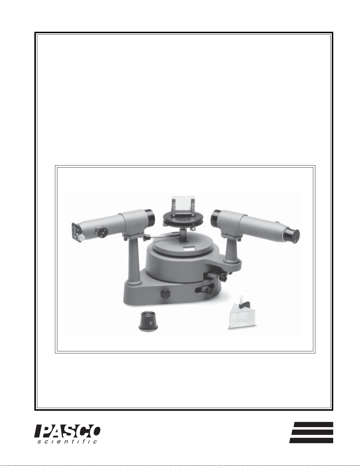
STUDENT
SPECTROMETER
012-02135F
10/03
Copyright © January 1991 $7.50
®
10101 Foothills Blvd. • P.O. Box 619011 • Roseville, CA 95678-9011 USA
Phone (916) 786-3800 • FAX (916) 786-8905 • email: techsupp@PASCO.com
better
ways to
teach science
Page 2

Model Name 012–0xxxxA
34
Page 3

012-02135F Spectrometer
T ab le of Contents
Section Page
Equipment Return .............................................................................................ii
Introduction ......................................................................................................1
Equipment ........................................................................................................2
Equipment Setup...............................................................................................3
Measuring Angles of Diffraction ....................................................................... 4
Using the Diffraction Grating ............................................................................5
Using the Prism.................................................................................................6
Maintenance .....................................................................................................8
Appendix: Using the Gaussian Eyepiece ...........................................................9
Technical Support ..................................................................................... back cover
®
i
Page 4
Spectrometer 012-02135F
Copyright, W arranty and Equipment Return
Please—Feel free to duplicate this manual
subject to the copyright restrictions below.
Copyright Notice
The PASCO scientific Model SP-9268A Student Spectrometer manual is copyrighted and all rights reserved.
However, permission is granted to non-profit educational
institutions for reproduction of any part of this manual
providing the reproductions are used only for their
laboratories and are not sold for profit. Reproduction
under any other circumstances, without the written
consent of PASCO scientific, is prohibited.
Limited Warranty
PASCO scientific warrants this product to be free from
defects in materials and workmanship for a period of one
year from the date of shipment to the customer. PASCO
will repair or replace, at its option, any part of the
product which is deemed to be defective in material or
workmanship. This warranty does not cover damage to
the product caused by abuse or improper use. Determination of whether a product failure is the result of a
manufacturing defect or improper use by the customer
shall be made solely by PASCO scientific. Responsibility for the return of equipment for warranty repair
belongs to the customer. Equipment must be properly
packed to prevent damage and shipped postage or freight
prepaid. (Damage caused by improper packing of the
equipment for return shipment will not be covered by the
warranty.) Shipping costs for returning the equipment,
after repair, will be paid by PASCO scientific.
Equipment Return
Should this product have to be returned to PASCO
scientific, for whatever reason, notify PASCO scientific
by letter or phone BEFORE returning the product. Upon
notification, the return authorization and shipping
instructions will be promptly issued.
NOTE: NO EQUIPMENT WILL BE
ACCEPTED FOR RETURN WITHOUT
AN AUTHORIZATION.
When returning equipment for repair, the units must be
packed properly. Carriers will not accept responsibility
for damage caused by improper packing. To be certain
the unit will not be damaged in shipment, observe the
following rules:
1. The carton must be strong enough for the item
shipped.
2. Make certain there is at least two inches of packing
material between any point on the apparatus and the
inside walls of the carton.
3. Make certain that the packing material can not shift
in the box, or become compressed, thus letting the
instrument come in contact with the edge of the box.
Address: PASCO scientific
10101 Foothills Blvd.
P.O. Box 619011
Roseville, CA 95678-9011
Phone: (916) 786-3800
FAX: (916) 786-8905
ii
®
Page 5
012-02135F Student Spectrometer
Introduction
In principle, a spectrometer is the simplest of scientific
instruments. Bend a beam of light with a prism or diffraction grating. If the beam is composed of more than
one color of light, a spectrum is formed, since the various colors are refracted or diffracted to different angles.
Carefully measure the angle to which each color of light
is bent. The result is a spectral "fingerprint," which carries a wealth of information about the substance from
which the light emanates.
In most cases, substances must be hot if they are to emit
light. But a spectrometer can also be used to investigate
cold substances. Pass white light, which contains all the
colors of the visible spectrum, through a cool gas. The
result is an absorption spectrum. All the colors of the visible spectrum are seen, except for certain colors that are
absorbed by the gas.
The importance of the spectrometer as a scientific instrument is based on a simple but crucial fact. Light is emitted or absorbed when an electron changes its orbit within
an individual atom. Because of this, the spectrometer is a
powerful tool for investigating the structure of atoms. It's
also a powerful tool for determining which atoms are
present in a substance. Chemists use it to determine the
constituents of molecules, and astronomers use it to determine the constituents of stars that are millions of light
years away.
In its simplest form, a spectrometer is nothing more than
a prism and a protractor. However, because of the need
for very sensitive detection and precise measurement, a
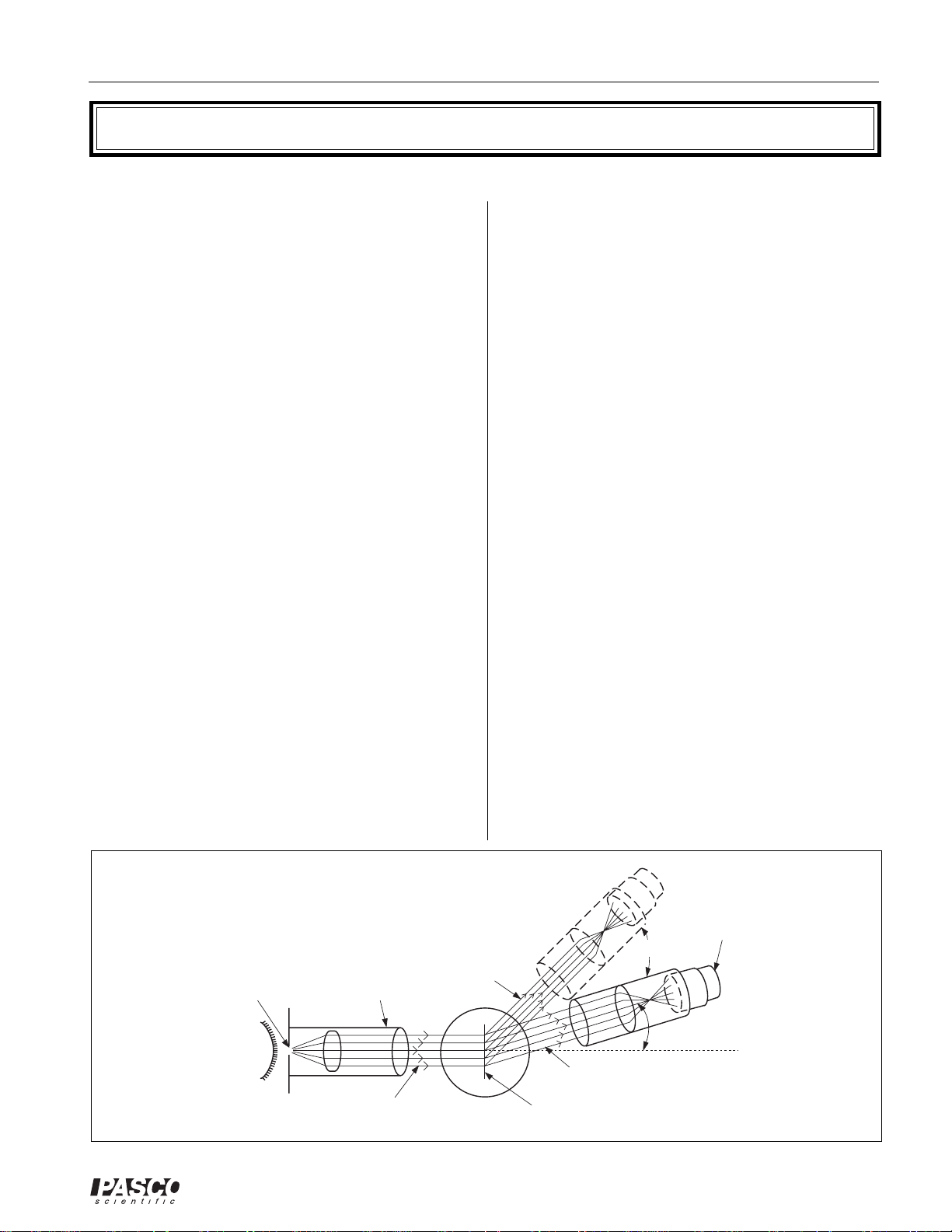
real spectrometer is a bit more complicated. As shown in
Figure 1, a spectrometer consists of three basic components; a collimator, a diffracting element, and a telescope.
The light to be analyzed enters the collimator through a
narrow slit positioned at the focal point of the collimator
lens. The light leaving the collimator is therefore a thin,
parallel beam, which ensures that all the light from the
slit strikes the diffracting element at the same angle of
incidence. This is necessary if a sharp image is to be
formed.
The diffracting element bends the beam of light. If the
beam is composed of many different colors, each color is
diffracted to a different angle.
The telescope can be rotated to collect the diffracted
light at very precisely measured angles. With the telescope focused at infinity and positioned at an angle to
collect the light of a particular color, a precise image of
the collimator slit can be seen. For example, when the
telescope is at one angle of rotation, the viewer might
see a red image of the slit, at another angle a green image, and so on. By rotating the telescope, the slit images
corresponding to each constituent color can be viewed
and the angle of diffraction for each image can be measured. If the characteristics of the diffracting element are
known, these measured angles can be used to determine
the wavelengths that are present in the light.
SOURCE
®
COLLIMATOR
SLIT
LIGHT
RED LIGHT
COLLIMATOR
PARALLEL BEAM
Figure 1 Spectrometer Diagram
DIFFRACTION GRATING
1
GREEN LIGHT
(OR PRISM)
TELESCOPE
ANGLE OF
DIFFRACTION
EYE PIECE
Page 6

Student Spectrometer 012-02135F
Equipment
The PASCO scientific Model SP-9268A Student Spectrometer provides precise spectroscopic measurements
using either a prism or a diffraction grating as the diffracting element. The spectrometer includes the following equipment (see Fig 2).
Collimator and Telescope
Both the collimator and the telescope have 178 mm focal length, achromatic objectives, and clear apertures
with 32 mm diameters. The telescope has a 15X
Ramsden eyepiece with a glass, cross-hair graticule. The
collimator is fitted with a 6 mm long slit of adjustable
width. Both the collimator and the telescope can be leveled. They can also be realigned (though this is rarely
necessary) so that their optical axes are square to the axis
of rotation.
Rotating Bases
The telescope and the spectrometer table are mounted on
independently rotating bases. Vernier scales provide
measurements of the relative positions of these bases to
within one minute of arc. The rotation of each base is
controlled with a lock-screw and fine adjust knob. With
the lock-screw released, the base is easily rotated by
hand. With the lock-screw tight, the fine adjust knob
can be used for more precise positioning.
Diffraction grating and
Mounting clamp
Slit plate
Collimator
Spectrometer table
Spectrometer Table
The spectrometer table is fixed to its rotating base with a
thumbscrew, so table height is adjustable. Three leveling screws on the underside of the table are used to adjust the optical alignment. (The table must be level with
respect to the optical axes of the collimator and telescope if the diffracting element is to retain its alignment
for all positions of the telescope.) Thumbscrews are
used to attach the prism clamp and the grating mount to
the table, and reference lines are etched in the table for
easy alignment.
Accessories
Accessories for the spectrometer include a dense flint
prism and two mounting clamps; a 300 line/mm diffraction grating and mounting clamp; two thumbscrews for
attaching the mounting clamps to the spectrometer table;
a magnifying glass for reading the vernier; three Allen
keys for leveling the telescope and collimator; and a polished hardwood case.
NOTE: A 600 line/mm diffraction grating is available from PASCO as an optional accessory.
Optional Equipment: Gaussian Eyepiece
The Gaussian eyepiece (SP-9285) is an optional component that simplifies the task of focusing and aligning the
spectrometer and aligning the diffraction grating. Its use
is described in the Appendix.
Slit width
adjust screw
Spectrometer
table base
Telescope
for reading Vernier
Focus knob
base
Magnifying glass
Table rotation:
Lock screw
Fine adjust knob
Focus knob
Eyepiece
Graticule lock
ring
Telescope
Telescope rotation:
Fine adjust knob
Vernier scale
Lock screw
Prism and
Mounting clamp
2
Figure 2
The Spectrometer
®
Page 7

012-02135F Student Spectrometer
Equipment Setup
NOTE: If you are using the optional Gaussian
Eyepiece (SP-9285), equipment setup is somewhat
simpler than described below. See the Appendix
for instructions.
Leveling the Spectrometer
For accurate results, the diffracting element must be
properly aligned with the optical axes of the telescope
and collimator. This requires that both the spectrometer
and the spectrometer table be level.
1. Place the spectrometer on a flat surface. If necessary
use paper or 3 X 5 cards to shim beneath the wood
base until the fixed-base of the spectrometer is level.
2. Level the spectrometer table by adjusting the three
thumbscrews on the underside of the table.
Focusing the Spectrometer
1. While looking through the telescope, slide the eye-
piece in and out until the cross-hairs come into sharp
focus. Loosen the graticule lock ring, and rotate the
graticule until one of the cross-hairs is vertical. Retighten the lock ring and then refocus if necessary.
2. Focus the telescope at infinity. This is best accom-
plished by focusing on a distant object (e.g.; out the
window).
3. Check that the collimator slit is partially open (use
the slit width adjust screw).
5. Looking through the telescope, adjust the focus of
the collimator and, if necessary, the rotation of the
telescope until the slit comes into sharp focus. Do not
change the focus of the telescope.
6. Tighten the telescope rotation lock-screw, then use
the fine adjust knob to align the vertical line of the
graticule with the fixed edge of the slit. If the slit is
not vertical, loosen the slit lock ring, realign the slit,
and retighten the lock ring. Adjust the slit width for a
clear, bright image. Measurements of the diffraction
angle are always made with the graticule line aligned
along the fixed edge of the slit, so a very narrow slit
is not necessarily advantageous.
NOTE: When the telescope and collimator are
properly aligned and focused, the slit should be
sharply focused in the center of the field of view of
the telescope, and one cross-hair should be perpendicular and aligned with the fixed edge of the slit.
If proper alignment cannot be achieved with the
adjustments just described, you will need to realign the spectrometer as follows.
Realigning the Spectrometer
Under normal circumstances, the spectrometer will maintain its alignment indefinitely. However, if the spectrometer can not be properly focused, as described above, it
may be necessary to adjust the optical axes of the collimator and telescope, as follows:
4. Align the telescope directly opposite the collimator
as shown in Figure 3.
TELESCOPE
Figure 3 Align the Telescope directly opposite
the Collimator
¨
COLLIMATOR
1. The telescope and collimator pivot about a fulcrum
on their respective mounting pillars (See Fig 4). Use
the aluminum rod provided with the accessory equipment to adjust the leveling screws. Loosen one as the
other is tightened until the unit is level and secure.
FULCRUM
LEVELING
SCREWS
MOUNTING
PILLAR
Figure 4 Leveling the Telescope and Collimator
3
Page 8

Student Spectrometer 012-02135F
2. The mounting pillars of the telescope and collimator
can be rotated by using an Allen wrench to loosen the
screws that attach the pillars to their respective bases.
To loosen the screw for the collimator, the spectrometer must be removed from the wood base.
Measuring Angles of Diffraction
When analyzing a light source, angles of diffraction are
measured using the vernier scales. However, the scales
only measure the relative rotational positions of the telescope and the spectrometer table base. Therefore, before
making a measurement, it's important to establish a vernier reading for the undeflected beam. All angles of diffraction are then made with respect to that initial reading
(see Fig 5).
To obtain a vernier reading for the undeflected beam,
first align the vertical cross-hair with the fixed edge of
the slit image for the undeflected beam. Then read the
θθ
vernier scale. This is the zero point reading (
=
VERNIER READING FOR
q
DIFFRACTED BEAM
ANGLE OF
DIFFRACTION
=
q
q
0
=
q
VERNIER
0
READING FOR
UNDIFFRACTED
BEAM
VERNIER SCALES
).
θ
θθ
0
LIGHT
SOURCE
3. To be sure both optical units are square to the axis of
rotation, follow the focusing procedure described
above, adjusting the mounting pillars as necessary so
the slit image is well centered in the viewing field of
the telescope.
Reading the Vernier Scales
To read the angle, first find
where the zero point of the
vernier scale aligns with
the degree plate and record
the value. If the zero point
is between two lines, use
the smaller value. In Figure 6, below, the zero
point on the vernier scale is between the 155 ° and 155 °
30' marks on the degree plate, so the recorded value is
155 °.
Now use the magnifying glass to find the line on the vernier scale that aligns most closely with any line on the
degree scale. In the figure, this is the line corresponding
to a measurement of 15 minutes of arc. Add this value to
the reading recorded above to get the correct measurement to within 1 minute of arc: that is, 155 ° + 15' = 155 °
15'.
Figure 5 Measuring an Angle of Diffraction
Now rotate the telescope to align the vertical cross-hair
with the fixed edge of a deflected image. Read the vernier scale again. If this second reading is
θθ
θθ
tual angle of diffraction is
. If the table base is ro-
θ –
θ
θθ
θθ
0
θθ
θ, then the ac-
θθ
tated for some reason, the zero point changes, and must
be remeasured.
VER I'
30
I70
Figure 6 Reading the Vernier Scales
4
20
I0
I60
155° + 15' = 155° 15'
0
I5
155° (on the degree scale)15' (on the vernier scale)
¨
Page 9

012-02135F Student Spectrometer
Using the Diffraction Grating
IMPORTANT: The Diffraction Grating is a delicate component. Be careful not to scratch the surface and always replace it in the protective foam
wrapping when it is not being used.
Aligning the Grating
To accurately calculate wavelengths on the basis of diffraction angles, the grating must be perpendicular to the
beam of light from the collimator.
1. Align and focus the spectrometer as described earlier.
The telescope must be directly opposite the collimator with the slit in sharp focus and aligned with the
vertical cross-hair.
TABLE ROTATION
SPECTROMETER TABLE
LOCK-SCREW
GRATING AND MOUNT
LOCK-SCREW
SPECTROMETER
TABLE BASE
ª 1 cm
LIGHT
SOURCE
5. Place a light source (preferably one with a discrete
spectrum, such as a mercury or sodium lamp) approximately one centimeter from the slit. Adjust the
slit width so the slit image is bright and sharp. If necessary, adjust the height of the spectrometer table so
the slit image is centered in the field of view of the
telescope.
IMPORTANT: Stray light can obscure the images. Use the spectrometer in a semi-darkened
room or drape a sheet of opaque material over the
spectrometer.
TABLE ROTATI ON FIN E
ANGLE OF
DIFFRACTION
ZERO
DIFFRACTION
ANGLE OF
DIFFRACTION
ADJUST KNOB
VERTICAL CROSS-HAIR
SLIT IMAGE
ª 1 cm
LIGHT
SOURCE
30
10
20
10
0
0
VERNIER
SCALES
0
180
10
20
190
30
Figure 7
Perform steps 2-5 with reference to Figure 7.
2. Loosen the spectrometer table lock-screw. Align the
engraved line on the spectrometer table so that it is,
as nearly as possible, colinear with the optical axes of
the telescope and the collimator. Tighten the lockscrew.
3. Using the thumbscrews, attach the grating mount so
it is perpendicular to the engraved lines.
4. Insert the diffraction grating into the clips of the
mount. To check the orientation of the grating, look
through the grating at a light source and notice how
the grating disperses the light into its various color
components. When placed in the grating mount, the
grating should spread the colors of the incident light
horizontally, so rotation of the telescope will allow
you to see the different colored images of the slit.
VIEW THROUGH
TELESCOPE
Figure 8
Perform steps 6-9 with reference to Figure 8.
6. Rotate the telescope to find a bright slit image. Align
the vertical cross-hair with the fixed edge of the image and carefully measure the angle of diffraction.
(See the previous section, Measuring Angles of Dif-
fraction.)
7. The diffraction grating diffracts the incident light into
identical spectra on either side of the line of the undiffracted beam. Rotate the telescope back, past the
zero diffraction angle, to find the corresponding slit
image. Measure the angle of diffraction for this image.
8. If the grating is perfectly aligned, the diffraction
angles for corresponding slit images will be identical.
If not, use the table rotation fine adjust knob to compensate for the difference (i.e.; to align the grating
perpendicular to the collimator beam so the two
angles will be equal).
¨
5
Page 10

Student Spectrometer 012-02135F
sin
n =
{ }
A+D
2
sin
A
2
9. Repeat steps 6-8 until the angles for the corresponding slit images are the same to within one minute of
arc.
Making the Reading
Once the grating is aligned, do not rotate the rotating
table or its base again. Diffraction angles are measured
as described in the previous section, Measuring Angles
of Diffraction. (Since the vernier scales were moved
when the spectrometer table was adjusted, the point of
zero diffraction must be remeasured).
Using the Prism
Advantages and Disadvantages
A prism can also be used as the diffracting element in a
spectrometer since the index of refraction of the prism
(and therefore the angle of refraction of the light) varies
slightly depending on the wavelength of the light.
A prism refracts the light into a single spectrum, whereas
the grating divides the available light into several spectra. Because of this, slit images formed using a prism are
generally brighter than those formed using a grating.
Spectral lines that are too dim to be seen with a grating
can often be seen using a prism.
Wavelengths are determined according to the formula:
a sin q
l =
n
where λ is the wavelength; a is the distance between
lines on the diffraction grating
(a = 3.3 x 10⁻³ mm for the 300 line/mm grating or
-3
1.66 x 10
mm for the optional 600 line/mm grating);
θ is the angle of diffraction; and n is the order of the diffraction spectrum under observation.
The Angle of Minimum Deviation
The angle of deviation for light traversing a prism is
shown in Figure 9. For a given wavelength of light traversing a given prism, there is a characteristic angle of
incidence for which the angle of deviation is a minimum.
This angle depends only on the index of refraction of the
prism and the angle (labeled A in Figure 8) between the
two sides of the prism traversed by the light. The relationship between these variables is given by the equation:
Unfortunately, the increased brightness of the spectral
lines is offset by a decreased resolution, since the prism
doesn't separate the different lines as effectively as the
grating. However, the brighter lines allow a narrow slit
width to be used, which partially compensates for the
reduced resolution.
With a prism, the angle of refraction is not directly proportional to the wavelength of the light. Therefore, to
measure wavelengths using a prism, a graph of wavelength versus angle of refraction must be constructed using a light source with a known spectrum. The wavelength of unknown spectral lines can then be interpolated from the graph.
Once a calibration graph is created for the prism, future
wavelength determinations are valid only if they are
made with the prism aligned precisely as it was when the
graph was produced. To ensure that this alignment can
be reproduced, all measurements are made with the
prism aligned so that the light is refracted at the angle of
minimum deviation.
where n is the index of refraction of the prism; A is the
angle between the sides of the prism traversed by the light;
and D is the angle of minimum deviation. Since n varies
with wavelength, the angle of minimum deviation also varies, but it is constant for any particular wavelength.
UNDEFLECTED
6
ANGLE OF
DEVIATION
RAY
DEFLECTED
RAY
Figure 9 Angle of Deviation
ANGLE A
LIGHT
SOURCE
¨
Page 11

012-02135F Student Spectrometer
To Measure the Angle of Minimum
Deviation:
1.
Align and focus the spectrometer as described earlier.
2. Use the two thumbscrews to attach the prism clamp
to the spectrometer table and clamp the prism in
place as shown in Figure 10.
3. Place the light source a few centimeters behind the
slit of the collimator. (It may be helpful to partially
darken the room, but when using the prism this is often not necessary.)
PRISM CLAMP
LIGHT
SOURCE
PRISM
Figure 10 Mounting the Prism
4. With the prism, it is generally possible to see the re-
fracted light with the naked eye. Locate the general
direction to which the light is refracted, then align the
telescope and spectrometer table base so the slit image can be viewed through the telescope.
5. While looking through the telescope, rotate the spec-
trometer table slightly back and forth. Notice that the
angle of refraction for the spectral line under observation changes. Rotate the spectrometer table until this
angle is a minimum, then rotate the telescope to align
the vertical cross-hair with the fixed edge of the slit
image. Use the fine adjust knobs to make these adjustments as precisely as possible, then measure the
telescope angle using the vernier scale.
6. Without changing the rotation of the spectrometer
table, remove the prism and rotate the telescope to
align the cross-hair with the fixed edge of the
undiffracted beam. Measure the angle on the vernier
scale. The difference between this angle and that recorded for the diffracted spectral line in step 5, is the
angle of minimum deviation. Notice that, since the
determination of the angle of minimum deviation for
each spectral line requires rotational adjustments of
the spectrometer table, the angle of the undeflected
beam must be remeasured for each line.
¨
7
Page 12

Student Spectrometer 012-02135F
Maintenance
Periodically clean the telescope aperture, the collimator
aperture, and the prism with a nonabrasive lens paper
(available at any camera store). No other regular maintenance is required.
IMPORTANT: Always handle the spectrometer
and its accessories with care to avoid scratching
the optical surfaces and throwing off the alignment. Also, when not in use, the spectrometer
should be stored in its hardwood case.
8
¨
Page 13

012-02135F Student Spectrometer
Appendix: Using the Gaussian Eyepiece
The optional Gaussian eyepiece (Model SP-9285) simplifies the task of aligning and focusing the spectrometer
and aligning the diffraction grating. One Gaussian eyepiece can be used to align and focus any number of spectrometers, so only one is generally needed per lab.
5. Looking through the telescope, rotate the table until a
patch of light is reflected back through the telescope
from the glass surfaces of the grating. The spectrometer table and the telescope must be at least
roughly level to achieve this reflection. If they are
not, see Realigning the Spectrometer, earlier in the
manual.
6. Adjust the focus of the telescope until the cross-hairs
and their reflected images are in sharp focus. The
glass slides of the grating are not efficient reflectors,
so you must look carefully to see them.
IMPORTANT: The grating is sandwiched between two glass slides so, depending on how parallel the slides are, you may see as many as four
reflected images of the cross-hairs. In the following steps, you will be instructed to superimpose
the graticule with its reflected image. If there is
more than one image, just center the cross-hairs as
accurately as possible between the images.
7. Use the table rotation fine adjust knob to align the
vertical cross-hair with its reflected image.
To Align and Focus the Spectrometer Using
the Gaussian Eyepiece:
1. Remove the telescope eyepiece and replace it with
the Gaussian eyepiece.
2. While looking through the telescope, slide the eye-
piece in and out until the cross-hairs come into sharp
focus. Loosen the graticule lock ring, and rotate the
graticule until one of the cross-hairs is vertical. Retighten the lock ring and then refocus if necessary.
3. Plug in the power supply of the Gaussian eyepiece.
The light from the eyepiece is reflected along the optical axis of the telescope by a half-silvered mirror.
Looking through the eyepiece, you'll see the crosshairs lighted up as they scatter some of the light back
into the eyepiece.
4. Mount the grating holder to the spectrometer table
and insert the diffraction grating.
8. Adjust the spectrometer table leveling screws until
the cross-hairs are superimposed on the reflected image.
9. Rotate the spectrometer table 180 ° and, using the
table rotation fine adjust knob, align the vertical
cross-hair with the reflected image.
10. Adjust the table leveling screws to remove half the
separation between the horizontal cross-hair and the
reflected image. Adjust the telescope leveling
screws to remove the remaining error, so the crosshairs and their reflected images are superimposed.
1
1. Repeat steps 9 and 10 until the cross-hairs and their
reflected images are superimposed from both sides
of the diffraction grating.
12. Unplug the Gaussian eyepiece. Adjust the slit of the
collimator so it is open and vertical.
¨
9
Page 14

Student Spectrometer 012-02135F
13. Illuminate the slit with an external light source. Ro-
tate the telescope directly opposite the collimator
and focus the collimator only (do not disturb the
telescope focus) until the illuminated slit is in sharp
focus. If the collimator slit is not vertical, loosen the
lock ring, align the slit vertically, and then retighten
the lock ring. Then align the fixed edge of the slit
with the vertical cross-hair.
14. Adjust the collimator leveling screws until the slit is
vertically centered in the field of view of the telescope. (As with the telescope, you may need to adjust the collimator so that its optical axis is square to
the axis of rotation.) The telescope, collimator, and
spectrometer table are now properly aligned.
15. If you are going to use the grating, plug the Gaussian
eyepiece back in and rotate the spectrometer table
until the vertical cross-hair is again aligned with its
reflected image. This insures that the grating is perpendicular to the optical axis of the spectrometer.
16. If you wish, you may replace the Gaussian eyepiece
with the original eyepiece. The focus of the telescope will be maintained if you slide in the original
eyepiece until the cross-hairs are in sharp focus.
Alignment Error
The multiple reflections from the glass slides of the grating introduce some error into the alignment procedure.
Normally, centering the cross-hairs between the reflected
images will reduce the error below the 1-minute resolution that is obtainable when reading the vernier scales.
To verify the alignment, use a light source with discrete
spectral lines such as a sodium or mercury vapor lamp.
If the alignment is correct, corresponding spectral lines
on opposite sides of the optical axis will have equal
angles of diffraction. If necessary, adjust the rotation of
the spectrometer table until the measurements are the
same.
10
¨
Page 15

012-02135F Student Spectrometer
Technical Support
FeedBack
If you have any comments about this product or this
manual please let us know. If you have any suggestions
on alternate experiments or find a problem in the manual
please tell us. PASCO appreciates any customer feedback. Your input helps us evaluate and improve our
product.
To Reach PASCO
For Technical Support call us at 1-800-772-8700 (tollfree within the U.S.) or (916) 786-3800.
email: techsupp@PASCO.com
Tech support fax: (916) 786-3292
WEB: http://www.pasco.com
Contacting Technical Support
Before you call the PASCO Technical Support staff it
would be helpful to prepare the following information:
• If your problem is computer/software related, note:
Title and Revision Date of software.
Type of Computer (Make, Model, Speed).
Type of external Cables/Peripherals.
• If your problem is with the PASCO apparatus, note:
Title and Model number (usually listed on the label).
Approximate age of apparatus.
A detailed description of the problem/sequence of
events. (In case you can't call PASCO right away, you
won't lose valuable data.)
If possible, have the apparatus within reach when calling. This makes descriptions of individual parts much
easier.
• If your problem relates to the instruction manual, note:
Part number and Revision (listed by month and year on
the front cover).
Have the manual at hand to discuss your questions.
¨
11
Page 16

Model Name 012–0xxxxA
34
 Loading...