

Page 1

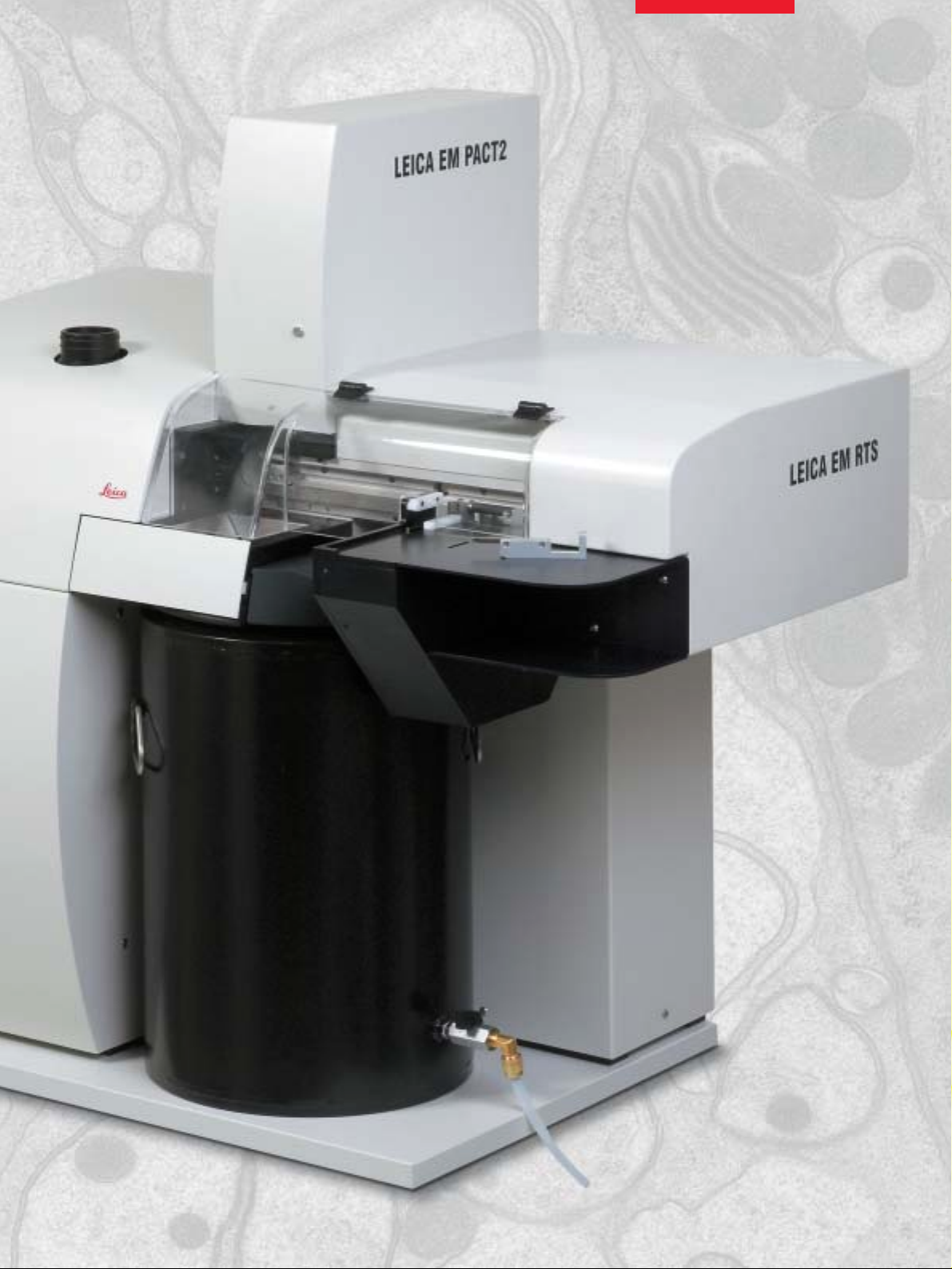
Leica EM PACT2
High Pressure Freezer
Leica EM RTS
Rapid Transfer System
Page 2
High Pressure Freezing for Everybody
2
The Leica EM PACT2 high pressure freezer serves the needs of
molecular and cell biologists and all researchers who want an “in
vivo” impression of their cellular structures and functions in question – without the artefacts of chemical fixation but with the
high resolution information of EM immunocytochemistry, frozen
hydrated sections and freeze fracturing.
The Rapid Transfer System EM RTS allows correlative LM/EM
experiments, taking a live specimen from a light microscope (e.g.
a confocal microscope) to freezing in less than 5 seconds. In the
same way, time resolved experiments are possible. Safety and
reproducibility for the specimen are increased while operator
mistakes are reduced.
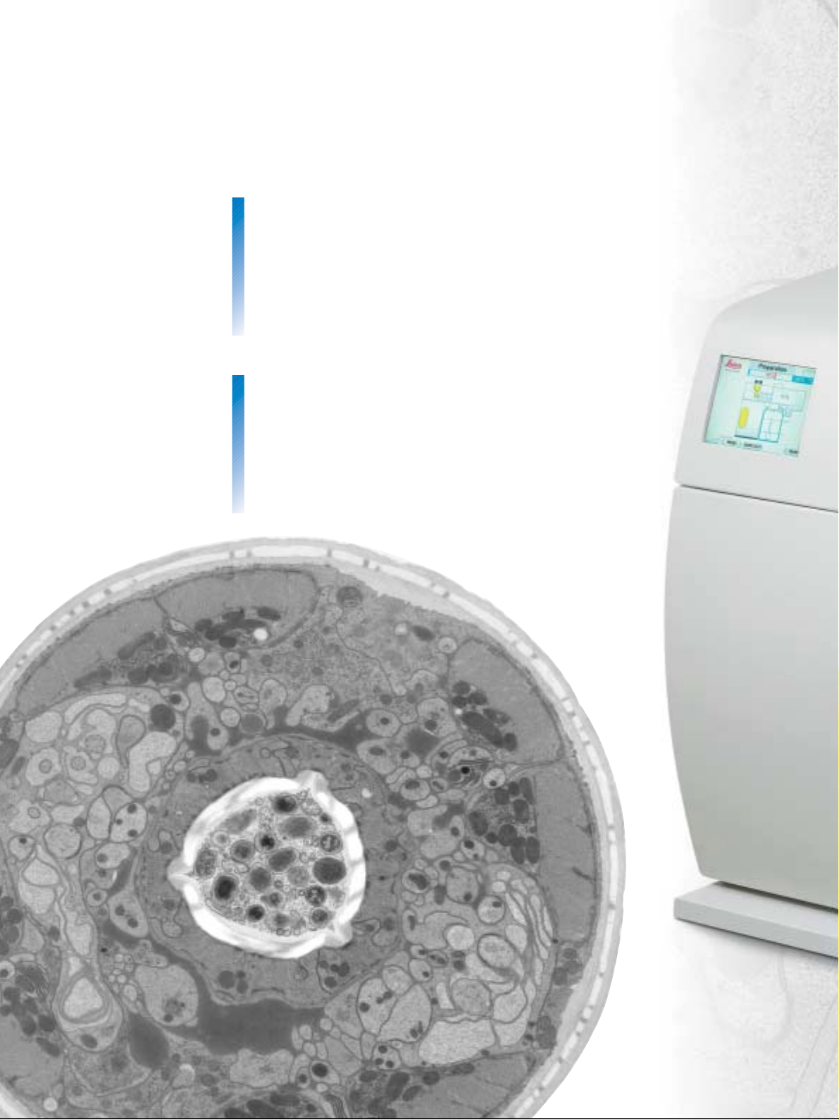
Structural details of the C. elegans
head in cross-section.
T. Müller-Reichert (MPI-CBG, Dresden,
Germany) and Kent McDonald
(University of California, Berkeley, USA)
Page 3
3
Leica Design by W. Hölbl
Page 4

Leica EM PACT2 –
High Pressure Freezer
Function: All You Need is LN2…
The specimen is locked (1) and set under pressure (2) just before
freezing (3) via the synchronization mechanism (4). Only the tissue
in the specimen holder is under pressure and not the complete
specimen chamber. As any fluid can be used in the universal
hydraulic system, toxic vapors (e.g. ethanol) are avoided.
Perfect results
– High cooling rates by strong jet of LN
2
– 120 samples can be frozen per hour
– Variety of specimen carriers available for all purposes
Easy to install
– Compact and mobile
– Standard electrical supply
– Compressor on EM PACT2 trolley
Easy to use
– Touch-sensitive color screen, menu prompts
– Bayonet loading device for automatic orientation
– Specimen ejection into LN2bath
– LED bath illumination
– 600 ms temperature/pressure curve displayed for each run
– Internal memory for 8 000 freezing runs
– Data download on memory stick
Safe and convenient
– Low LN2consumption
– Dewar with drain outlet
– Universal hydraulic system – can even use water
– Low noise
– Maintenance free “long life pods”
4
Page 5

5
Perfect results
– Specimen carrier loading in less than 5 secs
– Correlative LM/EM
– Rapid biopsy process
Easy to install
– Factory mounted on EM PACT2
– Control via touch sensitive screen
Easy to use
– Rapid loader for inserting specimen
– Automatic and reproducible specimen freezing
Safe and convenient
– Outlet Dewar
– Safety lid
– Low noise
– Funnel for filling LN
2
– Platform for cryopreparation, box, position for accessories
Leica EM RTS –
Rapid Transfer System
Schematic drawing showing the status of all parameters
Freezing data after ejection of the sample
Page 6

Specimen loading made simple
with the Rapid Transfer System:
insert specimen carrier into rapid
loader. Automatically freeze by
inserting rapid loader into RTS.
Prepare your sample (eg leaf or a
cell monolayer on a sapphire disc) …
… to fit into one of the various
specimen carriers (eg flat carrier
ø 1.2 mm, depth 200 µm or membrane carrier: ø 1.5 mm, depth
200 µm). Specimen and carrier are
held in the rapid loader. The long
life pod is connected to the loading
device with a bayonet lock.
Specimen and carrier are held in
the rapid loader.
The long life pod is connected to
the loading device with a bayonet
lock.
The primed loading device then
waits for freezing in the RTS.
By gently pushing the rapid loader
into the RTS the carrier is tightened
securely and then frozen automatically in less than 2.5 seconds.
After cryofixation the carriers are
collected in the LN2bath of the EM
PACT2 before freeze substitution in
the Leica EM AFS2.
Leica EM RTS
Application Solutions
6
Page 7

7
The Tube Holder for every fluid you
can think of … blood, milk, cell suspensions ... nematodes – and more!
Directly suck the suspension into
copper tubes (inner ø 350 µm)
already mounted in their holder.
Alternatively, take up sample into
cellulose microcapillary.
With the loading device the
specimen can be inserted into
the EM PACT.
After cryofixation the samples are
stored under LN2in the transfer
box.
Sample Punch: punching out of the
central part of the specimen tube
ready for Frozen Hydrated Sectioning
or ...
... peeling away of the top of the
copper tube for Freeze Substitution.
Storage of the copper tubes is
carried out under LN2.
The specimen tube holders are
recycled by reloading them with
copper tubes.
Leica Design by W. Hölbl
Page 8

8
Taking microbiopsies for EM is now
faster than ever before!
Prepare the Microbiopsy Transfer
Station for RTS under the Optical
Workstation…
… before taking a biopsy with
the biopsy gun.
Insert the biopsy gun into the
transfer station …
… and transfer the tissue into
the biopsy carrier.
The specimen and carrier are held
in the rapid loader …
… while the primed loading device
waits for freezing in the RTS.
By gently pushing the rapid loader
into the RTS the carrier is tightened
securely and then frozen automatically in less than 2.5 seconds.
After cryofixation the carriers are
collected in the LN2bath of the
EM PACT2 before freeze substitution in the Leica EM AFS2 or
cryosectioning in the EM FC6.
Leica EM RTS
Application Solutions
Page 9

9
Frozen Hydrated Sectioning
made easy.
Alternatively, the carriers can be
trimmed with a cutter ...
... for Frozen Hydrated Sectioning
in the cryoultramicrotome.
Preparation for follow-on
procedures.
All necessary steps for follow-on
procedures (eg. Freeze Substitution) can be conveniently performed in the cryopreparation box
supplied with the Leica EM PACT2.
The Freeze Fracture Holder for
everything that can be fractured ...
Prepare the Freeze Fracture Station
under the Optical Workstation ...
... and preload the freeze fracture
carrier. A copper ring is put on top
of the loaded carrier.
The carrier and copper ring are
sandwiched securely in the pod
with the supplied torque wrench.
With the loading device the
specimen can be inserted into
the instrument.
After cryofixation the carriers are
collected in the LN
2
bath of the
EM PACT2 before Freeze Fracturing.
Accessories are also available for loading EM PACT2 without EM RTS.
Page 10

10
Mouse embryonic fibroblast
grown on sapphire disc.
Soazig Lelay, Jana Maentler and
Paul Verkade, MPI-CBG, Dresden,
Germany
Page 11

11
Yeast frozen in the membrane carrier. Scale bar = 500 nm, 250 nm for insert.
Courtesy of Mark van Breugel, Jana Maentler and Paul Verkade, MPI-CBG, Dresden, Germany.
Insulin granules producing INS-1 cells. Scale bar = 5 µm.
Courtesy of Joke Ouwendijk, Jana Maentler, Melanie Jäger, Michele Solimena and Paul Verkade,
TUD and MPI-CBG, Dresden, Germany
Rat, Langerhans cells. Scale bar = 1 µm Courtesy of Joke Ouwendijk, Jana Maentler,
Melanie Jäger, Michele Solimena and Paul Verkade, TUD and MPI-CBG, Dresden, Germany
Page 12

@
www.em-preparation.com
Leica Mikrosysteme GmbH
Hernalser Hauptstrasse 219
A-1170 Vienna Austria
Tel. +43 1 48899-235
Fax +43 1 48899-350
Leica Microsystems –
the brand for outstanding products
Copyright
©
Leica Mikrosysteme GmbH
•
Vienna Austria
•
LEICA and the Leica Logo are registered trademarks of Leica IR GmbH.
Leica EM PACT2_EM RTS - E - 6/05 Order No 206002, Printed on chlorine-free bleached paper. Printed in Austria.
Leica Microsystems’ mission is to be the world’s first-choice provider of innovative
solutions to our customers’ needs for vision, measurement, lithography and analysis
of microstructures.
Leica, the leading brand for microscopes and scientific instruments, developedfrom
five brand names, all with a long tradition: Wild, Leitz, Reichert, Jung and Cambridge
Instruments. Yet Leica symbolizes innovation as well as tradition.
Leica Microsystems – an international company
with a strong network of customer services
Australia: Gladesville Tel. +61 2 9879 9700 Fax +61 2 9817 8358
Austria: Vienna Tel. +43 1 486 80 50 0 Fax +43 1 486 80 50 30
Canada: Richmond Hill/Ontario Tel. +1 905 762 2000 Fax +1 905 762 8937
Denmark: Herlev Tel. +45 4454 0101 Fax +45 4454 0111
France: Rueil-Malmaison Tel. +33 1 473 285 85 Fax +33 1 473 285 86
Germany: Bensheim Tel. +49 6251 136 0 Fax +49 6251 136 155
Italy: Milan Tel. +39 0257 486.1 Fax +39 0257 40 3273
Japan: Tokyo Tel. +81 3 5435 9600 Fax +81 3 5435 9615
Korea: Seoul Tel. +82 2 514 65 43 Fax +82 2 514 65 48
Netherlands: Rijswijk Tel. +31 70 4132 100 Fax +31 70 4132 109
People’s Rep. of China: Hong Kong Tel. +852 2564 6699 Fax +852 2564 4163
Portugal: Lisbon Tel. +351 21 388 9112 Fax +351 21 385 4668
Singapore Tel. +65 6779 7823 Fax +65 6773 0628
Spain: Barcelona Tel. +34 93 494 95 30 Fax +34 93 494 95 32
Sweden: Sollentuna Tel. +46 8 625 45 45 Fax +46 8 625 45 10
Switzerland: Glattbrugg Tel. +41 1 809 34 34 Fax +41 1 809 34 44
United Kingdom: Milton Keynes Tel. +44 1908 246 246 Fax +44 1908 609 992
USA: Bannockburn/lllinois Tel. +1 847 405 0123 Fax +1 847 405 0164
and representatives of Leica Microsystems
in more than 100 countries.
The companies of the Leica Microsystems
Group operate internationally in four business
segments, where we rank with the market
leaders.
•
Microscopy Systems
Our expertise in microscopy is the basis for all
our solutions for visualization, measurement
and analysis of microstructures in life sciences
and industry. With confocal laser technology
and image analysis systems, we provide threedimensional viewing facilities and offer new
solutions for cytogenetics, pathology and materials sciences.
•
Specimen Preparation
We provide comprehensive systems and services for clinical histo- and cytopathology
applications, biomedical research and industrial quality assurance. Our product range
includes instruments, systems and consumables for tissue infiltration and embedding,
microtomes and cryostats as well as automated stainers and coverslippers.
•
Medical Equipment
Innovative technologies in our surgical microscopes offer new therapeutic approaches in
microsurgery.
•
Semiconductor Equipment
Our automated, leading-edge measurement and
inspection systems and our E-beam lithography
systems make us the first choice supplier for
semiconductor manufacturers all over the world.
 Loading...