

Page 1

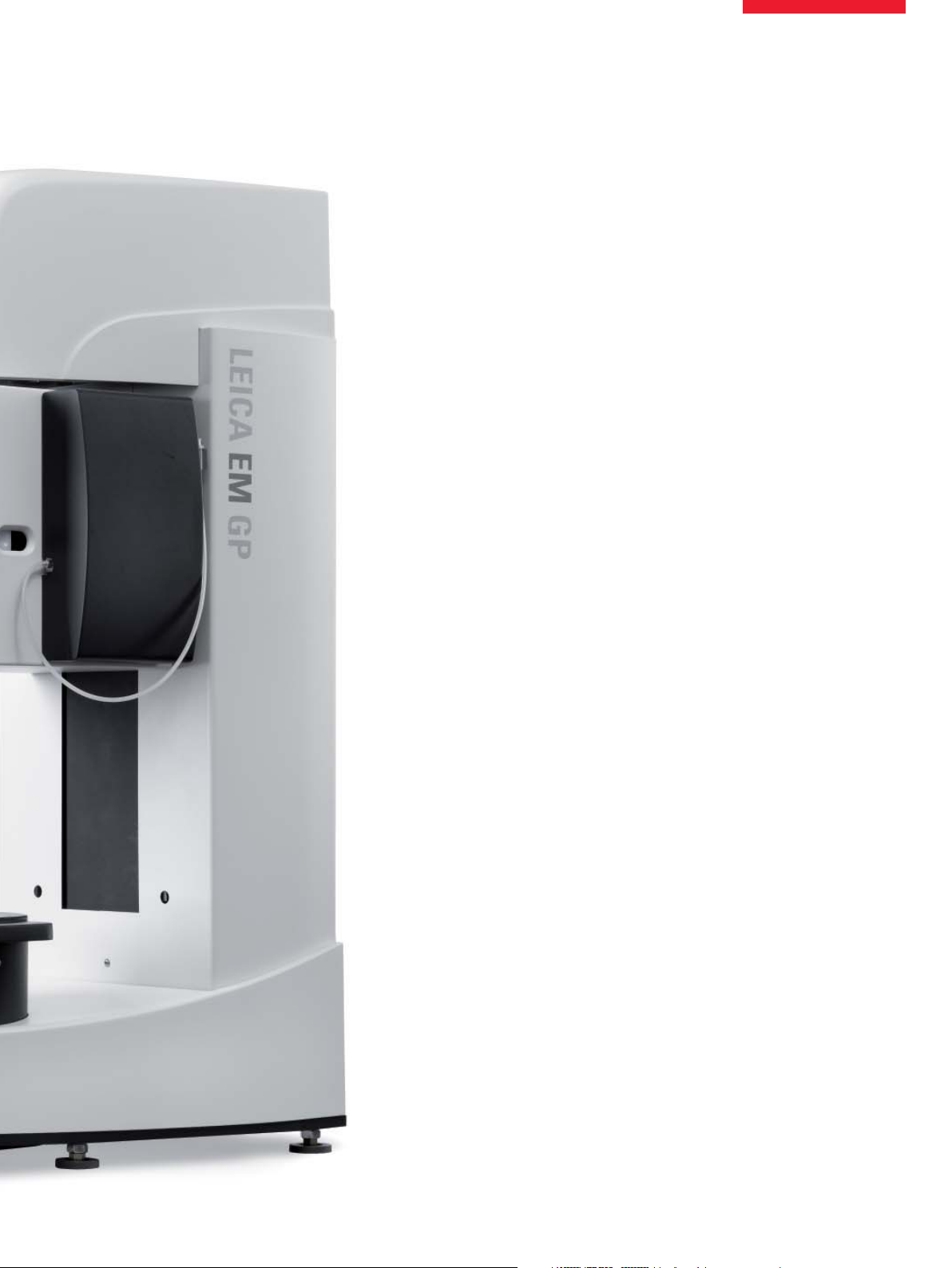
Leica EM GP
Automatic Plunge Freezer for the Bare Grid Technique
Page 2
The Bare Grid Technique
Many specimens for cryo-TEM can be prepared by immersion
freezing, where a fl uid sample is pipetted onto an EM grid (usually
coated) and the excess removed until a thin fi lm remains, before
plunging into a cryogen such as liquid ethane. The grid can then
be directly transferred under cryo conditions to the cryo electron
microscope (cryo-TEM) for observation. This is the bare grid technique.
The bare grid technique can be used for many types of sample
ranging from biological cell sub units to industrial emulsions.
Imaging macromolecular assemblies, viruses and cells in their
native, hydrated environment in the cryo-TEM is the state-of-theart technique in electron microscopy, providing maximum resolution with minimal specimen damage.
Although a simple method, it is imperative that the suspension
thickness on the grid can be reproduced and vitreous ice can be
formed, otherwise much time is wasted loading useless samples
into the cryo-TEM. The sample fi lm is only tens to hundreds of
nanometers thick and so can be easily infl uenced by temperature
shifts and humidity prior to freezing. If the humidity is too low then
the fi lm breaks due to it quickly drying. If the sample is adversely
affected by temperature, then the morphology may change before
freezing.
Leica Microsystems has developed a plunge freezer, in conjunction with Dr. Guenter Resch of the IMP/IMBA Electron Microscopy
Facility in Vienna, Austria, to standardize procedures and make
the bare grid technique more reproducible.
2
Page 3
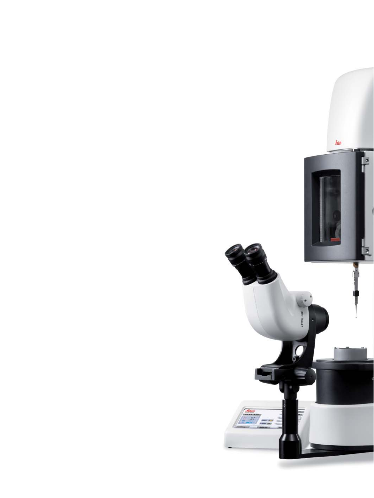
Leica EM GP Form and Function
The Leica EM GP plunge freezes samples into a secondary cryogen such as liquid ethane after removing excess fl uid by automatic blotting.
After connecting the forceps holding the grid to the Leica EM GP,
an environmental chamber lowers to surround the grid, providing
a protective temperature and humidity controlled environment.
Access ports on both sides of the environmental chamber allow
easy pipetting of solutions and suspensions for both left and right
handed users.
Excess fl uid is then removed by automatic blotting with fi lter paper from one side of the grid. Parallel, single sided blotting was
developed to prevent damage to delicate support fi lms. The blotter
touches the complete grid surface in one movement. The grid can
be programmed to automatically turn 180° prior to blotting to allow
fl uid removal from the correct side.
All parameters are displayed on the touch-screen control panel,
where numerous settings can be adjusted and monitored such as
blot time, grid/blotter positioning, temperature, humidity, LN2 level,
and secondary cryogen temperature.
After freezing, the grid is transferred to a pre-cooled grid box inside a transfer container fi lled with LN
can then be taken to the sample holder of the cryo-TEM to prepare
for loading into the electron microscope.
located in the Dewar. This
2
Leica Design by Werner Hölbl
3
Page 4

Leica EM GP Features
Environmental chamber
To provide the best conditions for pre-freezing of a suspension,
the environment surrounding the grid and sample has to be precisely controlled. The environmental chamber envelopes the grid
and forceps, providing a temperature and humidity controlled protective environment variable between +4° and +60°C and room
humidity to 99%.
To provide a clear view, an anti-fogging heater keeps the glass
window clear.
Pipette port
(also on right side)
Blotting
Excess fl uid is removed by automatic blotting which
can be initiated in two ways:
1. Autoblotting – after the suspension has been pipetted onto the
grid and the button pressed, the blotter automatically moves to
the pre-set blot position for the user-programmed time before
plunging of the grid.
2. Sensor control – the most automated method of plunging. After
pressing the plunge button the blotter moves towards the grid. A
photosensor detects the moment the droplet touches the grid and
the blotting time countdown begins. The grid is then either held
for the desired length of time (to allow redistribution of fl uid for
example) or immediately plunged into the secondary cryogen.
Blotting and plunging can be activated via the touch screen or
footswitch.
Magnetic holder
for blotting paper
Covered exit port
to ethane
Forceps with
grid in blotting
position
Blotter
4
Page 5

Viewing system
The Leica EM GP has an optional stereomicroscope to aid alignment and sample preparation. The LED illumination, fi xed both
inside the environmental chamber and beaming down onto the
Dewar, provides excellent high light levels for observation of the
complete preparation and plunging process.
The Dewar
After switching on the Leica EM GP, the 1 liter Dewar can be fi lled
with LN2 before liquefying the secondary cryogen, usually ethane. A full Dewar lasts for approximately 1 hour between refi lling.
Liquefying the secondary cryogen is fast, easy and safe with the
liquefying head. The head is connected to the secondary cryogen
regulator of the gas bottle and the gas slowly fed in. It condenses
within seconds, taking about one minute to fi ll the 2.5 ml container.
A cover is provided to prevent LN2 splashing into the ethane on
subsequent refi lling of the LN2. The temperature of the secondary cryogen can be controlled precisely from the control panel. A
container, fi lled with LN2, sits in the Dewar to hold a grid box for
transfer of prepared, vitrifi ed samples.
Liquefi er in place over ethane container in Dewar
After freezing the grid remains in or above the ethane (depending upon user settings) ready for transfer to the grid box
5
Page 6

Control Panel
Operation is via touch screen control with all adjustable parameters visible.
Program screen
All parameters can be adjusted and set for up to 10 programs.In
the Setup menu the positioning of the grid relative to the blotter
can be adjusted and also the transfer position after plunging.
Safety
The Leica EM GP operates under strict safety conditions. During
any movement of the environmental chamber a large red STOP
Main screen
Program in use
Current temperature of
environmental chamber
Current humidity of
environmental chamber
Current temperature of
ethane container
LN2 level
button appears on the control panel. Touching this button will immediately stop any movement. An alarm signals when either the
secondary cryogen is too warm and may evaporate or the LN2
level is too low.
Bake-out
At the end of a run the bake-out cycle takes 60 minutes to dry the
Dewar and environmental chamber, which allows a second run
within a short time if the user does not wish to maintain the LN2
level in the Dewar.
Sets start position to
accept forceps
Chamber moves downwards
ready for dispensing suspension
Blotting/plunging sequence
Press to raise grid in ethane and
increase Dewar GN2 production
to keep out moisture
Program screen
Program number
Rotate grid after
applying suspension
Rotate grid before
applying suspension
Countdown for blotter exchange
Plunge automatically
after blotting
Blotting with blot
sensor activated
Delay time
before blotting
Blotting
time
Scroll through
programs 1-5 or 6-10
Delay before
freezing after blotting
6
Page 7

Applications
The Leica EM GP is designed for all EM laboratories with a need
to view vitrifi ed fl uid samples or extremely thin samples in the
cryo-TEM, including biological research, virology, protein crystallography, pharmaceutical research, cosmetics and industrial
laboratories.
Samples that can be prepared vary for example from suspensions of
viruses, liposomes, microtubules, proteins and other cellular components to paint or solutions and emulsions in both aqueous and
inorganic solvents. The Leica EM GP can be used to plunge freeze
samples not only on EM grids for the Bare Grid Technique, but also
sapphire discs and samples in freeze fracture planchettes.
Montage overview of plunge frozen grid.
Note the homogeneity of the fi lm thickness.
Micrographs courtesy of Dr. Guenter Resch, IMP/IMBA Electron Microscopy Facility, Vienna, Austria
Liposomes
Micrographs courtesy of Angela Pickl-Herk, MFPL, Vienna, Austria
Microtubules
Rhinovirus particles on holey carbon fi lm
7
Page 8

“With the user, for the user”
Leica Microsystems
Leica Microsystems operates globally in four divi sions,
where we rank with the market leaders.
Life Science Division
•
The Leica Microsystems Life Science Division supports the
imaging needs of the scientifi c community with advanced
innovation and technical expertise for the visualization,
measurement, and analysis of microstructures. Our strong
focus on understanding scientifi c applications puts Leica
Microsystems’ customers at the leading edge of science.
Industry Division
•
The Leica Microsystems Industry Division’s focus is to
support customers’ pursuit of the highest quality end result.
Leica Microsystems provide the best and most innovative
imaging systems to see, measure, and analyze the microstructures in routine and research industrial applications,
materials science, quality control, forensic science investigation, and educational applications.
Biosystems Division
•
The Leica Microsystems Biosystems Division brings histopathology labs and researchers the highest-quality,
most comprehensive product range. From patient to pathologist, the range includes the ideal product for each
histology step and high-productivity workfl ow solutions
for the entire lab. With complete histology systems featuring innovative automation and Novocastra™ reagents,
Leica Microsystems creates better patient care through
rapid turnaround, diagnostic confi dence, and close customer collaboration.
Surgical Division
•
The Leica Microsystems Surgical Division’s focus is to
partner with and support surgeons and their care of patients with the highest-quality, most innovative surgi cal
microscope technology today and into the future.
The statement by Ernst Leitz in 1907, “with the user, for the user,” describes the fruitful collaboration
with end users and driving force of innovation at Leica Microsystems. We have developed fi ve
brand values to live up to this tradition: Pioneering, High-end Quality, Team Spirit, Dedication to
Science, and Continuous Improvement. For us, living up to these values means: Living up to Life.
Active worldwide
Australia: North Ryde Tel. +61 2 8870 3500 Fax +61 2 9878 1055
Austria: Vienna Tel. +43 1 486 80 50 0 Fax +43 1 486 80 50 30
Belgium: Groot Bijgaarden Tel. +32 2 790 98 50 Fax +32 2 790 98 68
Canada: Richmond Hill/Ontario Tel. +1 905 762 2000 Fax +1 905 762 8937
Denmark: Herlev Tel. +45 4454 0101 Fax +45 4454 0111
France: Nanterre Cedex Tel. +33 811 000 664 Fax +33 1 56 05 23 23
Germany: Wetzlar Tel. +49 64 41 29 40 00 Fax +49 64 41 29 41 55
Italy: Milan Tel. +39 02 574 861 Fax +39 02 574 03392
Japan: Tokyo Tel. +81 3 5421 2800 Fax +81 3 5421 2896
Korea: Seoul Tel. +82 2 514 65 43 Fax +82 2 514 65 48
Netherlands: Rijswijk Tel. +31 70 4132 100 Fax +31 70 4132 109
People’s Rep. of China: Hong Kong Tel. +852 2564 6699 Fax +852 2564 4163
Portugal: Lisbon Tel. +351 21 388 9112 Fax +351 21 385 4668
Singapore Tel. +65 6779 7823 Fax +65 6773 0628
Spain: Barcelona Tel. +34 93 494 95 30 Fax +34 93 494 95 32
Sweden: Kista Tel. +46 8 625 45 45 Fax +46 8 625 45 10
Switzerland: Heerbrugg Tel. +41 71 726 34 34 Fax +41 71 726 34 44
United Kingdom: Milton Keynes Tel. +44 1908 246 246 Fax +44 1908 609 992
USA: Bannockburn/lllinois Tel. +1 847 405 0123 Fax +1 847 405 0164
and representatives in more than 100 countries
LEICA and the Leica Logo are registered trademarks of Leica Microsystems IR GmbH.
•
XII/09/??/???
•
Vienna Austria
•
Leica Mikrosysteme GmbH
©
Copyright
Leica EM GP - E - 7/09 Order No. 16222002
www.leica-microsystems.com
 Loading...