

Page 1
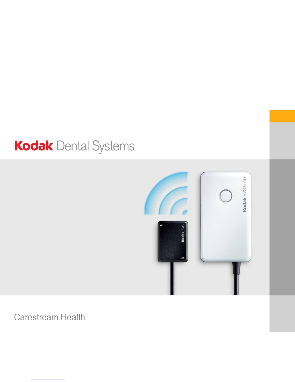
Introducing the new
KODAK RVG 6500
System
Page 2
2
Innovation
New features
• Wireless image transmission
– First Wi-Fi enabled sensor delivering
the same film–quality images as our
best wired sensor
• Intelligent Positioning System
• iPhone / iPod touch compatible
Page 3
Page 4
4
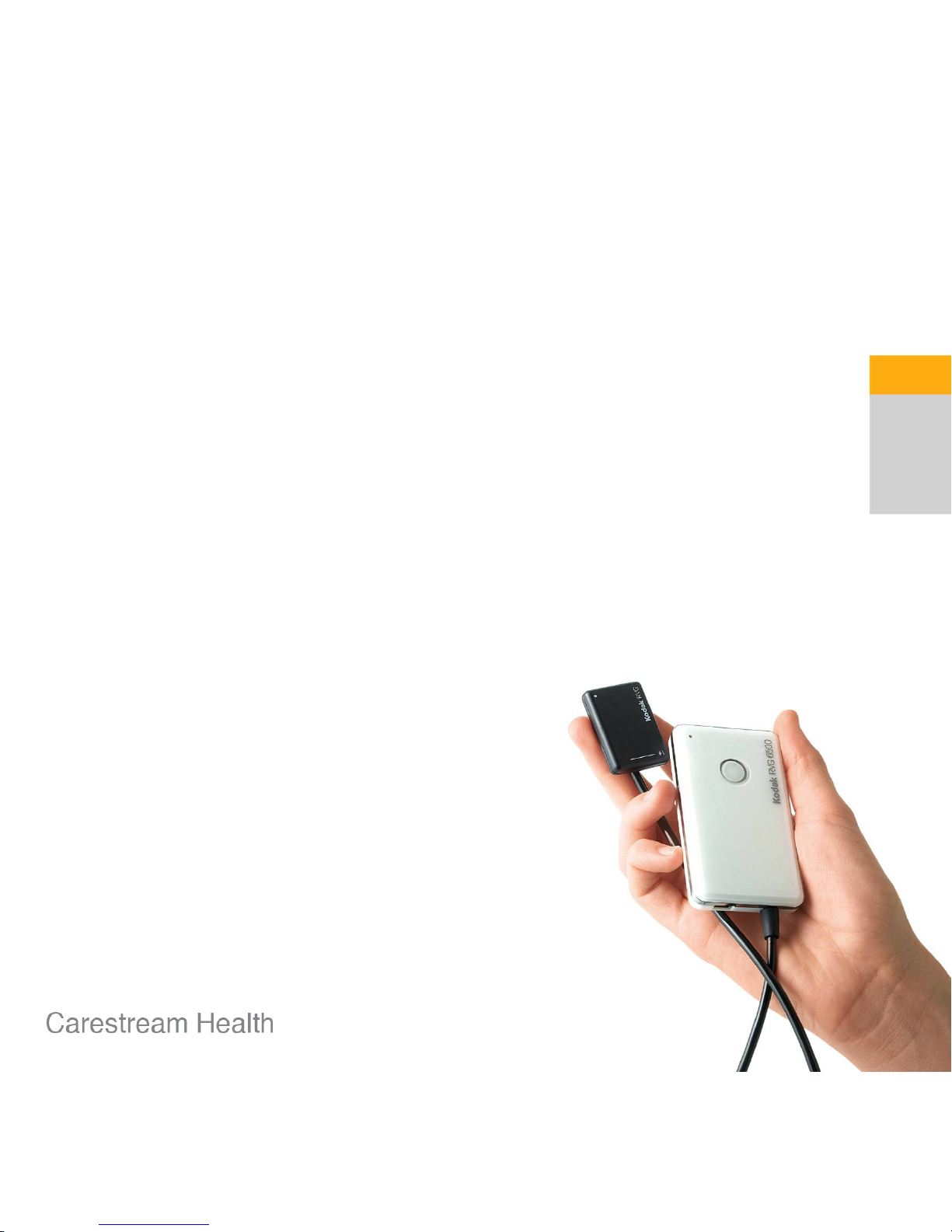
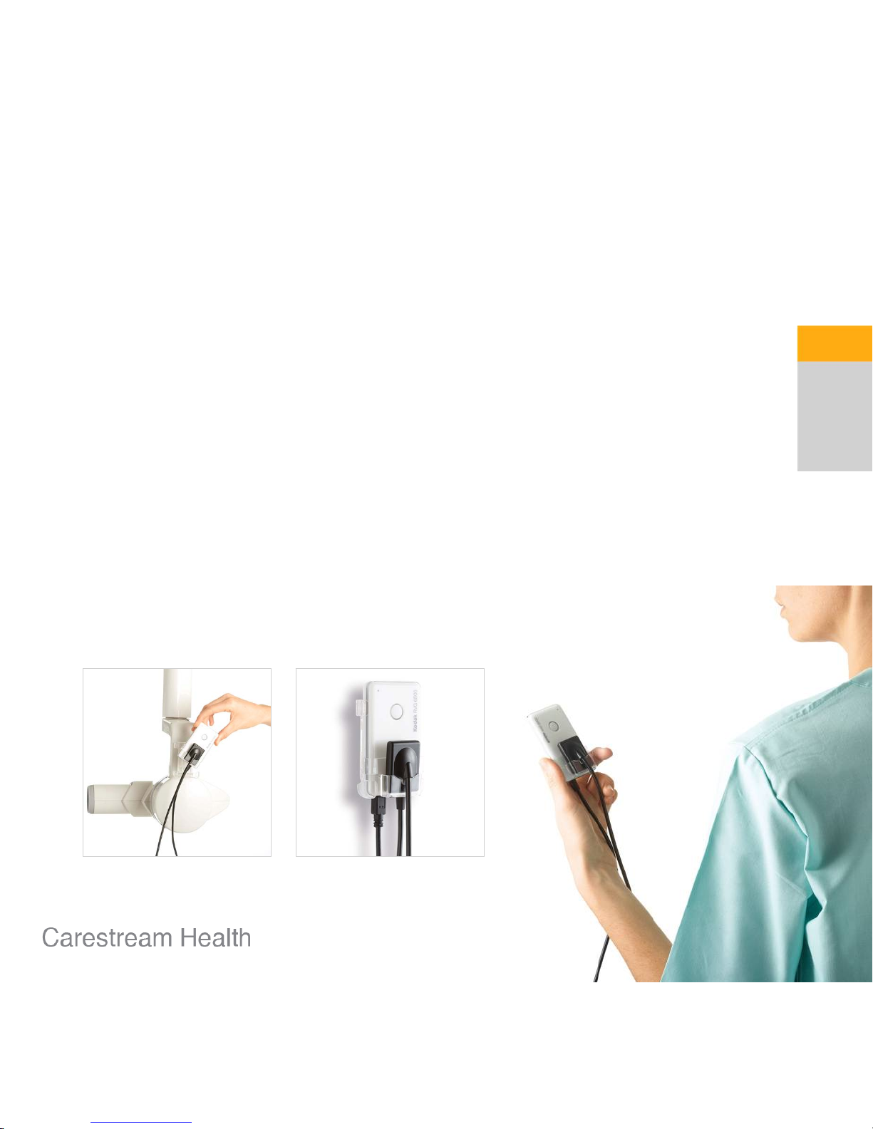
Optimized portability and integration
• No wired connection between the sensor
and the computer
• Sensors can be easily moved from
one operatory to the other
• Eliminates cumbersome cable
improving comfort during exams
• Creates a clean, cable-free working
environment
Compact wireless solution
Page 5
5
Optimized portability and integration
• A full set of holders lets you place the sensor
exactly where it is most convenient for the user
Compact wireless solution
Page 6
6
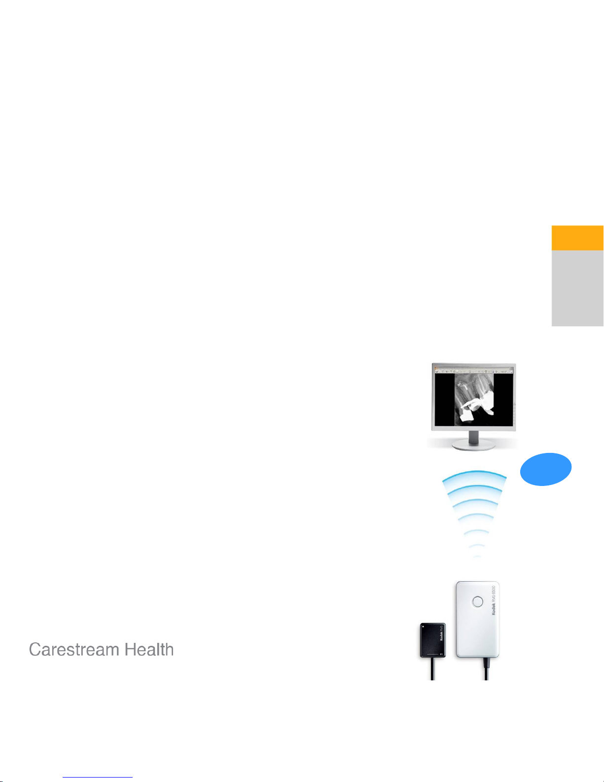
Proven Wi-Fi technology
Fast, reliable, user-friendly
• Faster image transfer for improved productivity
• Secured image transfer with no interference
• Wi-Fi network provides complete mobility for the
entire practice
• Sensors are automatically recognized by the Wi-Fi
network without interrupting workflow
• Sensors can be used while charging, ensuring an
uninterrupted workflow even when battery is empty
05”
Page 7
7
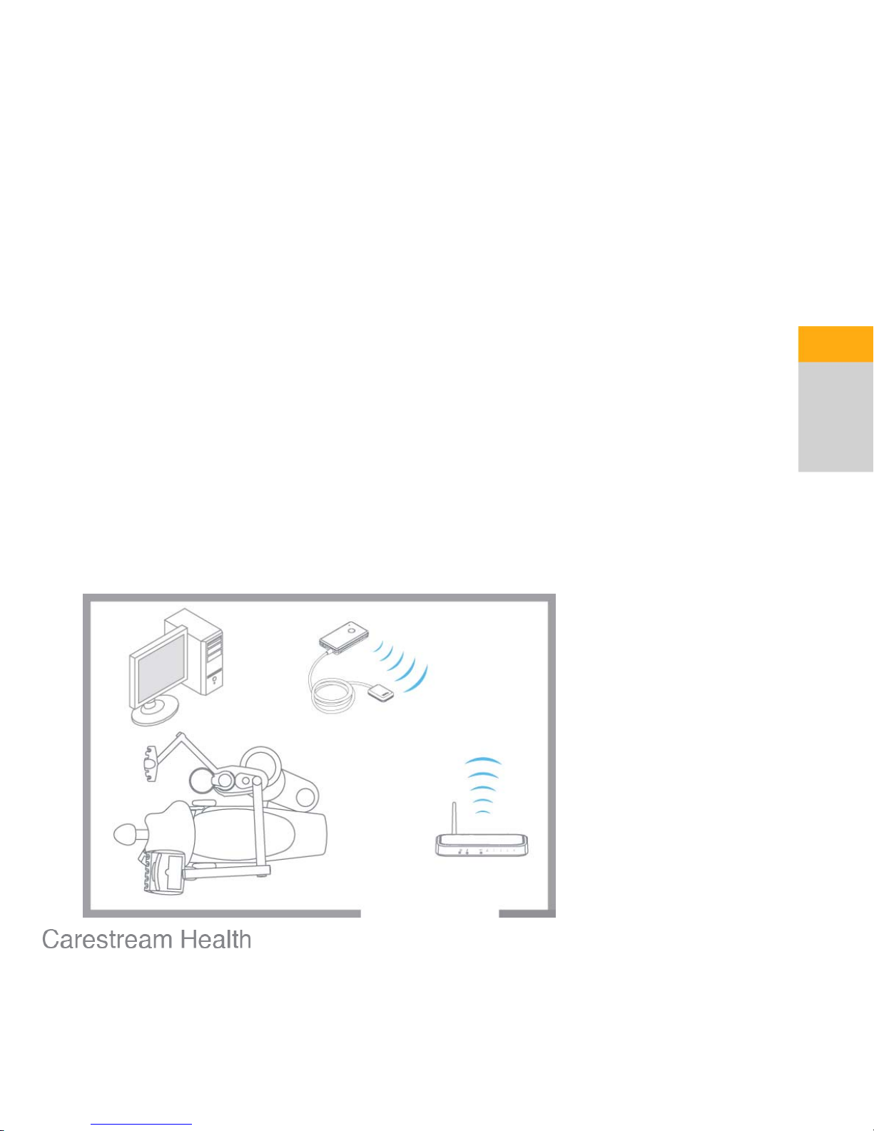
Proven Wi-Fi technology
Easy to integrate, easy to share
• Single chair practice
• 1 dental chair
• 1 sensor
• 1 WiFi access point
Page 8
8
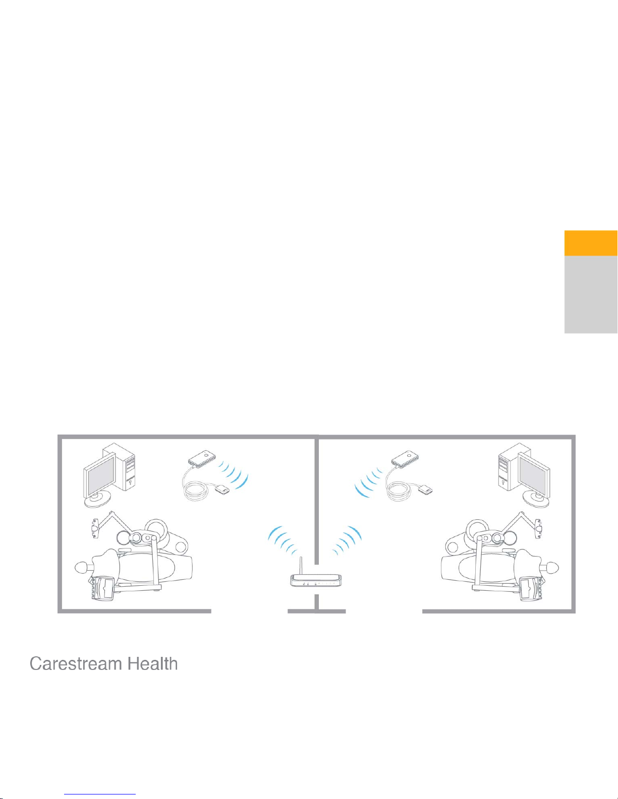
Proven Wi-Fi technology
Easy to integrate, easy to share
• Multiple chairs practice
• 2 dental chairs
• 1 sensor
• 1 Wi-Fi access point
Page 9
9
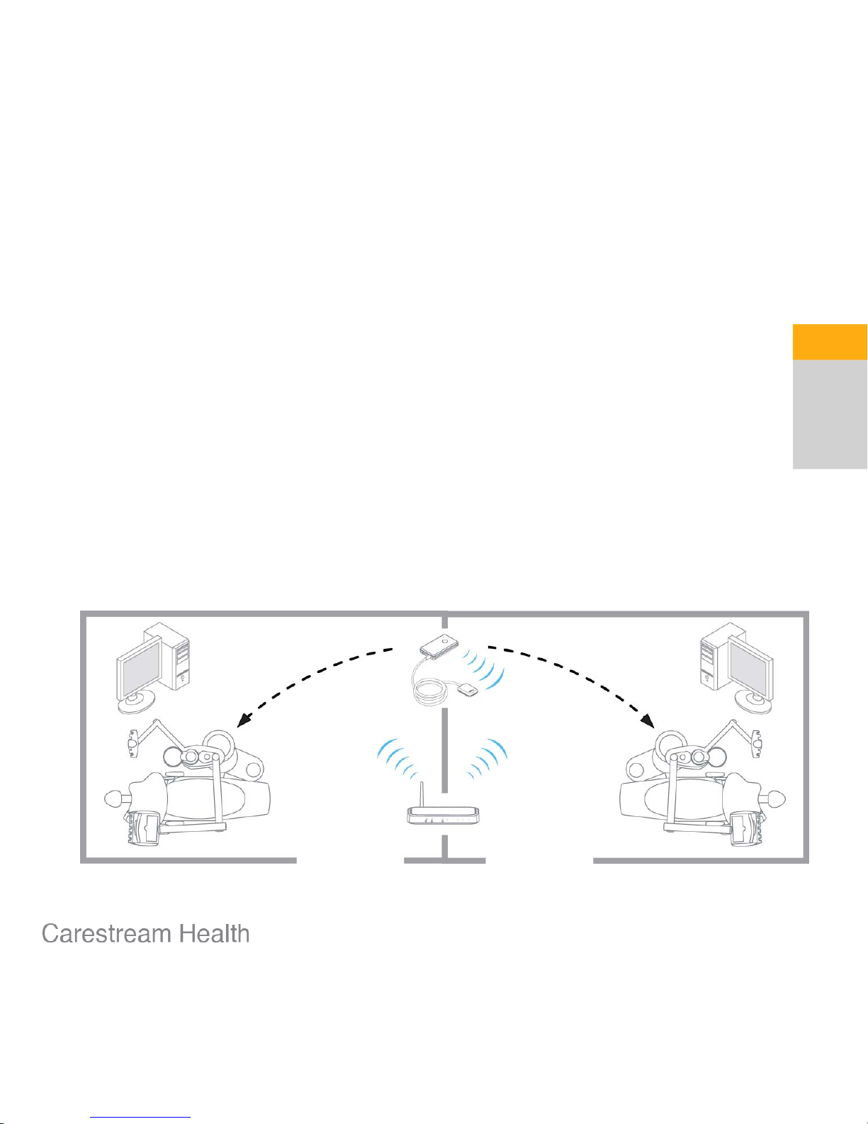
Proven Wi-Fi technology
Easy to integrate, easy to share
• Multiple chairs practice
• 2 dental chairs
• 2 sensors
• 1 Wi-Fi access point
Page 10
Page 11
11
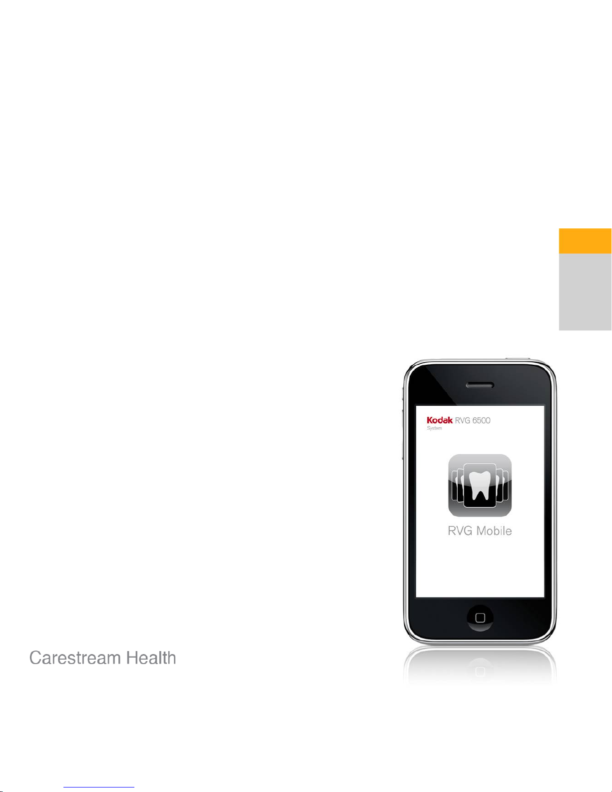
iPhone / iPod touch compatible
Redefining mobility
• Direct RVG acquisition on iPod / iPhone
• Acquire and review images through a
dedicated RVG Mobile application
• RVG Mobile application downloadable
from the Apple Store
Page 12
12
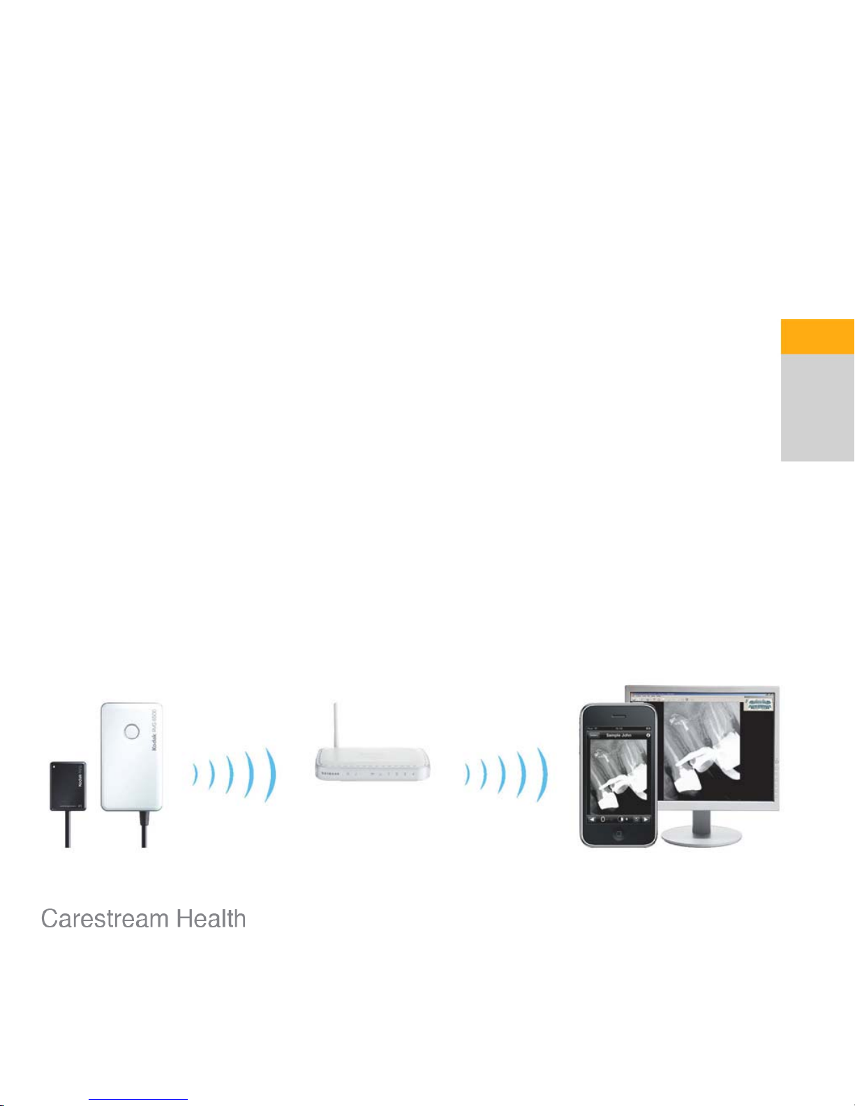
iPhone / iPod touch compatible
Direct acquisition on a iPod / iPhone as well as a
standard computer*
Transfer images from iPod / iPhone to PC for
diagnostic review and archiving
* Only images on the computer screen
are suitable for diagnostic purposes.
Page 13

13
iPhone / iPod touch compatible
Download and upload images on a PC with our specific
transfer application
Download
and upload
images
Page 14

14
iPhone / iPod touch compatible
Image portability
• Eliminates the need for a computer
in each operatory
• Improves portability in and outside
the practice
• Maintains modern practice image
Page 15

15
iPhone / iPod touch compatible
Image portability
• Image review on
iPod / iPhone:
– Zoom in/out
– Brightness and contrast
enhancements
– Perio/Endo/DEJ
enhancements
– Edit comments
– Tooth numbering
Page 16

Page 17

Intelligent Positioning System
Get the right image every time
• Exclusive technology providing an unique
assistance for proper sensor alignment
and angulation
• Guides the operator during positioning
and displays when perfect positioning
has been achieved
• Controls the X-ray beam alignment
with the sensor through a control
interface
17
Page 18

Y
X
Intelligent Positioning System
How does it work?
The software displays realtime notifications regarding
the position of the X-ray beam
in relation to the sensor
Software
X-ray generator
Sensor
The IPS ring localizes the
position of the sensor
during positioning
IPS ring
18
Page 19

Intelligent Positioning System
Example: Step 1
The X-ray tube and the sensor
are not properly aligned
IPS detects the position
of the sensor (X and Y)
The software displays a realtime notification if sensor is
not properly centered
19
Page 20

Intelligent Positioning System
Example: Step 2
The sensor is shown in the
center of the target, so the
alignment is correct.
The X-ray tube and the sensor
are now properly aligned
20
Page 21

Intelligent Positioning System
Display: Sensor in the « target » / green cross
State
: X-ray beam centered on sensor’s active sensor
X-ray beam perpendicular to active surface
Benefit:
No cone cut
Proper angulation to achieve a good
paralelling alignment
Display:
Sensor in the « target » / red cross
State:
X-ray beam centered on sensor’s active sensor
Benefit
: No cone cut
Paralelling alignment not achieved
21
Page 22

Intelligent Positioning System
Benefits
• Can be used for both paralleling and bisectingangle techniques:
– For paralleling technique, IPS allows the user to
achieve parallel alignment and X-ray beam centering
– For bisecting-angle technique, IPS allows the user to
center the X-ray beam on the active surface
• Eliminates the need for cumbersome positioning
devices
• Minimizes the risk of cone cuts, missed apices,
angulations errors and most other alignment
mistakes
• Provides a great educational tool for the staff
Elongation
Cone cut
Missed apices
22
Page 23

Intelligent Positioning System
• Electro-magnetic technology
• Built-in receptors in the sensor’s head,
– no extra bulkiness in the mouth
– no additional electronics visible
• Universal ring compatible with most of
the X-ray generators, even short cones
or portable systems
(cylindric or conic cones - diameter up to 70,7 mm)
• No cable to mount on the generator’s
arm (powered with 4 AAA-type batteries)
• No interference with wireless networks
23
Page 24

IPS Compatibility with Kodak Generators
IPS ring compatible with all Kodak generators
(Irix, Elitys, Kodak 2100, Kodak 2200)
• Ring supplied with 3 sets of mounting brackets:
– Short brackets
– Long brackets
– Brackets for conic cones
On High Frequency generators (Elitys, Kodak 2100, 2200):
Need to upgrade the scissor arm with a new jack to better
counterweight the ring. An Jack Upgrade Kit will be supplied on
demand with the RVG6500 IPS sensor.
(CAT# 5174370)
x4
B
x4
C
30 mm
24
Page 25

RVG technology
Hermetically sealed
sensor housing
Shock-protective
layer
High sensibility
CSI scintillator
Fiber optics
protects from X-ray
High resolution
CMOS sensor
Electronic
Safe, sound and superior
• Technology designed to
deliver the highest possible
image quality— at the lowest
possible dose.
• Sensor’s elements work in
perfect harmony to deliver
the best results.
• Robust design ensures
maximum durability and a
long lifespan—protecting from
water, x-ray, and other
damages.
25
Page 26

26
Best-in-class image quality & resolution
Highest diagnostic value
• First Wi-Fi enabled sensor to deliver the
same image quality as our best wired
sensor
• 20 lp/mm true resolution for higher
clinical details
• Clear film-quality images
Page 27

27
Best-in-class image quality & resolution
Why is true resolution critical?
• Magnify clinical details with no loss of quality
Page 28

28
Robust design
Built to last
• Flexible and robust cable supports tens of thousands of torsions
• Waterproof to allow for surface disinfection
• Shock-resistant casing protects from falls, bites, and other damage
Page 29

29
3 sensor sizes available
Perfectly sized for each application
• For periapical, vertical or horizontal:
Size 1 / Size 1 with Intelligent Positioning System
• For bitewing, occlusal:
Size 2 / Size 2 with Intelligent Positioning System
• For pediatric applications:
Size 0
Vertical anterior
Size 1
Horizontal posterior
Size 1
Bitewing
Size 2
Occlusal
Size 2
Page 30

30
Accurate and comfortable positioning
• A full set of holders included
– Toothbrush-type holders
– KODAK RINN-type bite blocks
– RINN XCP-ORA™ kit
(1 bar, 1 ring, 4 bite blocks)
Positioning accessories
Holders available as an option
Page 31

Kit Content
RVG 6500 sensor box:
-RVG sensor
- Mounting & charging accessories (charger, USB cable, control box &
sensor holder, stickers…)
- Printed Regulatory guide
- Printed Quick Start Guide
- CD-ROM with User & Installation guides
RVG Access point box
:
- access point, CD-ROM, charger and Ethernet cable
RVG Positioning Accessories box:
- RINN XCP-ORA kit
- Kodak RINN-type bite blocks
- Kodak toothbrush holders
- Protection sleeves (x 50)
IPS Aiming Ring box (optional):
- IPS ring including a set of mounting brackets
- 4 AAA-type batteries
- 2 extra sets of mounting brackets
(one set forconic cones, one set of smaller
diameter cylindric cones)
-Cableties
31
Page 32

Pre-sale Check-List
; Evaluate the Wi-Fi practice’s environment
Determine the total number of Wi-Fi networks in the environment using the wireless
troubleshooting inSSIDer tool
(free download on http://www.metageek.net/products/inssider) or
using the KDIS Trouble-shooting tool.
If the environment is saturated, a USB solution may be more appropriate.
; Review the practice workflow and the number of operatory rooms
where the sensor will be shared.
This helps anticipate the access point in the practice and to identify how many Wi-Fi
adapters will be required at installation.
(Board or USB dongle compliant with 802.11g standard
, type NETGEAR Wireless
G54 for instance)
; For an RVG6500 sensor with IPS, check the brand of the X-ray
generator to ensure the IPS ring will mount on the cone.
In case of a Kodak high frequency generator (manufactured before July 2010), order
the Front Jack Upgrade kit.
32
Page 33

How to get the RVG Mobile application?
The application, called
RVG Mobile, is free of charge.
You can access the Apple Store from
ITunes on your PC or directly from
your IPhone or IPod Touch.
Don’t hesitate to use the search tool
to find it!
33
Page 34

What does this application do? (RVG Mobile v.1.2.3)
This application can be either used for:
• Demonstration purpose
• Real X-ray acquisition (using the KODAK RVG6500 and the transfer application)
34
To set the Acquisition Mode:
1) Tap Settings on the iPod/ iPhone.
2) Tap RVG Mobile .
3) Select the X-ray mode.
Slide I button. This mode enables you to connect to the RVG
sensor to acquire live X-ray images.
Important:
In this mode, the « Demo » mode is automatically inactive.
• Select the Demo mode:
– Demo with sensor connected:
With an RVG 6500 sensor
connected to the iPod/ iPhone. X-ray image is displayed on the iPod/ iPhone.
– Demo with sensor simulated: Without an RVG 6500 sensor
connected to the iPod/ iPhone. X-ray image is displayed on the iPod/ iPhone.
– Demo with Sensor IPS simulated: Without an RVG 6500 sensor
connected to the iPod/ iPhone. X-ray image is displayed on the iPod/ iPhone.
Page 35

More to read…
• The Product News on the New Kodak RVG 6500 and 6500 IPS
Sensors
– PN 229 for EAMER
– PN 230 for LAR-GAR-JAPAN
• The Product News PN 231 on the IPS Compatibility List with
Competitive Generators
• The FAQs
• The Product News on the Available Accessories and
Consumables for Kodak RVG 6500 Sensors
– PN 224 for EAMER
– PN 228 for LAR-GAR-JAPAN
35
Page 36

Technical specifications
SENSOR SIZE 0 SENSOR SIZE 1 SENSOR SIZE 2
Sensor technology
CMOS, scintillator, optical fiber,
shock-resistant protective layer
True image resolution 15 lp/mm >20 lp/mm >20 lp/mm
Pixel size 18.5 µm 18.5 µm 18.5 µm
Outside dimensions 22.2 x 30.8 mm 27.5 x 37.7 mm 32.2 x 44.1 mm
Dimensions of active area 17 x 22 mm 22 x 30 mm 27 x 36 mm
Matrix dimensions (pixels) 900 x 1200 1200 x 1600 1440 x 1920
Wireless technology Wi-Fi 802.11g
Intelligent Positioning System
y y
Battery Lithium
Charging Time 4 hours
Battery autonomy (w/o IPS) Approx. 180 images
Battery autonomy (with IPS) Approx. 100 images
Control box dimensions (w/o IPS) 83 (H) x 47 (W) x 16 (D)
Control box dimensions (with IPS) 83 (H) x 47 (W) x 21 (D)
Control box weight (w/o IPS) 65 g
Control box weight ((with IPS) 90 g
Waterproof (sensor)
y y y
36
Page 37

Imaging Software
and Recommended PC Requirements
Imaging Software Kodak Dental Imaging Software 6.12.9.0-D or higher
Operating System:
XP SP2/SP3 with DirectX 9.0c or higher, Windows Vista (Home
Premium, Business, Ultimate Editions) or Windows 7 32 bits or 64 bits
(Home Premium, Professional, Ultimate)
Microprocessor: 1 GHz (Pentium type or equivalent – Intel chipset)
CPU: 2 GHz Intel Dual Core
RAM: 2 GB
Hard disk drive:
1,2 GB for software installation
80 GB free space to use the software
Graphics card:
Graphic board supporting OpenGL
(Nvidia or Ati recommended)
Screen configuration:
17” or larger
1280 x 880 – 16 million colors (32 bits color mode)
Ethernet interface: 1 wired or Wi-Fi(g) ethernet interface
DVD drive: DVD drive
Backup media: Removable / portable, external disk drive
37
Page 38

©Carestream Health, Inc., 2007.
“Kodak” and the Kodak trade dress are trademarks
of Eastman Kodak Company used under license.
 Loading...