

Well REF 89000 User Manual

®
REVOLUTION
45MHz ROTATIONAL
IMAGING CATHETER
REF 89000
English Page 1
Nederlands Page 4
Français Page 7
Deutsch Page 10
Italiano Page 13
Español Page 16
REVOLUTION
ENGLISH
45MHz ROTATIONAL IMAGING CATHETER
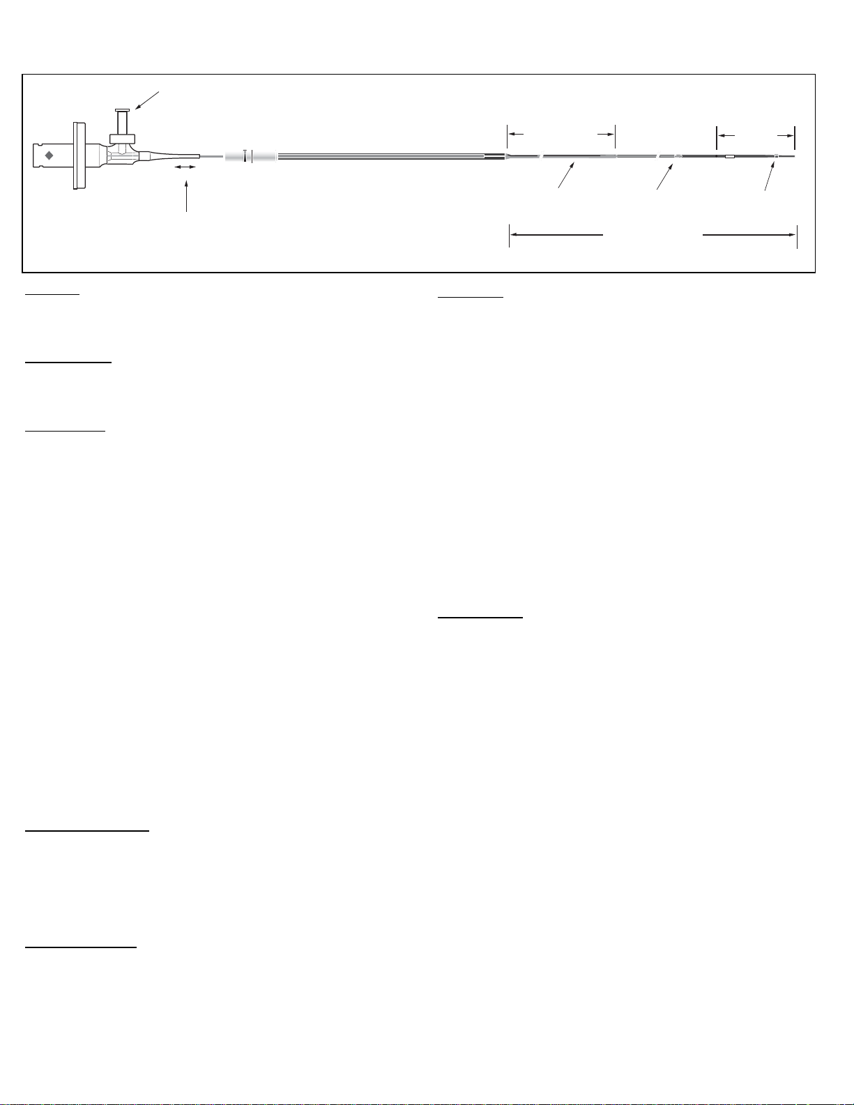
One Way Luer
Telescoping Section
Fig. 1
CAUTION:
1. U.S. Federal Law restricts this device to sale by or on the order of a
physician.
2. Prior to use, read this entire Instructions For Use.
INTENDED USE:
The Revolution
examination of coronary arteries. Intravascular ultrasound imaging is indicated in
patients who are candidates for transluminal interventional procedures.
DESCRIPTION:
The Revolution 45MHz rotational IVUS imaging catheter consists of two main
assemblies; the imaging core and the catheter body. The catheter body
comprises of three sections; distal section w/ .014" compatible F/X port,
proximal section (single lumen), telescope section.
The distal section and proximal (single lumen) sections comprise the "working
length" of the catheter, the telescoping section remains outside of the guiding
catheter. The telescoping shaft (section) allows the imaging core to be advanced
and retracted for up to 150 mm of linear movement. The corresponding movement
of the transducer occurs from the proximal end of the guidewire exit port to the
proximal end of the window portion of the distal section.
The imaging core is composed of a hi-torque, flexible, rotating drive cable with a
distal outward looking 45MHz ultrasonic transducer. An electromechanical
connector interface at the proximal end makes the connection to the patient
interface module (PIM). The PIM-catheter interface consists of an integrated
mechanical drive assembly and electrical connection.
A flushing port with a one-way valve (Fig. 1) is used to displace the air initially
present within the catheter. The catheter must be flushed with heparinized
saline prior to use, as this provides the acoustic coupling media required for
ultrasonic imaging. The one-way valve helps retain saline in the catheter
during use.
The catheter body has a distal guidewire lumen with a proximal exit port
located 2 cm from the distal end (Fig 1). A radiopaque (RO) marker is
embedded in the catheter body at 0.5 cm from the tip. In addition, an insertion
depth indicator is located on the catheter body at 100 cm, corresponding to
femoral insertions.
The catheter is for use with the In-Vision Gold imaging system with software
V5.0 or higher or the Volcano s5 and Volcano s5i imaging system. Consult
your System Operator’s Manual.
CONTRAINDICATIONS:
This device is not currently indicated for use in cerebral or peripheral vessels.
Use of IVUS Imaging Catheters is contraindicated where introduction of any
catheter would constitute a threat to patient safety. Contraindications include:
bateremia or sepsis, major coagulation system abnormalities, patients
disqualified for CABG surgery, patients disqualified for PTCA, severe
hemodynamic instability or shock, patients diagnosed with coronary artery
spasm, and total occlusion.
ADVERSE EFFECTS:
Bleeding at the entry puncture site, injury to the vascular wall, thrombosis of
the vessel, and peripheral embolization has occurred with the use of
percutaneous intravascular catheter devices.
®
Pullback Length
150 mm
catheter is intended for the intravascular ultrasound
Proximal Shaft
109 cm
3.5F
3.2F
Monorail
23mm
RO
Marker
Usable Length
135 cm
WARNINGS:
Use of the Revolution catheters is restricted to specialists who are familiar
with, and have been trained to perform, the procedures for which this device
is intended.
DO NOT advance the catheter if resistance is encountered. The catheter
should never be forcibly inserted into lumens narrower than the catheter
body or forced through a tight stenosis.
Care should be taken when utilizing devices that comprise a short monorail;
in such instances advancement of the device distal to a deployed stent can
result in exposure of the guidewire to the stent struts.
In instances where the device has crossed a deployed stent, care should be
taken when retracting the device to ensure that entanglement does not
occur. Fluoroscopy should be used to monitor guidewire position with the
respect to the imaging catheter and the stent; at no time should the imaging
catheter be retracted if there is evidence of guidewire prolapse or if
significant resistance to withdrawal is experienced. If either of these events
occur, advance the imaging catheter distal of the stent and then carefully
remove the whole system under the guidance of fluoroscopy.
Care should be taken when re-advancing a guide wire after stent deployment. A
guide wire may exit between stent struts when re-crossing a stent that is not fully
apposed to the vessel wall. Subsequent advancement of the catheter could
cause entanglement between the catheter and the stent. Care should be taken
to slowly remove the catheter from a stented vessel.
PRECAUTIONS:
The Revolution device is a delicate scientific instrument and should be treated
as such. Always observe the following precautions:
Contents supplied STERILE using an EtO (ethylene oxide) process. Do not
use if sterile barrier is damaged. If damage is found call your Volcano
Corporation representative.
To maintain optimal patient safety, inspect the product prior to use. Do not
use if saline leaks from any location other than the vent port in the monorail
section.
For single use only. Do not re-use, reprocess or re-sterilize. Re-use,
reprocessing or re-sterilization may compromise the structural integrity of
the device and/or lead to device failure which in turn may result in patient
injury, illness or death.
Re-use, reprocessing or re-sterilization may also create a risk of
contamination of the device and/or cause patient infection or cross-infection,
including, but not limited to the transmission of infectious disease(s) from
one patient to another. Contamination of the device may lead to injury,
illness or death of the patients.
The catheter has no user serviceable parts. Do not attempt to repair or to
alter any component of the catheter assembly.
Do not attempt to connect the catheter to electronic equipment other than
the designated systems.
Never attempt to attach or detach the catheter while the PIM motor is
running. To do so may damage the connector.
Avoid any sharp bends, pinching, or crushing of the catheter.
Do not kink or sharply bend the catheter at any time. This can cause drive
cable failure. An insertion angle greater than 45º is considered excessive.
Turn the PIM "OFF" before withdrawing the imaging catheter.
1

INSTRUCTIONS FOR USE:
Materials and Equipment
Revolution Catheter
Sterile PIM cover
10 in. extension tubing
3 cc and 10 cc syringes
3-way stop cock
Pre-formed guide catheter [0.064 in. (1.63 mm) I.D. min.] with Y-adapter
assembly*
In-Vision Gold imaging system with software V5.0 or higher, Volcano s5 and
Volcano s5i imaging system*
Heparinized, physiologic saline solution*
Guide wire, 0.014 in. (0.36 mm) max. diameter*
*not packaged with catheter
Inspection Prior to Use
Carefully inspect the package prior to use for any breach of the sterile barrier
or damage to the contents. If the sterile barrier integrity is compromised or the
contents damaged, contact your Volcano Corporation representative.
Preparation For Use
Refer to the operator’s manual or user’s guide for instrumentation and PIM
Setup.
Using sterile technique, remove the catheter from its sterile packaging.
Remove the Packaging Coil protecting the catheter. Retract the movable
Imaging Core completely to the proximal position via the telescope shaft.
Connect the 3 cc and 10 cc syringes to the 3-way stopcock, then connect the
assembly to the extension tube and fill both syringes with heparinized saline.
Ensure that all air is expelled from the system. Do not use if saline leaks from
any location other than the vent port in the monorail section.
Connect the extension tube to the one-way valve on the catheter hub. The 10
cc syringe is to be used as a reservoir for refilling the 3 cc flushing syringe.
Flush the imaging catheter TWICE continuously with 3 cc volume each time.
DO NOT USE EXCESSIVE PRESSURE. Advance the imaging core to its fully
distal position; via the telescope shaft.
Connect the imaging catheter to the PIM by inserting the proximal end of the
connector through the opening in the Sterile PIM cover, gently twisting the
connector until it locks into place. To ensure that the hub is fully seated in the
PIM, gently tug on the catheter hub.
Begin imaging by pressing the IMAGE button on the PIM long enough to
ensure proper function of the catheter by observing a pattern of partial bright
concentric rings on the monitor. Refill the 10 cc syringe as needed and
reattach to the stopcock without introducing air into the line.
Place Guide Catheter
Prepare the entry site with a sheath introducer according to the standard practice.
Prior to inserting the imaging catheter, ensure the patient has been prepared
using standard procedure for interventional treatment.
Backload the guide wire into the distal end of the catheter. Advance the
guidewire into the catheter until the guidewire exits from the wire exit port. Place
the guide catheter and Y-adapter. Introduce the guidewire and advance it to the
region of interest. Introduce imaging catheter into guide catheter.
Note: Guidewires that supply more stiffness near the distal tips are recommended.
Note: Always wipe down the guidewire with heparinized saline prior to loading
the catheter onto the guidewire.
CAUTION: Never advance the imaging catheter without guidewire support.
CAUTION: Never advance or withdraw the imaging catheter without the
imaging core assembly in the most distal position.
CAUTION: Never advance or withdraw the imaging catheter without direct,
fluoroscopic visualization.
CAUTION: Never advance the distal tip of the imaging catheter near the very
floppy end of the guidewire. This part of the guidewire will not adequately
support the catheter. A catheter advanced to this portion may not follow the
guidewire when it is retracted and cause the guide wire to buckle into a loop.
The catheter may then drag along the inside of vessel and catch on the guide
catheter tip. If this occurs, remove the catheter assembly, guidewire and the
guide catheter together. If the catheter is advanced too near the end of the
guidewire, advance the guidewire while holding the imaging catheter steady. If
this fails, withdraw the catheter and guidewire together.
Continue to advance the imaging catheter into the guide catheter, up to the
femoral marker. Tighten the hemostasis valve on the guide catheter’s Yadapter. Tighten only enough to prevent fluid/blood leakage.
Note: AN EXCESSIVELY TIGHTENED HEMOSTASIS VALVE MAY DISTORT
THE IMAGE DUE TO BINDING OF THE ROTATING DRIVE CABLE.
Catheter Placement and Imaging
With the PIM image "OFF" and using fluoroscopy, advance the imaging
catheter over the guide wire until the distal marker crosses a minimum of 3 cm
beyond the region of interest in the vessel/lesion.
Keeping the catheter body and guidewire fixed, turn PIM image "ON" and
retract the imaging core slowly along its 150 mm travel, imaging any region of
interest.
Note: Always turn the PIM image "OFF" before advancing the imaging core
within the catheter.
When finished, stop imaging by pressing the IMAGE button on the PIM, in
manual mode, advance the imaging core to its most distal position. Maintain
the position of the wire and remove the catheter.
Troubleshooting
If your system menu does not include "Revolution Catheters", contact your
Volcano Corporation representative before proceeding. If the images fade
during use, flush the catheter with heparinized saline. If shadowed areas
persist after flushing in situ, the distal lumen or catheter body may contain air
bubbles.
STORAGE AND HANDLING:
Products should be stored in a dry place with the temperature not exceeding
54 degrees Celsius (54˚ C) in their original cardboard box.
PRODUCT SPECIFICATIONS:
Model Revolution
Catalog number 89000
Crossing profile at transducer 3.2F
Maximum guide wire 0.014” (0.36 mm)
Minimum guide catheter 6F (1.63 mm)
Usable length 135 cm
Acoustic Output Parameter B-Mode
I
(mW/cm2)* 70.778
SPTA.3
I
(W/cm2)* 95.533
SPPA.3
Pr.3 (MPa)** 1.901
PD (μs) 0.048
PRF (Hz) 15360
Center Freq (MHz) 42.3
MI** 0.281
TI* 0.010
Uncertainty
* +/- 29.1%
** +/- 14.6%
TI: Thermal Index defined as TI = W
210
: Bounded-square Output (mW)
W
01x1
: Center Frequency (MHz)
f
c
MI: Mechanical Index defined as MI= Pr.3/(f
I
: Derated Intensity, Spatial Peak Pulse Average (W/cm2)
SPPA.3
I
: Derated Intensity, Spatial Peak Temporal Average (mW/cm2)
SPTA.3
Pr.3: Derated Peak Negative Pressure at a location of the maximum
derated pulse intensity integral (MPa)
: Total Power (mW)
W
0
PD: Pulse Duration (μs)
PRF: Pulse Repetition Frequency (Hz)
01x1fc
1/2
)
c
2
LIMITED WARRANTY:
Subject to the conditions and limitations on liability stated herein, Volcano
Corporation (“VOLCANO”) warrants that the Revolution catheter (the
“Catheter”), as so delivered, shall materially conform to VOLCANO’S then
current specification for the Catheter upon receipt for a period of one year from
the date of delivery. ANY LIABILITY OF VOLCANO WITH RESPECT TO THE
CATHETER OR THE PERFORMANCE THEREOF UNDER ANY
WARRANTY, NEGLIGENCE, STRICT LIABILITY OR OTHER THEORY WILL
BE LIMITED EXCLUSIVELY TO CATHETER REPLACEMENT OR, IF
REPLACEMENT IS INADEQUATE AS A REMEDY OR, IN VOLCANO’S
OPINION, IMPRACTICAL, TO REFUND OF THE FEE PAID FOR THE
CATHETER. EXCEPT FOR THE FOREGOING, THE CATHETER IS
PROVIDED “AS IS” WITHOUT WARRANTY OF ANY KIND, EXPRESSED OR
IMPLIED, INCLUDING WITHOUT LIMITATION, ANY WARRANTY OF
FITNESS, MERCHANTABILITY, AND FITNESS FOR A PARTICULAR
PURPOSE OF NONINFRINGEMENT. FURTHER, VOLCANO DOES NOT
WARRANT, GUARANTEE, OR MAKE ANY REPRESENTATIONS
REGARDING THE USE, OR THE RESULTS OF THE USE, OF THE
CATHETER OR WRITTEN MATERIALS IN TERMS OF CORRECTNESS,
ACCURACY, RELIABILTY, OR OTHERWISE. Licensee understands that
VOLCANO is not responsible for and will have no liability for any items or any
services provided by any persons other than VOLCANO. VOLCANO shall
have no liability for delays or failures beyond its reasonable
control.Additionally, this warranty does not apply if:
1. The Catheter is used in a manner other than described by VOLCANO in the
Instructions For Use supplied with the Catheter.
2. The Catheter is used in a manner that is not in conformance with purchase
specifications or specifications contained in the Instructions For Use.
3. The Catheter is re-used or re-sterilized.
4. The Catheter is repaired, altered, or modified by other than VOLCANO
authorized personnel or without VOLCANO authorization.
If claims under this warranty become necessary, contact VOLCANO for
instructions and issuance of a Return Material Authorization number if the
Catheter is to be returned. Equipment will not be accepted for warranty
purposes unless the return has been authorized by VOLCANO.
This product, and the use thereof, may be covered by one or more of the
following U.S. and international patents: 5081993; 5183048; 5257629;
5453575; 5601082; 5921931; 6036650; 6165128; 6200268; 6213950;
6381350; DE69031685.2; EP0386058; EP0473665; EP0611291; EP0707453;
EP0871043; GB2233094; GB2246632; JP2905489; JP3188470; JP3194582.
Other U.S. and international patents pending.
This product is licensed to the customer for single use only. Any re-sterilization
or subsequent re-use is an unlicensed use and therefore constitutes patent
infringement.
Revolution is a trademark of Volcano Corporation and is registered in the
United States and other countries.
Volcano and the Volcano logo are trademarks of Volcano Corporation and are
registered in the United States and other countries.
ADDITIONAL QUESTIONS REGARDING THIS PRODUCT SHOULD BE
DIRECTED TO:
Manufactured By:
Volcano Corporation
2870 Kilgore Road
Rancho Cordova, CA 95670 USA
(800) 228-4728 (916) 638-8008 (916) 638-8112 fax
Authorized European Representative:
Volcano Europe SA/NV
Excelsiorlaan 41
B-1930 Zaventem Belgium
+32.2.679.1076 +32.2.679.1079 fax
805007-001/008
3
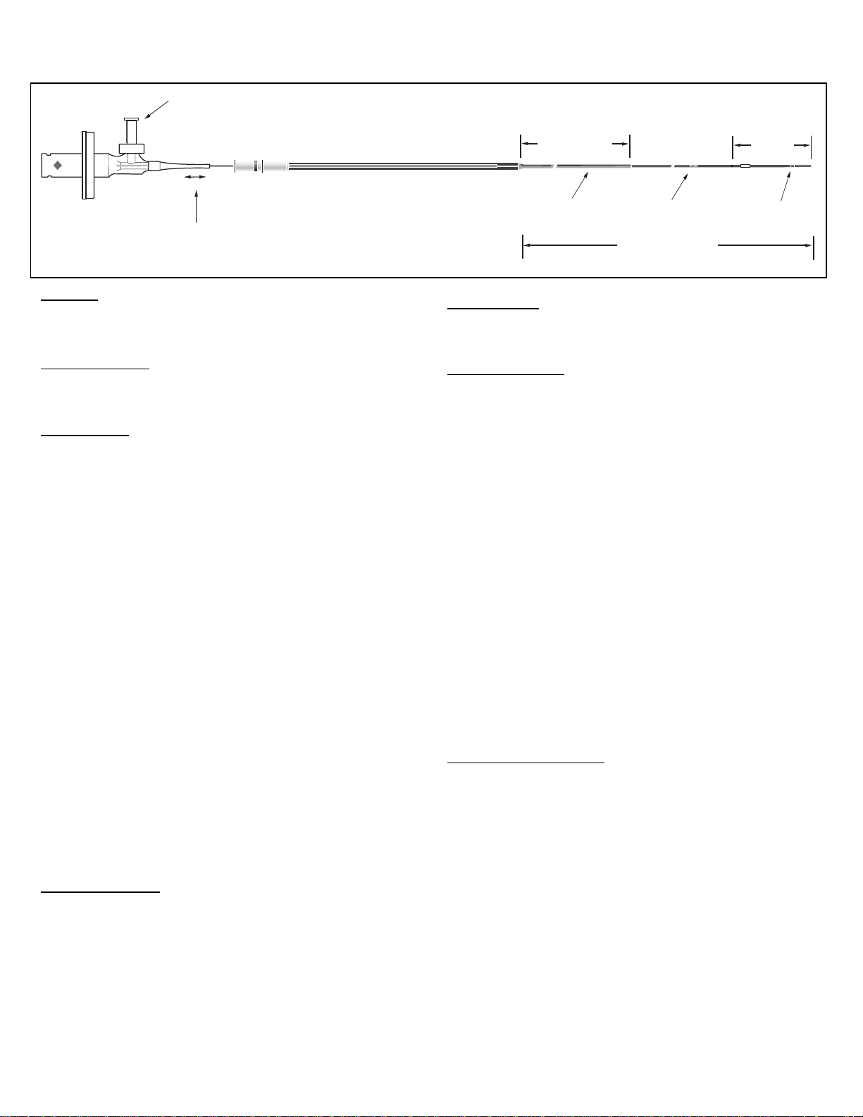
Eenweg-Luer
REVOLUTION
NEDERLANDS
45MHz ROTERENDE IMAGINGKATHETER
Telescopische deel,
Afb. 1
OPGELET:
1. Volgens de federale wetgeving van de VS mag dit product alleen
2. Lees vóór gebruik deze volledige gebruiksaanwijzing.
BEOOGD GEBRUIK:
De Revolution-katheter is bedoeld voor gebruik bij intravasculaire
echoscopie van kransslagaderen. Intravasculaire echografie is geïndiceerd
bij patiënten die kandidaat zijn voor transluminale ingrepen.
BESCHRIJVING:
De Revolution 45MHz roterende IVUS-imagingkatheter bestaat uit twee
hoofddelen: de “Imaging Core” en het katheterdeel. Het katheterdeel bestaat
uit drie onderdelen: het distale deel met een compatibele F/X-poort van
0,014 inch, een proximaal deel (enkelvoudige lumen) en een telescoopdeel.
Het distale deel en de proximale delen (enkelvoudige lumen) vormen samen
de “werklengte” van de katheter, het telescoopdeel blijft buiten de
geleidekatheter. De telescoopschacht (het telescoopdeel) maakt het mogelijk
de “Imaging Core” tot maximaal 150 mm lineair op te voeren en terug te
trekken. De corresponderende beweging van de transducer treedt op vanaf
het proximale uiteinde van de uitgangspoort van de voerdraad tot aan het
proximale uiteinde van het vensterdeel van het distale deel.
De “Imaging Core” bestaat uit een hi-torque, flexibele, roterende drive-kabel
met een distale naar buiten gerichte ultrasone transducer van 45 MHz. Een
elektromechanische connectorinterface aan het proximale uiteinde vormt de
verbinding met de interfacemodule voor de patiënt (PIM). De interface van
de PIM-katheter bestaat uit een geïntegreerde mechanische drive en een
elektrische verbinding.
Een spoelpoort met een eenwegventiel (afb. 1) wordt gebruikt om de lucht te
verdringen die zich aanvankelijk in de katheter bevindt. De katheter moet
voorafgaand aan gebruik met een gehepariniseerde zoutoplossing worden
gespoeld, omdat dit het beste akoestische hechtmiddel biedt dat voor
echo-grafie noodzakelijk is. Het eenwegventiel zorgt ervoor dat het
fysiologisch zout tijdens gebruik in de katheter blijft.
Het katheterdeel is voorzien van een distaal voerdraadlumen met een
proximale uitgangspoort die zich 2 cm van het distale uiteinde bevindt (afb.
1). Op 0,5 cm van de tip is een radiopake marker in het katheterdeel
ingebouwd. Daarnaast bevindt zich op 100 cm een indicator voor de
inbreng-diepte, overeenkomend met femorale insertie.
De katheter is bedoeld voor gebruik met het In-Vision Goldbeeldvormingssysteem met software V5.0 of hoger of met het Volcano s5- en
Volcano- s51-beeldvormingssysteem. Raadpleeg de bedieningshandleiding
van uw systeem.
CONTRA-INDICATIES:
Dit apparaat wordt momenteel niet geïndiceerd voor gebruik in cerebrale “of
perifere” bloedvaten. Het gebruik van de IVUS-imagingkatheter is gecontraindiceerd in die gevallen waarbij introductie van een katheter een bedreiging
kan vormen voor de veiligheid van de patiënt. Onder de contra-indicaties
vallen onder meer: bacteriëmie en sepsis, belangrijke afwijkingen van het
stollingssysteem, patiënten die niet in aanmerking komen voor CABGchirurgie, patiënten die niet in aanmerking komen voor PTCA, ernstige
hemodynamische instabiliteit of shock en patiënten die zijn gediagnosticeerd
met coronariaspasmen in geval van totale occlusies.
door of op voorschrift van een arts worden verkocht.
uittreklengte
150 mm
Proximale schacht
109 cm
3,5 F
3,2 F
Monorail
23mm
RO-markering
Bruikbare lengte
135 cm
BIJWERKINGEN:
Tijdens gebruik van percutane intravasculaire kathetersystemen zijn de
volgende bijwerkingen opgetreden: bloeding bij de toegangsincisie, letsel
aan de vaatwand, trombose in het bloedvat en perifere embolisatie.
WAARSCHUWINGEN:
• Het gebruik van de Rvolution- katheters is beperkt tot specialisten die
vertrouwd zijn met en zijn opgeleid in de procedures waarvoor dit systeem
is bedoeld.
• Voer de katheter NIET op als u enige weerstand ondervindt. De katheter
mag nooit met kracht worden ingebracht in lumina die nauwer zijn dan het
katheterdeel of door een nauwe stenose.
• Wees voorzichtig bij het gebruik van hulpmiddelen die een korte monorail
omvatten; in dergelijke gevallen kan het opvoeren van het hulpmiddel
distaal van een ontplooide stent ertoe leiden dat de voerdraad contact
maakt met de stentribben.
• Wanneer het hulpmiddel een ontplooide stent gepasseerd heeft, dient
zorgvuldigheid betracht te worden bij het terugtrekken van het hulpmiddel
om ervoor te zorgen dat er geen verstrikking optreedt. Gebruik
röntgendoorlichting om de positie van de voerdraad ten opzichte van de
imagingkatheter en de stent te controleren; de imagingkatheter mag nooit
teruggetrokken worden als duidelijk is dat de voerdraad weggegleden is of
wanneer beduidende weerstand gevoeld wordt tijdens het terugtrekken.
Als zich een van beide situaties voordoet, voert u de imagingkatheter
distaal van de stent op en verwijdert vervolgens het hele systeem onder
röntgendoorlichting.
• Bij het opnieuw opvoeren van een voerdraad na het ontplooien van de
stent moet voorzichtigheid betracht worden. Een voerdraad kan bij het
opnieuw passeren van een stent die niet volledig aan de vaatwand is
gehecht tussen de stentribben naar buiten steken. Als de katheter
vervolgens wordt opgevoerd, kunnen de katheter en de stent hierdoor in
elkaar verstrikt raken. Verwijder de katheter voorzichtig en langzaam uit
een gestent vat.
VOORZORGMAATREGELEN:
Het Revolution-apparaat is een kwetsbaar wetenschappelijk instrument en
moet als zodanig worden behandeld. Neem altijd de volgende
voorzorgs-maatregelen in acht:
• De inhoud wordt met behulp van een EtO-proces (ethyleenoxide)
STERIEL geleverd. Gebruik het product niet als de steriele barrière is
beschadigd. Neem contact op met een vertegenwoordiger van Volcano
Corporation als beschadigingen worden aangetroffen.
• Voor een optimale veiligheid van de patiënt moet het product voor gebruik
gecontroleerd worden. Gebruik het toestel niet wanneer er zoutoplossing
lekt op andere plaatsen dan de ventilatiepoort in het monorailgedeelte.
• Uitsluitend bestemd voor eenmalig gebruik. Niet opnieuw gebruiken,
-verwer-ken of steriliseren. Opnieuw gebruiken, verwerken of steriliseren
kan de struc-turele integriteit van het product aantasten en/of leiden tot
falen van het pro-duct wat vervolgens voor de patiënt kan leiden tot letsel,
ziekte of overlijden.
• Opnieuw gebruiken, verwerken of steriliseren kan ook het risico van
besmetting van het product met zich meebrengen en/of een infectie of
kruisinfectie bij de patiënt veroorzaken, inclusief, maar niet beperkt tot, het
overbrengen van een (of meerdere) besmettelijke ziekte(n) van de ene
patiënt op de andere. Besmetting van het product kan leiden tot letsel,
ziekte of overlijden van de patiënt.
4

• De katheter bevat geen onderdelen die voor onderhoud door de gebruiker
in aanmerking komen. Probeer geen onderdelen van de katheter te
repa-reren of te wijzigen.
• Probeer niet om de katheter aan te sluiten op andere elektrische
appara-tuur dan de hiervoor bestemde systemen.
• Probeer de katheter nooit te bevestigen of los te koppelen terwijl de PIMmotor draait. Hierdoor kan de connector beschadigd raken.
• Voorkom het ontstaan van scherpe hoeken, afklemmen of indeuken van
de katheter.
• Zorg ervoor dat op geen enkel moment knikken of scherpe hoeken in de
katheter ontstaan. Dit kan resulteren in een storing van de drive-kabel. Een
inbrenghoek van meer dan 45º wordt als te scherp beschouwd.
Bij gebruik van een voerdraad in een gestent vat moet voorzichtigheid
worden betracht. Katheters waarbij de voerdraad niet is ingekapseld,
kunnen tussen de verbinding van de katheter en de voerdraad vast komen
te zitten aan de stent.
Bij het opnieuw opvoeren van een voerdraad na het ontplooien van de
stent moet voorzichtigheid worden betracht. Een voerdraad kan bij het
opnieuw passeren van een stent die niet volledig aan de vaatwand is
gehecht tussen de stentribben naar buiten steken. Als de katheter
vervolgens wordt opgevoerd, kunnen de katheter en de stent hierdoor in
elkaar verstrikt raken. Verwijder de katheter voorzichtig en langzaam uit
een gestent vat.
• Zet de PIM “UIT” (OFF) voordat de imagingkatheter wordt teruggetrokken.
GEBRUIKSAANWIJZING:
Materiaal en apparatuur
Revolution-katheter
Steriele PIM-hoes
Verlengslang van 25 cm (10")
Injectiespuiten van 3 cc en 10 cc
3-weg plugkraan
Voorgevormde geleidekatheter [binnendiameter minimaal 1,63 mm (0,064")]
met Y-adapter*
In-Vision Gold-beeldvormingssysteem met software V5.0 of hoger, Volcano
s5 en Volcano s5i-beeldvormingssysteem.*
Gehepariniseerde fysiologische zoutoplossing*
Voerdraad met een maximale diameter van 0,36 mm (0,014")*
*wordt niet bij de katheter meegeleverd
Controle vóór gebruik
Inspecteer de verpakking vóór gebruik zorgvuldig op beschadiging van de
steriele barrière of de inhoud. Als de integriteit van de steriele barrière is
aangetast of als de inhoud is beschadigd, dient u contact op te nemen met
uw vertegenwoordiger bij Volcano Corporation.
Voorbereiding voor het gebruik
Raadpleeg de bedieningshandleiding of de gebruikershandleiding voor de
apparatuur en de set-up van de PIM.
Verwijder de katheter met behulp van een steriele techniek uit de steriele
verpakking. Verwijder de verpakkingsspoel die de katheter beschermt. Trek
de beweegbare “Imaging Core” via de telescoopschacht volledig naar de
proximale positie.
Verbind de injectiespuiten van 3 cc en 10 cc met de 3-weg plugkraan, sluit
de set vervolgens aan op de verlengslang en vul beide injectiespuiten met
gehepariniseerde fysiologisch zoutoplossing. Zorg ervoor dat alle lucht uit
het systeem wordt verdreven. Niet gebruiken als de zoutoplossing uit andere
plaatsen lekt dan de ontluchtingspoort in het monorailgedeelte.
Sluit de verlengslang aan op het eenwegventiel op de naaf van de katheter.
De injectiespuit van 10 cc wordt gebruikt als reservoir voor het opnieuw
vullen van de spoelspuit van 3 cc.
Spoel de imagingkatheter TWEEMAAL continu door, met telkens 3 cc
volume. OEFEN GEEN OVERMATIGE DRUK UIT. Voer de “Imaging Core”
met behulp van de telescoopschacht op naar de volledige distale positie.
Sluit de imagingkatheter aan op de PIM door het proximale uiteinde van de
connector in te brengen via de opening in de steriele PIM-hoes, waarbij de
connector voorzichtig wordt gedraaid tot deze op zijn plaats klikt. Trek
zachtjes aan de naaf van de katheter om er zeker van te zijn dat de naaf
volledig en juist in de PIM is geplaatst.
Begin de beeldvorming door net zo lang op de IMAGE-knop op de PIM te
drukken totdat duidelijk is dat de katheter op de juiste wijze functioneert
doordat op de monitor een patroon van gedeeltelijk heldere concentrische
ringen kan worden waargenomen. Vul de 10 cc injectiespuit zonodig
opnieuw en bevestig de plugkraan opnieuw zonder lucht in de lijn te
brengen.
Plaatsing van de geleidekatheter
Maak de toegangsplaats op gangbare wijze met een invoerhuls gereed.
Zorg er voordat de imagingkatheter wordt ingebracht voordat de patiënt op
de ingreep is voorbereid volgens de standaardprocedure voor chirurgische
ingrepen.
Plaats de voerdraad op de achterkant van het distale uiteinde van de
katheter. Voer de voerdraad op in de katheter totdat de voerdraad weer uit
de uitgangspoort voor de draad komt. Plaats de geleidekatheter en de Yadapter. Breng de voerdraad in en voer hem op naar de gewenste plaats.
Plaats de imagingkatheter in de geleidekatheter.
Opmerking: het verdient de aanbeveling voerdraden te gebruiken die aan de
distale tip wat stijver zijn.
Opmerking: veeg de voerdraad altijd eerst met gehepariniseerd fysiologisch
zout af voordat de katheter op de voerdraad wordt geladen.
OPGELET: voer de imagingkatheter nooit op zonder ondersteuning van een
voerdraad.
OPGELET: voer de imagingkatheter nooit op en trek deze nooit terug zonder
dat de “Imaging Core” zich in de meest distale positie bevindt.
OPGELET: voer de imagingkatheter nooit op zonder directe visualisatie
onder röntgendoorlichting.
OPGELET: voer de distale tip van de imagingkatheter nooit op in de buurt
van het zeer slappe uiteinde van de voerdraad. Dit deel van de voerdraad
kan de katheter niet op adequate wijze ondersteunen. Een katheter die tot
aan dit deel is opgevoerd volgt de voerdraad mogelijk niet wanneer deze
wordt teruggetrokken, waardoor de voerdraad zich in een lus kan omkrullen.
De katheter kan dan langs de binnenkant van een bloedvat slepen en
vastraken aan de tip van de geleidekatheter. Als dit gebeurt, dient u de
katheter, voerdraad en geleidekatheter in zijn geheel te verwijderen. Als de
katheter tot te dicht bij het einde van de voerdraad wordt opgevoerd, dient u
de voerdraad verder op te voeren terwijl de imagingkatheter vast wordt
gehouden. Als dit niet lukt, dient u de katheter en de voerdraad tegelijkertijd
terug te trekken.
Ga door met het opvoeren van de imagingkatheter in de geleidekatheter tot
aan de femorale marker. Draai de hemostaseklep op de Y-adapter van de
geleidekatheter aan. Draai deze slechts strak genoeg om lekken van
vocht/bloed te voorkomen.
Opmerking: DOOR EEN OVERMATIG STRAK AANGEDRAAIDE
HEMOSTASEKLEP KAN HET BEELD VERVORMD RAKEN ALS GEVOLG
VAN HET VASTZITTEN VAN DE ROTERENDE DRIVE-KABEL.
Plaatsing van de katheter en beeldvorming
Voer de imagingkatheter op over de voerdraad onder röntgendoorlichting
terwijl het PIM-beeld “UIT” (OFF) staat, totdat de distale marker zich
minimaal 3 cm voorbij het beoogde gebied in het bloedvat of de laesie
bevindt.
Houd het katheterdeel en de voerdraad vast, draai het PIM-beeld “AAN”
(ON) en trek de “Imaging Core” langzaam langs het traject van 150 mm,
waarbij van elk belangwekkend gebied opnamen worden gemaakt.
Opmerking: zet het PIM-beeld altijd “UIT” (OFF) voordat de “Imaging Core”
binnen de katheter wordt opgevoerd.
Wanneer u klaar bent, stopt u met het maken van opnamen door, in
handmatige modus, de IMAGE-knop op de PIM in te drukken, waarna u de
“Imaging Core” opvoert tot aan de meest distale positie. Handhaaf de positie
van de draad en verwijder de katheter.
Problemen oplossen
Als in het menu van uw systeem “Revolution Catheters” (Revolutionkatheters) niet is terug te vinden, neem dan voordat u verder gaat contact op
met uw vertegenwoordiger bij Volcano Corporation. Als de beelden tijdens
het gebruik vager worden, spoelt u de katheter door met een
gehepariniseerde fysio-logische zoutoplossing. Als er na het doorspoelen in
situ schaduwplekken blijven voorkomen, is het mogelijk dat het distale lumen
of het katheterdeel luchtbelletjes bevat.
5
 Loading...