

Page 1
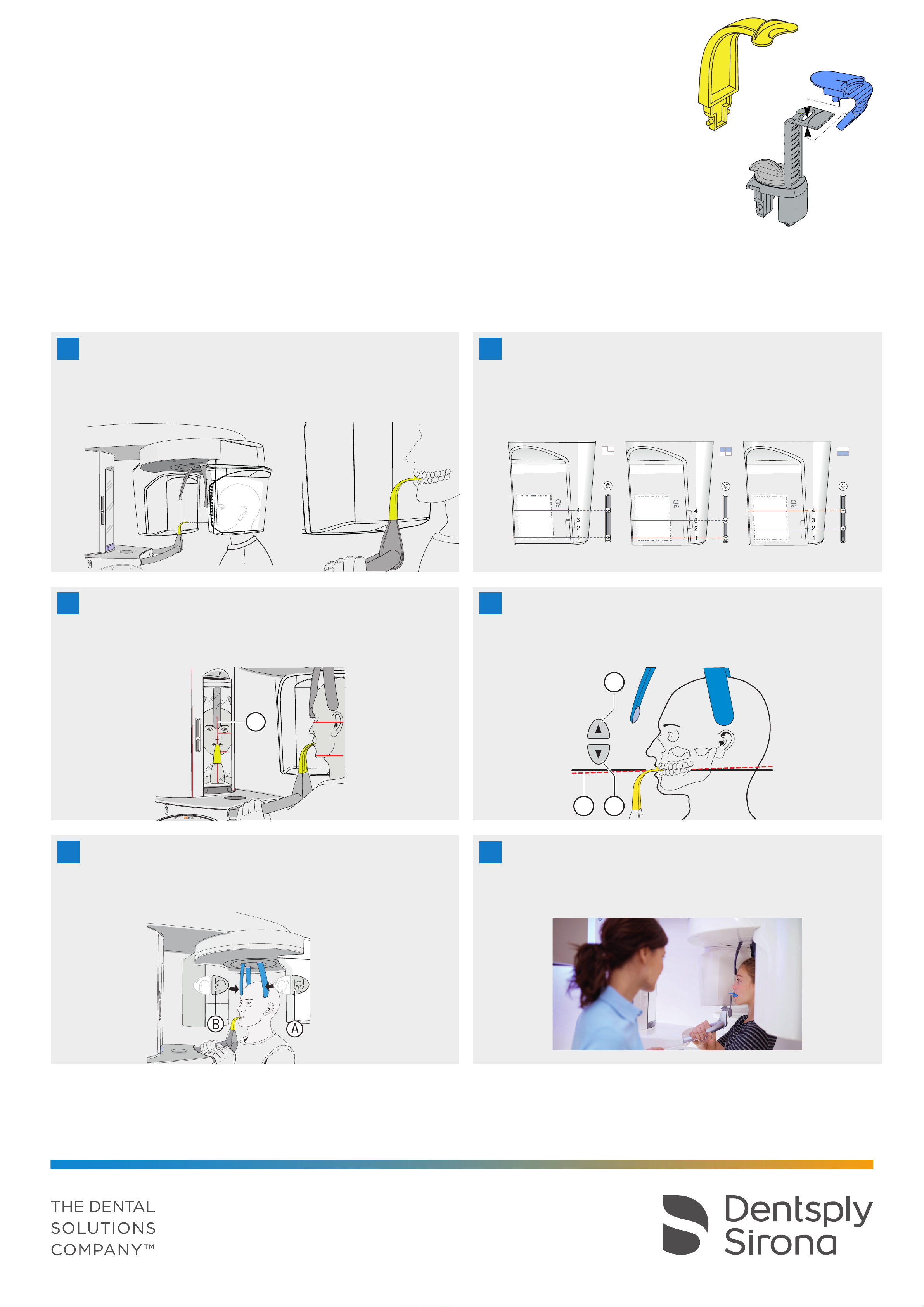
3D X-ray with Orthophos XG 3D
Patient positioning
(1)
for the perfect 3D image
Notes:
• Please use the 3D bite block (1) or the universal bite block (2)
• All volumes can be imaged with the 3D bite block (whole volumes with approx. 8 cm height and upper/lower jaw volumes with collimation to approx.
5.5 cm height). Use the blue mark of the universal bite block for volume images in the sinus area.
Preparation: Remove glasses, piercings, jewelry, and any removable dental prostheses, then put on an X-ray apron.
Hygienic protection: The appropriate hygienic protection is to be used prior to every image. This must be disposed of immediately after use.
(2)
Front teeth of the upper and lower jaw
1
Position the front teeth of the upper and lower jaw in the marked
notch of the bite block. The front teeth are then positioned exactly
in the sharp slice. Ask the patient to hold the hand grips firmly with
both hands.
2
Checking the volume
Align the light localizer with the marks on the 3D sensor.
Vol 1: Ø 8 cm, Vol 2: Ø 5 cm
• Full volume: 1
• Lower jaw: 1st and 3rd mark, collimated for Vol 1 and Vol 2
• Upper jaw: 2nd and 4th mark , collimated for Vol 1 and Vol 2
Full volume
Ø 8 cm x h 8 cm
st
and 4th mark, only for Vol 1
Lower jaw
Ø 8 cm x h 5,5 cm
Upper jaw
Ø 8 cm x h 5,5 cm
3
Checking the midsagittal plane
The light localizer is used to ensure symmetry. Align the patient
(nasal root) with central light line (A) (midsagittal line). Then swivel
the control mirror outwards.
A
4
Checking the occlusal plane
Align the patient‘s head as much as possible along the occlusal
plane (C). Correct the patient‘s head inclination as necessary.
Press the height adjustment buttons, up (A) and down (B).
A
C
B
5
Head fixation
Check the midsagittal plane again. First close the temple supports
(A) and then move the forehead support (B) until it just contacts
the patient. Swivel the mirror back.
6
Before taking X-ray
While taking the X-Ray you will hear an accustic signal. Instruct the
patient to hold this position until the end of the exposure in order to
avoid artefacts.
Please read the instructions for use for a more detailed description.
Caution: Strong disinfectants can corrode the surface of the device. Therefore do not use substances containing phenol, peracetic acid, peroxide or other
oxygen-splitting agents, sodium hypochlorite or iodine-splitting agents. Use only cleaning and disinfecting agents approved by Dentsply Sirona! You can
find more information at dentsplysirona.com/careagents
Subject to technical changes and errors in the text, Order No. A91100-M47-C115-01-7600, Printed in Germany, Dispo-No. 04602, OEW 1117.V0
 Loading...