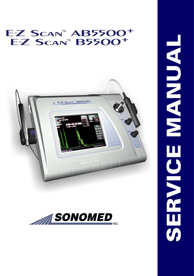
Sonomed E-Z Scan 5500 Plus User manual

1979 MARCUS AVENUE, LAKE SUCCESS, NY 11042 USA Tel 800-227-1285 or 516-354-0900
Fax 516-354-5902 www.sonomedinc.com

Table of Contents
1.Preface…………………………………………………………………………………….. 3
1.1EZ-Scan B5500+……………………………………………………………………….3
1.2EZ-Scan A/B5500+…………………………………………………………………… 3
2.Theory of Operation……………………………………………………………………… 4
2.1A-Scan Function…………………………………………………………………….… 4
2.2B-Scan Function…..………………………………………………………………….. 4
3.System Components……………………………………………………………………….5
4.System Specifications……………………………………………………………………...6
4.1Regulatory Requirements………………………………………………………………6
4.2Safety Regulations…………………………………………………………………….. 6
4.3Environmental Conditions…………………………………………………………….. 6
4.4Physical………………………………………………………………………………...6
4.5Electrical………………………………………………………………………………. 6
4.6Interface……………………………………………………………………………….. 6
4.7Probes…………………………………………………………………………………..6
4.8Display………………………………………………………………………………… 6
4.9Printer…………………………………………………………………………………..7
5.A-Scan System Specifications……………………………………………………………. 8
5.1Calibration…………………………………………………………………………….. 8
5.2Capture Modes…………………………………………………………………………8
5.3Examination Modes…………………………………………………………………… 8
5.4Typical Ultrasound Intensities in Tissues……………………………………………...8
5.5A-Scan Waveform Processing………………………………………………………… 8
5.6Statistical Analysis……………………………………………………………………. 8
5.7IOL Analysis………………………………………………………………………….. 8
5.8IOL Calculations……………………………………………………………………….8
5.9Measurement Accuracy……………………………………………………………….. 8
5.10A-Scan Amplifier……………………………..………………………………………8
5.11User Profiles………………………………………………………………………… 8
5.12General Specifications/Features………………………………………………………8
6.B-Scan System Specifications……………………………………………………………. 10
6.1Examination Modes.………………………………………………………………….. 10
6.2Display Scale………………………………………………………………………….. 10
6.3Probe……………………………………………………………………………………10
6.4Measurements……………..…………………………………………………………... 10
6.5Amplifier……………………………………………………………………………….10
6.6Magnification…………………………………………………………………………..10
6.7Display Resolution……………………………………………………………………..10
6.8Processing……………………………………………………………………………... 10
6.9Freeze…………………………………………………………………………………..10
6.10Image………………………………………………………………………………….10
6.11Gray Scale…………………………………………………………………………….10
6.12Display……………………………………………………………………………….. 10
6.13Typical Ultrasound Intensities in Tissues…………………………………………….10
6.14General Specifications/Features………………………………………………………10
7.Trouble Shooting B5500+…………………………………………………………………11
7.1Factor Defaults Reset………………………...……………………………………….. 11
7.2Touch Screen Calibration...…………………………………………………………… 13
7.3RGB Fill………………………………………………………………………………..14
7.4Video Output…………………………………………………………………………...14
7.5Joystick Alignment……………………………………………………………………. 15
7.6Rotary Encoder………………………………………………………………………... 15
7.7System Information…………………………………………………………………….16
7.8Mixed Pattern Test……………………………………………………………………..16
5550-A-1903-2A |
page 1 |

Table of Contents, con’t.
7.9Cal Pattern Distance Test………………………………………………………………19
7.10Cal Pattern Area Test…………………………………………………………………20
7.11Losing date, time and user information…..………………………………………….. 22
7.12Unit does not turn on………………………………………………………………….23
7.13Rear Connector Discriptions………………………………………………………….23
7.14LCD Image is Upside Down………………………………………………………….23
8.Trouble Shooting A/B5500+……………………..………………………………………. 24
8.1A-Scan Calibration………….……………………………………………………….…24
8.2Measure………………………………………………………………………………...25
8.3Losing date, time and user information…………………………………………….…. 25
8.4A-Scan PCB……………………………………………………………………………26
8.5A-Scan Probe Cable replacement……………………………………………………... 27
9.Routine Maintenance……………………………………………………………………...29
9.1System General Inspection………………………………………………………….… 29
9.2Enclosure Cleaning…………………………………………………………………... 29
9.3Probes Cleaning……………………………………………………………………… 29
9.4Storage……………………………………………………………………………….. 30
9.5Probe general Inspection……………………………………………………………... 30
10.Unit Assembly…………………………………………………………………………….. 31
10.1Top cover Bill of Materials…………………………………………………………...31
10.2Top Cover Assembly Drawing………………………………………………………. 32
10.3Bottom Cover Bill of Materials……………………………………………………… 33
10.4Bottom Cover Assembly Drawing……………………………………………………34
10.5Accessories Bill of Materials…………………………………………………………35
10.6Accessories Drawings………………………………………………………………...36
11.Schematics………………………………………………………………………………… 37
11.1Clock Generator………………………………………………………………………37
11.2Front End Interface.…………………………………………………………………. 38
11.3Power Supply………..………………………………………………………………. 39
11.4Pulser Receiver………………………………………………………………………. 40
11.5Servo Controller………………………………………………………………………41
11.6System Processor…………………………………………………………………….. 42
11.7Video Interface………………………………………………………………………..43
11.8Xilinx Configuration………………………………………………………………….44
11.9Xilinx Power/Decouple……………………………………………………………….45
11.10Xilinx RAM Interface……………………………………………………………….46
11.11Xilinx Test Points…………………………………………………………………... 47
5550-A-1903-2A |
page 2 |

Preface
This manual is intended to provide assistance in maintaining and repairing the E-Z Scan 5500+ series of instruments manufactured by Sonomed, Inc. For additional technical assistance, please contact Sonomed customer service at 1-800-227-1285.
General Description
The E-Z Scan 5500+ series is the latest general ophthalmic biometry instrument introduced by the industry leading Sonomed. The series consists of two different models:
1.1 E-Z Scan B5500+
This B-Scan system allows for measuring a two-dimensional image of an eye along one plane.
1.2 E-Z Scan A/B5500+
This A-Scan system allows for measuring the axial length (AXL), anterior chamber depth (ACD) and lens thickness of an eye and for calculating the associated IOL power for an implanted lens.
Both systems utilize a high-resolution backlit touch screen liquid crystal display (LCD) by which the user can enter information and view data and calculations. Adjustable legs can be tilted to several angles for user comfort. After completion of measurements and calculations, a hardcopy may be obtained of the results using the video printer.
5550-A-1903-2A |
page 3 |

Theory of Operation
2.1 A-Scan Function
One of the major applications of A-Scan is to measure the axial length of the eye in order to calculate the necessary power of an intraocular lens replacement (such as required in the treatment of cataracts).
In A-Scan, a beam of ultrasound is transmitted along a fixed line through the eye and the reflected echoes are displayed. An echo is produced whenever the ultrasound beam encounters a boundary between two media having different values of acoustic impedance. The acoustic impedance is a function of density and elasticity and the amount of energy reflected depends upon the difference in the acoustic impedance of the two media. A-Scan is equipped with software that calculates the distance between the echoes.
The probe acts as the interface between the instrument and the patient. The probe contains a piezoelectric crystal, which converts electrical energy into ultrasound when transmitting and then acts as a detector to convert the received ultrasound echoes into electrical signals for display and measurement. The A-Scan probe contains a red LED fixation light in its center. The fixation light provides a target, which assists the operator to align the visual axis of the eye being examined. To minimize indentation of the eye, which is caused by the operator applying excessive pressure while holding the probe on the eye, A-scan probes feature "Soft-Touch". This feature minimizes the compression effect by allowing the probe handle to float on bushings located inside the probe handle.
Doc# 0300-1903-3A
2.2 B-Scan Function
The operation of the B-Scan is similar to a radar operation. A short ultrasound wave is produced by a transducer and sent to the eye. Part of the energy of that wave is reflected back to the transducer from various structures of the eye (lens, retina, etc.). The reflected energy is converted into electrical signals that are amplified and displayed on the LCD as an intensity. Since the reflected energy is proportional to the reflected properties of the different eye structures, one can examine the eye by observing the resulting grey or color scale image.
The ultrasound transducer is located in the B-Scan probe and is driven by two (2) solenoids to produce a 60° sector scan. The sector scan is divided into 256 rays. The data acquired along each ray is digitized at 512 discrete time intervals (pixels) and each sample voltage is encoded into 7 bits of information.
The acquired information is stored in one (1) of the two (2) B-Scan memory buffers. At any time during the acquisition, the content on one memory buffer is displayed while the other is updated with new data from the amplifier.
The display control maps data in the memory buffer onto the LCD screen, thus showing sector scans. The microprocessor periodically checks touch screen controls and calculates mapping coefficients, to pan the image.
There is a simultaneous vector display that shows the signal along a selected ray, represented as an amplitude waveform. This supplemental information enables the doctor to differentiate between the two (2) signals that have the closest intensities of grey.
page 4

System Components
LCD Backlit Display with Touch Screen Overlay
The touch screen is a highly sensitive device that enables selections to be made and recorded on the color liquid crystal display. On screen selections should only be made by gently using a finger or the provided Stylus pen (do not use pencil, pen or other sharp objects). The LCD display and touch screen mount on the front cover.
Bail Stand
The bail stand consists of two adjustable legs and two neoprene pads. When shipped, the legs will lie flat. The legs can be tilted to one of three angles for user comfort.
Note: Do not remove the neoprene pads.
Video Out
Connector located on the rear of the instrument. The signal is black and white. Connects with the video printer provided.
Power On - Off Switch
Two position slide switch located on the rear of the instrument. The LED on the front panel will illuminate green when the switch is in the Power On position. In the Off position the LED will not illuminate.
DC Input Port
Interfaces to the Sonomed AC Adapter, 15 VDC @ 2.0 A, Part No. 9200-1409-1A
RS 232 Port
Connector located on the rear of the unit. Connects with PC (software required).
A Scan Probe Port (A/B5500+)
5-pin Lemo connector located on the side of the instrument. Interfaces to the A-Scan probe.
Doc# 0300-1903-3A
B-Scan Probe Port (B5500+)
10-pin Lemo connector located on the side of the instrument. Interfaces to the B-Scan probe.
Foot Switch Port
1-pin jack connector located on the rear of the instrument.
AC Adapter
Desktop regulated AC/DC power supply.
Video Printer
SONY Black &White Video Graphic Printer
A-Scan Probes
Styles: Standard and Soft-Touch
B-Scan Probe
Coupling Gel
The coupling gel is provided with the B5500+ and A/B5500+ units.
Stylus Touch Pen
Instruction Manual
An instruction manual is supplied with each unit.
page 5

System Specifications
4.1 Regulatory Requirements
The E-Z Scan 5500+ series of instruments shall meet and comply with the following regulatory requirements:
*European Medical Device Directive (MDD 93/42/EEC)
*CE mark including EMC "residential class"
*United States FDA regulations for ultrasonic medical devices.
4.2 Safety Regulations
The E-Z Scan 5500+ series shall comply with the safety requirements of IEC 60601-1 and the European norm EN 60601 which include:
*IEC 60601-1-1, Safety requirements for medical electrical systems
*IEC 60601-1-2 Electromagnetic compatibility - requirements and tests
*EN 60601-1-2:1993, EMC for EU Medical Devices Directive
*IEC 60601-1-4, Programmable electrical medical systems
4.3 Environmental Conditions
Temperature, Operating: 5 to 40° C (41 to 104° F)
Temperature, Storage: -40 to 70° C (-40 to 158° F)
Humidity, Operating: 10 to 90%, non-condensing
Humidity, Storage: 10 to 90%, non-condensing
4.4 Physical
Dimensions:
Width 31.7 cm (12.5") Depth 25.4 cm (10.0") Height 8.2 cm (3.25")
Doc# 0300-1903-3A
Weight: |
2.4 kg (5.25 lbs) |
Enclosure: |
Plastic, off-white color, |
internal EMC shielded
4.5 Electrical
The E-Z Scan 5500+ series shall be powered from the AC mains using an external "brick" power supply.
Power Consumption: 10.0 W (typical)
Line Voltage: 100/120/220 Volts AC ±10%
Line Frequency: 50/60 Hz
DC Output: 15 VDC
2.0 A
4.6 Interface |
|
DC Input: |
1-pin jack connector |
Foot Pedal: |
1-pin jack connector |
Probe Input: |
|
A-Probe: |
5-pin with insertion key |
B-Probe: |
10-pin with insertion key |
Printer Output: BNC connector
4.7 Probes
A-Scan Probes: Direct contact Styles: Standard Solid Tip
Soft-Touch
Immersion
Frequency: 10 MHz ±10%
Focal Length: Standard: 25 mm ± 3 mm Soft-Touch: 25 mm ± 3mm
Fixation Light: Internal red LED Pulse repetition frequency: 19.2 Hz
B-Scan Probes: Mechanical sector scan Frequency: 10 MHz ±10%
Focal Length: 24 mm ± 2mm Probe Tip Diameter: 1.75 mm Pulse repetition frequency: 3800 Hz ISO 9000 Certified
page 6

System Specifications, con’t
4.8 Display
Type: TFT Active Matrix Color LCD with touch screen overlay (262,144 colors) Resolution: 640 pixels (H) x 480 pixels (V) Dimensions: 6.5” (17cm) Diagonal
High Luminance (250:1)
4.9 Printer
Model: Sony UP-890MD Type: Video printer Paper Size: Roll paper
Width: 110 mm (4.3") Diameter: 50 mm (2.0")
Video Output: RS-170 BNC for B/W Video Printer, VCR and remote viewing
Doc# 0300-1903-3A |
page 7 |

A-Scan System Specifications
5.1 Calibration
Calibration Mode: User invoked mode, will confirm proper functioning using a test target
Calibration Distance: 10.0 mm ± 0.1 mm Gain Control: Manual or automatic gain selection.
5.2 Capture Modes
Automatic Capture: Normal Cataract Dense Cataract Aphakic Pseudophakic
A-scan will automatically freeze when an acceptable waveform has been acquired. Manual Capture: User activated using touch screen or foot pedal.
5.3 Examination Modes
Contact: Direct contact of probe tip onto cornea.
Immersion: Water bath standoff of probe tip.
5.4 Typical Ultrasonic Intensities in
Tissue
ISPTA: 0.0068 mW/cm2
ISPPA: 3.11 W/cm2
MI: 0.085
Ultrasonic Power: .00134 mW
5.5A-Scan Waveform Processing
Waveform Display: Real-time update Display Scale:
Minor markers at 1.0 mm intervals Major markers at 5.0 mm intervals
Display Range: 49 mm
Storage: Up to five waveforms with associated statistics
5.6Statistical Analysis
Average, Standard Deviation, Range and Maximum Difference from average
Doc# 0300-1903-3A
5.7 IOL Analysis
Standard Programs: Binkhorst
Regression-II
Theoretic-T
Holladay
Hoffer-Q
Haigis (optional)
5.8 IOL Calculations
2 Tables of 9 Operator AOL Displayed in 1/2 Diopter steps Selectable Refractions Customizable IOL Constants
Measurement Limits (in Automatic mode @ 1550 m/s)
AXL: 18 - 40 mm Lens: 2 - 6 mm ACD: 2 - 6 mm Vitreous
5.9 Measurement Accuracy
Electrical: ±0.023 mm Clinical: ±0.1 mm Measurement Range: 18-40 mm
Calibration: Automatic with built-in calibration cylinder
5.10A-Scan Amplifier
Nominal Gain: 40-80 dB Control: Variable gain set by user Low Noise
5.11User Profiles
Permanent storage of settings for up to five users.
User Profiles: Preferred IOL formula Personal Surgeon Factor
5.12 General Specifications / Features
Printing mode: Standard Video Out Review: Stored A-scan patterns, A-scan measurements and statistics
Displays: Multiple screens available for tabled, summarized and compared calculations.
page 8

A-Scan System Specifications Con’t
Memory: Stores 5 scans and measurements, selected formula, IOL constants and user name.
Report Data: Patients Name, ID#, Eye examined, K-readings, User Name, Date & Time.
Live A-Scan Display
Axial length, anterior chamber depth, and lens thickness measurements for each scan.
Axial length average and standard deviation for up to 5 scans.
Data Entry: Full alpha-numeric via touchscreen.
Adjustable legs for angled viewing for 0-60º
Doc# 0300-1903-3A |
page 9 |

B-Scan System Specifications
6.1 Examination Modes: B-only
B/a
A/b
6.2 Display Scale: Electronic Markers @
2.0mm Intervals
Measurement Accuracy
Electronic: ±0.0484 mm
Clinical: ±0.1mm
6.3Probe: 10MHz, Focused Transducer, 30 frames/sec
6.4Measurements: Distance and area
6.5Amplifier: 100 dB Gain, Logarithmic / Linear / S-Curve, Gain and TVG controls
6.6Magnification: Continuous Zoom (0.5x – 2.0x) with Pan (joystick controlled)
6.7Display Resolution: 640 x 480 Pixels, color VGA with optimal tissue resolution of
0.15mm
6.8Processing: Reject below level, enhance contour and texture.
6.9Freeze: Foot pedal or touch screen activated
6.10Image: B-Scan with simultaneous selectable vector A-Scan
6.11Gray Scale: 256 levels
6.12Display: 60º sector fan, 128 lines, Grey Scale, B/a presentation (B emphasized) or A/b (A emphasized), Gain/TVG, Electronic Scale, Amplifier, OD/OS, Velocity, Probe Orientation, Patient and User Names, Date/Time.
Doc# 0300-1903-3A
6.13 Typical Ultrasonic Intensities in
tissue: |
|
|
B-scan: |
(autoscanning) |
|
ISPTA: |
0.881 mW/cm2 |
|
ISPPA: |
11.45 W/cm2 |
|
MI: |
0.151 |
|
Ultrasonic Power |
0.133 mW |
|
6.14 General Specifications / Features
Maintains high resolution at all magnifications.
Pan feature using built-in “joystick” control Gain and TVG controls for optimal diagnostic capabilities.
Selectable Color or Grey Scale image. Software enhancement capability of frozen image.
Selectable, simultaneous A-scan vector. Sealed B-scan probe provides smooth scanning with virtually no audible sound Five user selections.
page 10

Trouble Shooting B5500+
7.1 Factory Defaults Reset
It is recommended that the unit be turned OFF before starting this procedure to ensure that the RAM is clear and the microprocessor is reset.
Turn the unit on. The unit will display the screen as shown in figure #1.
Touch the B-SCAN field, the unit will display the screen as shown in figure #2.
Fig. 1
Touch the USER 1 field, the unit will display the screen as shown in figure #3.
Type in the letters SET and then the ENTER field, the unit will display the Calibration & Test screen as shown in figure #4.
Touch the FACTORY RESET field, the unit will display the screen as shown in figure #5.
Touch the RESET field, the unit will display the screen as shown in figure #6.
Fig. 2
Touch the DONE field, the unit will display the Calibration & Test screen (figure #4).
WARNING! Once the system is reset, all entered data and scans are erased.
Fig. 3
Doc# 0300-1903-3A |
page 11 |

Trouble Shooting B5500+ con’t.
Fig. 4
Fig. 5
Fig. 6
Doc# 0300-1903-3A |
page 12 |

Trouble Shooting B5500+ con’t.
Fig. 7
1 |
4 |
3
2 |
5 |
|
Fig. 8
Fig. 9
7.2 Touch Screen Calibration
To verify that the Touch Screen is calibrated, repeat the first several steps in the Factory Defaults Restore section until the unit displays the Calibration & Test screen (figure #4).
Touch the TOUCH TEST field , the unit will display the screen as shown in figure #7.
Touch the CALIBRATE field, the unit will display the screen as shown in figure #8.
Press all the red crosshairs in the sequence illustrated in figure #8 until they all beep. The screen will automatically go to the screen as shown in figure #9.
Press in several random points on the screen to test calibration. The response points displayed on the screen will turn green. The response points should be within 3mm of the pressed points. If the green response points are not displayed beneath the pressed points, contact the Sonomed service department.
Touch the DONE field, to return to the Calibration & Test screen (figure #4).
Doc# 0300-1903-3A |
page 13 |

Trouble Shooting B5500+ con’t.
Fig. 10
7.3 RGB Fill
To Verify that the three basic colors on the LCD are working, repeat the first several steps in the Factory Defaults Restore section until the unit displays the Calibration & Test screen (figure #4).
Touch the RGB FILL field.
The LCD display will scroll the colors Red, Green and Blue continuously. All the pixels on the screen should be the same color ( not spots of mixed colors or black pixels) If the screens are not completely Red, Green or Blue, contact the Sonomed service department.
Press the touch screen in any spot for approximate 2 seconds, the unit will return to the Calibration and Test Screen (figure #4).
7.4 Video Output
To verify that the Video output is correct, repeat the first several steps in the Factory Defaults Restore section until the unit displays the Calibration & Test screen (figure #4).
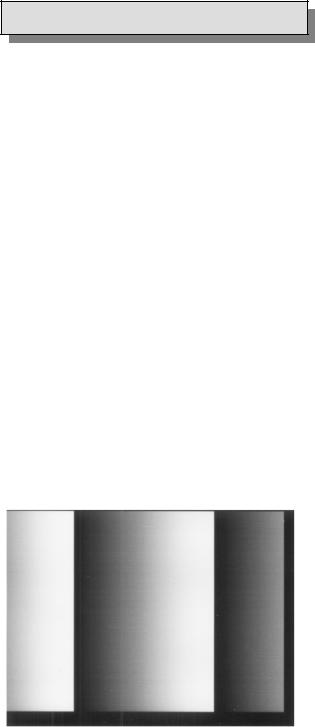
Touch the GREY SCALE field, the unit will display the screen as shown in figure #10.
Using a scope, measure and verify peak to peak voltage is 1.0VppVDC+/- 10mVpp. This measurement should be taken at the video output on the rear of the instrument.
Doc# 0300-1903-3A |
page 14 |
 Loading...